

Arbiters of endogenous opioid analgesia: role of CNS estrogenic and glutamatergic systems
- PMID: 33567346
- PMCID: PMC8217383
- DOI: 10.1016/j.trsl.2021.02.002
Arbiters of endogenous opioid analgesia: role of CNS estrogenic and glutamatergic systems
Abstract
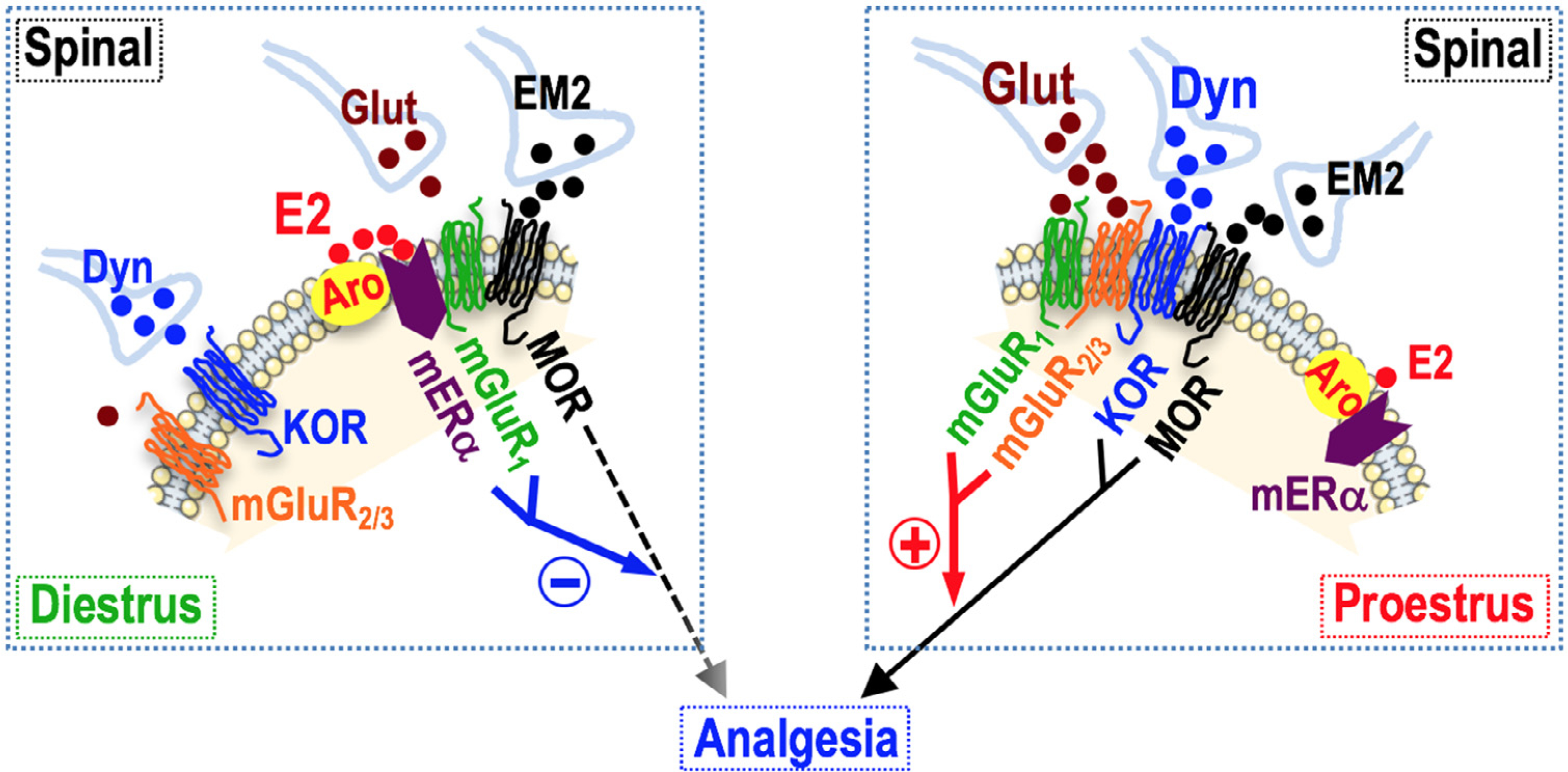
Nociception and opioid antinociception in females are pliable processes, varying qualitatively and quantitatively over the reproductive cycle. Spinal estrogenic signaling via membrane estrogen receptors (mERs), in combination with multiple other signaling molecules [spinal dynorphin, kappa-opioid receptors (KOR), glutamate and metabotropic glutamate receptor 1 (mGluR1)], appears to function as a master coordinator, parsing functionality between pronociception and antinociception. This provides a window into pharmacologically accessing intrinsic opioid analgesic/anti-allodynic systems. In diestrus, membrane estrogen receptor alpha (mERα) signals via mGluR1 to suppress spinal endomorphin 2 (EM2) analgesia. Strikingly, in the absence of exogenous opioids, interfering with this suppression in a chronic pain model elicits opioid anti-allodynia, revealing contributions of endogenous opioid(s). In proestrus, robust spinal EM2 analgesia is manifest but this requires spinal dynorphin/KOR and glutamate-activated mGluR1. Furthermore, spinal mGluR1 blockade in a proestrus chronic pain animal (eliminating spinal EM2 analgesia) exacerbates mechanical allodynia, revealing tempering by endogenous opioid(s). A complex containing mu-opioid receptor, KOR, aromatase, mGluRs, and mERα are foundational to eliciting endogenous opioid anti-allodynia. Aromatase-mERα oligomers are also plentiful, in a central nervous system region-specific fashion. These can be independently regulated and allow estrogens to act intracellularly within the same signaling complex in which they are synthesized, explaining asynchronous relationships between circulating estrogens and central nervous system estrogen functionalities. Observations with EM2 highlight the translational relevance of extensively characterizing exogenous responsiveness to endogenous opioids and the neuronal circuits that mediate them along with the multiplicity of estrogenic systems that concomitantly function in phase and out-of-phase with the reproductive cycle.
Copyright © 2021 Elsevier Inc. All rights reserved.
Conflict of interest statement
Conflicts of interest: Authors declare that there are no conflicts of interest or competing financial/nonfinancial interests to disclose. We confirm that this work is original and has not been published as peer reviewed material elsewhere, nor is it currently under consideration for publication elsewhere.
All authors have read the journal’s policy on disclosure of potential conflicts of interest, and declare no potential conflicts of interest for this work.
Figures
Similar articles
-
Plasticity of Signaling by Spinal Estrogen Receptor α, κ-Opioid Receptor, and Metabotropic Glutamate Receptors over the Rat Reproductive Cycle Regulates Spinal Endomorphin 2 Antinociception: Relevance of Endogenous-Biased Agonism.J Neurosci. 2017 Nov 15;37(46):11181-11191. doi: 10.1523/JNEUROSCI.1927-17.2017. Epub 2017 Oct 12. J Neurosci. 2017. PMID: 29025923 Free PMC article.
-
Estrogens synthesized and acting within a spinal oligomer suppress spinal endomorphin 2 antinociception: ebb and flow over the rat reproductive cycle.Pain. 2017 Oct;158(10):1903-1914. doi: 10.1097/j.pain.0000000000000991. Pain. 2017. PMID: 28902684 Free PMC article.
-
Pharmacological Modulation of Endogenous Opioid Activity to Attenuate Neuropathic Pain in Rats.J Pain. 2019 Feb;20(2):235-243. doi: 10.1016/j.jpain.2018.10.003. Epub 2018 Oct 23. J Pain. 2019. PMID: 30366152 Free PMC article.
-
Opioids in chronic pain.Eur J Pharmacol. 2001 Oct 19;429(1-3):79-91. doi: 10.1016/s0014-2999(01)01308-5. Eur J Pharmacol. 2001. PMID: 11698029 Review.
-
Spinal opioid systems in inflammation.Inflamm Res. 1995 Jun;44(6):231-41. doi: 10.1007/BF01782974. Inflamm Res. 1995. PMID: 7583517 Review.
Cited by
-
Our Life and Times….Alan R. Gintzler, Ph.D. (1947-2021) Analgesics, Endogenous Opioids and the Variable of Sex.Front Pain Res (Lausanne). 2021 Sep 7;2:753792. doi: 10.3389/fpain.2021.753792. eCollection 2021. Front Pain Res (Lausanne). 2021. PMID: 35295441 Free PMC article. No abstract available.
-
Estrogen metabolites increase nociceptor hyperactivity in a mouse model of uterine pain.JCI Insight. 2022 May 23;7(10):e149107. doi: 10.1172/jci.insight.149107. JCI Insight. 2022. PMID: 35420999 Free PMC article.
-
Emerging Evidence on Membrane Estrogen Receptors as Novel Therapeutic Targets for Central Nervous System Pathologies.Int J Mol Sci. 2023 Feb 17;24(4):4043. doi: 10.3390/ijms24044043. Int J Mol Sci. 2023. PMID: 36835454 Free PMC article. Review.
-
Recent Advances in the Modulation of Pain by the Metabotropic Glutamate Receptors.Cells. 2022 Aug 21;11(16):2608. doi: 10.3390/cells11162608. Cells. 2022. PMID: 36010684 Free PMC article. Review.
References
-
- Fillingim RB, Gear RW. Sex differences in opioid analgesia: clinical and experimental findings. Eur J Pain 2004;8:413–25. - PubMed
-
- Craft RM. Sex differences in opioid analgesia: “from mouse to man”. Clin J Pain 2003;19:175–86. - PubMed
-
- Teepker M, Peters M, Vedder H, et al. Menstrual variation in experimental pain: correlation with gonadal hormones. Neuropsychobiology 2010;61:131–40. - PubMed
-
- Ibironke GF, Aji KE. Pain threshold variations in female rats as a function of the estrus cycle. Niger J Physiol Sci 2011;26:67–70. - PubMed
Publication types
MeSH terms
Substances
Grants and funding
LinkOut - more resources
Full Text Sources
Other Literature Sources
Research Materials
