

Autofluorescence Imaging of the Skin Is an Objective Non-Invasive Technique for Diagnosing Pseudoxanthoma Elasticum
- PMID: 33567497
- PMCID: PMC7915757
- DOI: 10.3390/diagnostics11020260
Autofluorescence Imaging of the Skin Is an Objective Non-Invasive Technique for Diagnosing Pseudoxanthoma Elasticum
Abstract
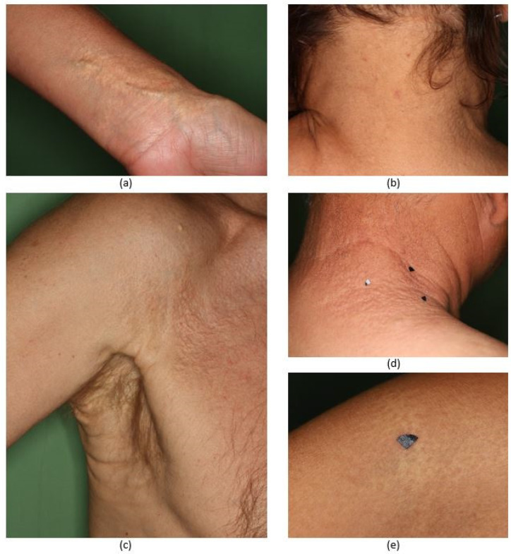
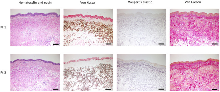
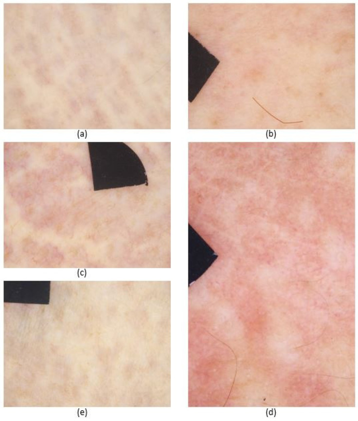
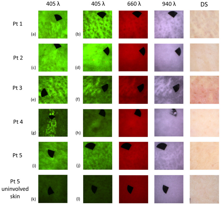
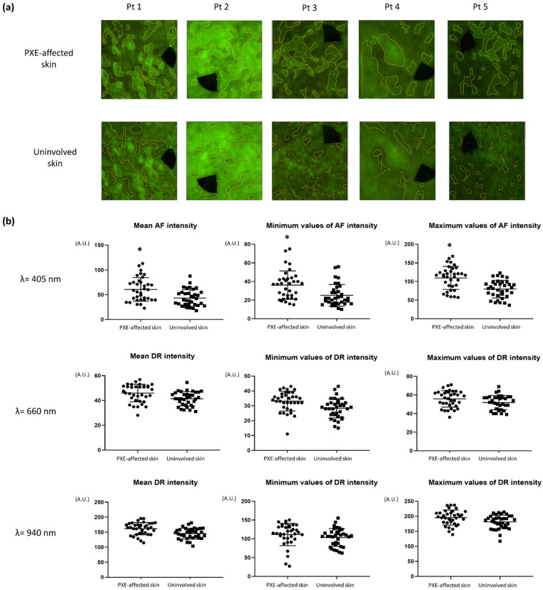
Pseudoxanthoma elasticum (PXE) is a rare multisystemic autosomal recessive connective tissue disease. In most cases, skin manifestations of PXE are the first to develop, followed later by severe ocular and cardiovascular complications. In our present study, in addition to dermoscopy, we introduced novel techniques, autofluorescence (AF) and diffuse reflectance (DR) imaging for the assessment of affected skin sites of five PXE patients. PXE-affected skin areas in most skin sites showed a previously observed pattern upon dermoscopic examination. With the novel imaging, PXE-affected skin lesions displayed high AF intensity. During our measurements, significantly higher mean, minimum and maximum AF intensity values were found in areas of PXE-affected skin when compared to uninvolved skin. Conversely, images acquired with the use of 660 and 940 nm illumination showed no mentionable difference. Our results demonstrate that AF imaging may be used in the in vivo diagnostics and quantification of the severity of the skin lesions of PXE patients. In addition, it is a safe, fast and cost-effective diagnostic method. AF imaging may be also used to objectively monitor the efficacy of the possible novel therapeutic approaches of PXE in the future.
Keywords: LED; autofluorescence imaging; calcification; dermoscopy; diagnosis; diffuse reflectance imaging; pseudoxanthoma elasticum; quantitative analysis.
Conflict of interest statement
The authors declare no conflict of interest. The funders had no role in the design of the study; in the collection, analyses, or interpretation of data; in the writing of the manuscript, or in the decision to publish the results.
Figures
Similar articles
-
Dermoscopic features of pseudoxanthoma elasticum.Clin Exp Dermatol. 2018 Mar;43(2):175-179. doi: 10.1111/ced.13308. Epub 2017 Dec 22. Clin Exp Dermatol. 2018. PMID: 29271496
-
Nonlinear optical microscopy is a novel tool for the analysis of cutaneous alterations in pseudoxanthoma elasticum.Lasers Med Sci. 2020 Oct;35(8):1821-1830. doi: 10.1007/s10103-020-03027-w. Epub 2020 May 6. Lasers Med Sci. 2020. PMID: 32372237 Free PMC article.
-
[From gene to disease; pseudoxanthoma elasticum and the ABCC6 gene].Ned Tijdschr Geneeskd. 2004 Aug 7;148(32):1586-9. Ned Tijdschr Geneeskd. 2004. PMID: 15382558 Review. Dutch.
-
The spectrum of ocular alterations in patients with β-thalassemia syndromes suggests a pathology similar to pseudoxanthoma elasticum.Ophthalmology. 2014 Mar;121(3):709-18. doi: 10.1016/j.ophtha.2013.10.016. Epub 2013 Dec 4. Ophthalmology. 2014. PMID: 24314836
-
Pseudoxanthoma elasticum and skin: Clinical manifestations, histopathology, pathomechanism, perspectives of treatment.Intractable Rare Dis Res. 2015 Aug;4(3):113-22. doi: 10.5582/irdr.2015.01014. Intractable Rare Dis Res. 2015. PMID: 26361562 Free PMC article. Review.
Cited by
-
Multispectral Imaging Algorithm Predicts Breslow Thickness of Melanoma.J Clin Med. 2021 Dec 30;11(1):189. doi: 10.3390/jcm11010189. J Clin Med. 2021. PMID: 35011930 Free PMC article.
-
A Cross-Sectional Study of the Dermatological Manifestations of Patients with Fabry Disease and the Assessment of Angiokeratomas with Multimodal Imaging.Diagnostics (Basel). 2023 Jul 14;13(14):2368. doi: 10.3390/diagnostics13142368. Diagnostics (Basel). 2023. PMID: 37510112 Free PMC article.
-
Serum Calcification Propensity T50 Associates with Disease Severity in Patients with Pseudoxanthoma Elasticum.J Clin Med. 2022 Jun 28;11(13):3727. doi: 10.3390/jcm11133727. J Clin Med. 2022. PMID: 35807012 Free PMC article.
-
[Dermoscopy of genodermatoses].Dermatologie (Heidelb). 2023 Apr;74(4):256-261. doi: 10.1007/s00105-023-05124-7. Epub 2023 Mar 7. Dermatologie (Heidelb). 2023. PMID: 36882583 Free PMC article. Review. German.
-
Quantitative Multispectral Imaging Differentiates Melanoma from Seborrheic Keratosis.Diagnostics (Basel). 2021 Jul 22;11(8):1315. doi: 10.3390/diagnostics11081315. Diagnostics (Basel). 2021. PMID: 34441250 Free PMC article.
References
-
- Jansen R.S., Duijst S., Mahakena S., Sommer D., Szeri F., Váradi A., Plomp A., Bergen A.A., Oude Elferink R.P., Borst P., et al. ABCC6-mediated ATP secretion by the liver is the main source of the mineralization inhibitor inorganic pyrophosphate in the systemic circulation-brief report. Arterioscler. Thromb. Vasc. Biol. 2014;34:1985–1989. doi: 10.1161/ATVBAHA.114.304017. - DOI - PMC - PubMed
Grants and funding
- FK_131916, 2019/National Research, Development and Innovation Office
- K_132695, 2019/National Research, Development and Innovation Office
- 16-2017-00009/EFOP- 3.6.3-VEKOP
- "Time-resolved autofluorescence methodology for non-invasive skin cancer diagnostics" No. 1.1.1.2/16/I/001, agreement No. 1.1.1.2/VIAA/1/16/014/European Regional Development Fund projects
- "Development and clinical validation of a novel cost effective multi-modal methodology for early diagnostics of skin cancers" No. 1.1.1.2/16/I/001 agreement No. 1.1.1.2/VIAA/1/16/052/European Regional Development Fund projects
- ÚNKP-20-4-II-SE-7/New National Excellence Program of the Ministry For Innovation and Technology from the source of the National Research, Development and Innovation Fund of Hungary
- ÚNKP-20-3-I-SE-24/New National Excellence Program of the Ministry For Innovation and Technology from the source of the National Research, Development and Innovation Fund of Hungary
- CA16115/EuroSoftCalcNet COST action
LinkOut - more resources
Full Text Sources
Other Literature Sources
