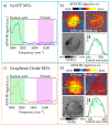
Infrared Nanospectroscopy of Individual Extracellular Microvesicles
- PMID: 33567597
- PMCID: PMC7915346
- DOI: 10.3390/molecules26040887
Infrared Nanospectroscopy of Individual Extracellular Microvesicles
Abstract
Extracellular vesicles are membrane-delimited structures, involved in several inter-cellular communication processes, both physiological and pathological, since they deliver complex biological cargo. Extracellular vesicles have been identified as possible biomarkers of several pathological diseases; thus, their characterization is fundamental in order to gain a deep understanding of their function and of the related processes. Traditional approaches for the characterization of the molecular content of the vesicles require a large quantity of sample, thereby providing an average molecular profile, while their heterogeneity is typically probed by non-optical microscopies that, however, lack the chemical sensitivity to provide information of the molecular cargo. Here, we perform a study of individual microvesicles, a subclass of extracellular vesicles generated by the outward budding of the plasma membrane, released by two cultures of glial cells under different stimuli, by applying a state-of-the-art infrared nanospectroscopy technique based on the coupling of an atomic force microscope and a pulsed laser, which combines the label-free chemical sensitivity of infrared spectroscopy with the nanometric resolution of atomic force microscopy. By correlating topographic, mechanical and spectroscopic information of individual microvesicles, we identified two main populations in both families of vesicles released by the two cell cultures. Subtle differences in terms of nucleic acid content among the two families of vesicles have been found by performing a fitting procedure of the main nucleic acid vibrational peaks in the 1000-1250 cm-1 frequency range.
Keywords: atomic force microscopy; extracellular vesicles; infrared nanoscale spectroscopy.
Conflict of interest statement
The authors declare no conflict of interest.
Figures


References
-
- Lötvall J., Hill A.F., Hochberg F., Buzás E.I., Di Vizio D., Gardiner C., Gho Y.S., Kurochkin I.V., Mathivanan S., Quesenberry P., et al. Minimal Experimental Requirements for Definition of Extracellular Vesicles and their Functions: A Position Statement from the International Society for Extracellular Vesicles. J. Extracell. Vesicles. 2014;3:26913. doi: 10.3402/jev.v3.26913. - DOI - PMC - PubMed
-
- Théry C., Witwer K.W., Aikawa E., Alcaraz M.J., Anderson J.D., Andriantsitohaina R., Antoniou A., Arab T., Archer F., Atkin-Smith G.K., et al. Minimal information for studies of extracellular vesicles 2018 (MISEV2018): A position statement of the International Society for Extracellular Vesicles and update of the MISEV2014 guidelines. J. Extracell. Vesicles. 2018;7:1535750. doi: 10.1080/20013078.2018.1535750. - DOI - PMC - PubMed
-
- Van Deun J., EV-TRACK Consortium. Mestdagh P., Agostinis P., Akay Ö., Anand S., Anckaert J., Martinez Z.A., Baetens T., Beghein E., et al. EV-TRACK: Transparent reporting and centralizing knowledge in extracellular vesicle research. Nat. Methods. 2017;14:228–232. doi: 10.1038/nmeth.4185. - DOI - PubMed
MeSH terms
LinkOut - more resources
Full Text Sources
Other Literature Sources

