. 2021 Jul 1;106(7):1910-1922.
doi: 10.3324/haematol.2020.264085.

Crucial role of hematopoietic JAK2 V617F in the development of aortic aneurysms

Affiliations

Crucial role of hematopoietic JAK2 V617F in the development of aortic aneurysms

Tetsuro Yokokawa et al. Haematologica. .

Abstract

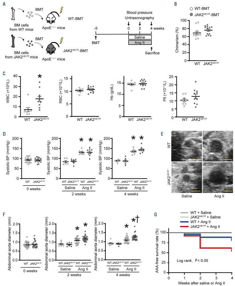
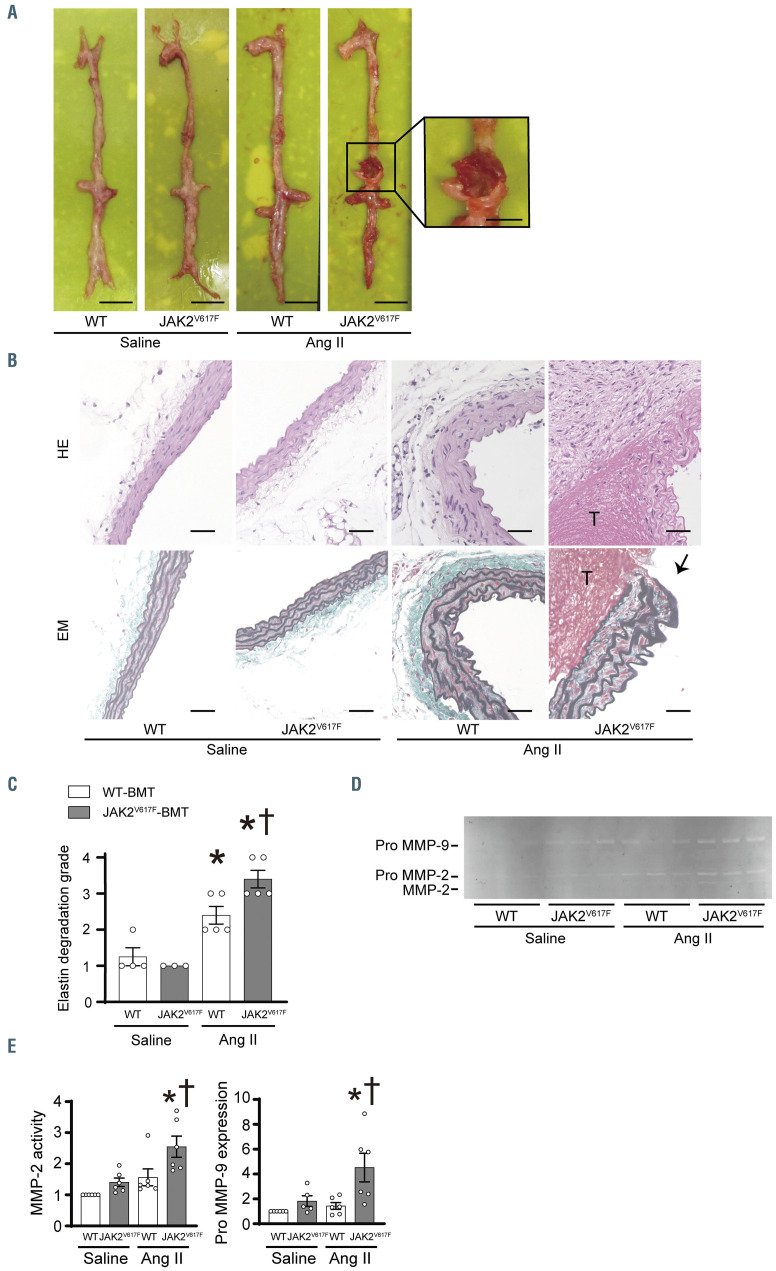
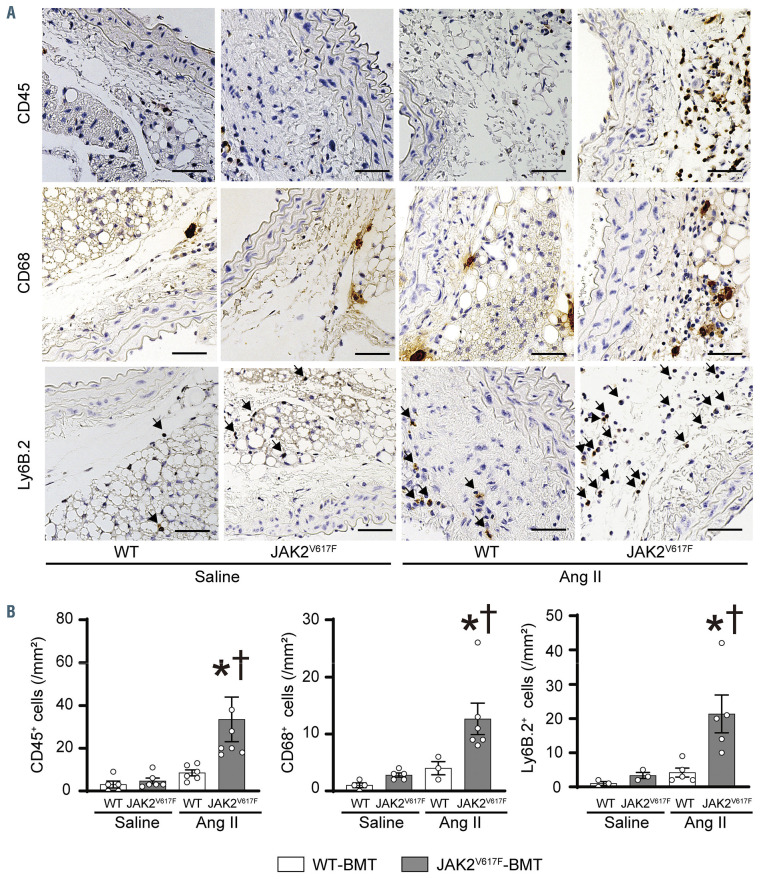
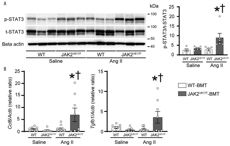
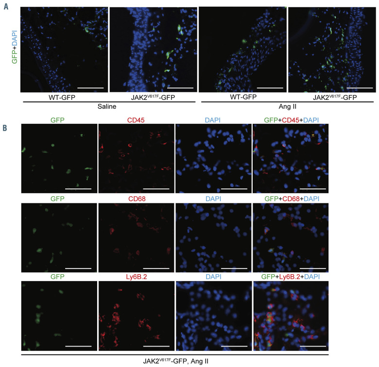
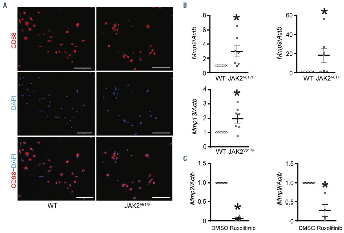
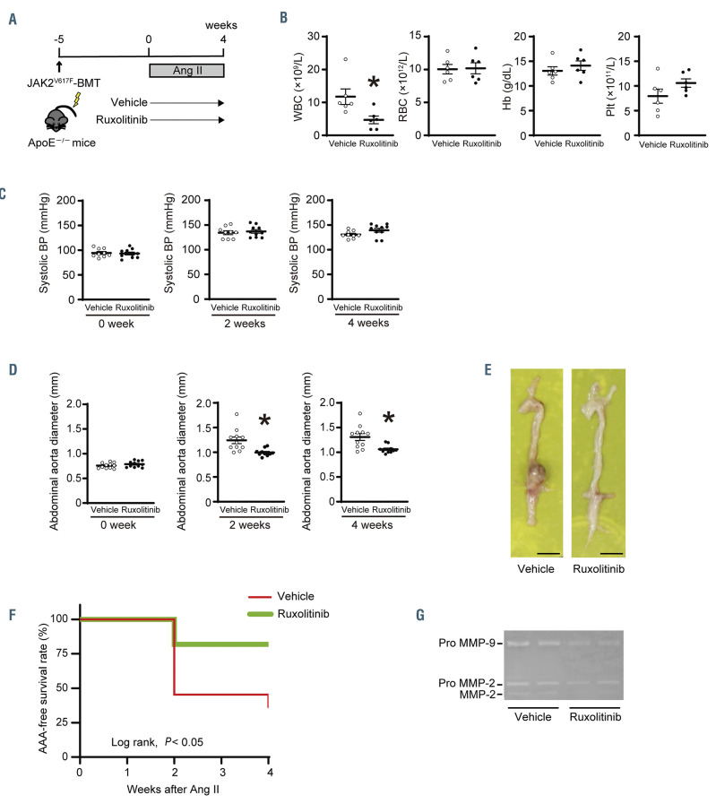
JAK2V617F is the most frequent driver mutation in myeloproliferative neoplasms (MPNs) and is associated with vascular complications. However, the impact of hematopoietic JAK2V617F on the aortic aneurysms (AAs) remains unknown. Our cross-sectional study indicated that 9 (23%) out of 39 MPN patients with JAK2V617F exhibited the presence of AAs. Next, to clarify whether the hematopoietic JAK2V617F contributes to the AAs, we applied a bone marrow transplantation (BMT) with the donor cells from Jak2V617F transgenic (JAK2V617F) mice or control wild-type (WT) mice into lethally irradiated apolipoprotein E-deficient mice. Five weeks after BMT, the JAK2V617F-BMT mice and WT-BMT mice were subjected to continuous angiotensin II infusion to induce AA formation. Four weeks after angiotensin II infusion, the abdominal aorta diameter in JAK2V617F-BMT mice was significantly enlarged compared to that in the WT-BMT mice. Additionally, the abdominal AA-free survival rate was significantly lower in the JAK2V617F-BMT mice. Hematopoietic JAK2V617F accelerated aortic elastic lamina degradation as well as activation of matrix metalloproteinase (MMP)-2 and MMP-9 in the abdominal aorta. The numbers of infiltrated macrophages were significantly upregulated in the abdominal aorta of the JAK2V617F-BMT mice accompanied by STAT3 phosphorylation. The accumulation of BM-derived hematopoietic cells carrying JAK2V617F in the abdominal aorta was confirmed by use of reporter GFP-transgene. BM-derived macrophages carrying JAK2V617F showed increases in mRNA expression levels of Mmp2, Mmp9, and Mmp13. Ruxolitinib decreased the abdominal aorta diameter and the incidence of abdominal AA in the JAK2V617F-BMT mice. Our findings provide a novel feature of vascular complications of AAs in MPNs with JAK2V617F.


Figures

Figure 1.
Figure 1.
Hematopoietic JAK2 V617F contributes to abdominal aortic aneurysm formation. (A) Schematic diagram of the experimental design of bone marrow (BM) transplantation (BMT). BM cells from control wild-type (WT) mice or JAK2V617F mice were injected into the lethally irradiated ApoE−/− recipient mice. Five weeks after BMT, the ApoE−/− recipient mice transplanted with WT BM cells (WT-BMT mice) or JAK2V617F BM cells (JAK2V617F-BMT mice) were subjected to saline or angiotensin II (Ang II, 1900 ng/kg per min) infusion for 4 weeks. (B) Chimerism of donor cells in the peripheral leukocytes (n=11–13) and (C) blood cell counts (n=9–11) in the WT-BMT mice and JAK2V617F-BMT mice at 5 weeks after BMT. *P<0.05 versus the WT-BMT mice by the unpaired Student’s t-test. (D) Systolic blood pressure (BP) at the baseline (n=17 each), 2 weeks (n =7–10) and 4 weeks (n=7–8) after saline or Ang II infusion. (E) Representative ultrasound images of abdominal aorta under saline or Ang II infusion for 4 weeks. Scale bars, 1.0 mm. (F) Abdominal aorta diameter at the baseline (n= 33–34), 2 weeks (n=7–24) and 4 weeks (n=7–24) after saline or Ang II infusion. (G) Abdominal aortic aneurysm (AAA)-free survival rate in the WT-BMT mice and JAK2V617F-BMT mice after saline or Ang II infusion (n=7–26) by log-rank test. All data are presented as mean ± standard error of the mean. *P<0.05 vs. the corresponding saline-infused mice and P<0.05 vs. the corresponding WT-BMT mice by one-way ANOVA with Tukey post-hoc analysis. WT, ApoE−/−: mice transplanted with bone marrow cells from wild-type mice; JAK2V617F, ApoE−/−: mice transplanted with bone marrow cells from JAK2V617F mice; Ang II: angiotensin II; WBC: white blood cell count; RBC: red blood cell count; Hb: hemoglobin concentration; Plt: platelet count.
Figure 2.
Figure 2.
Hematopoietic JAK2 V617F induces elastin degradation and rupture of aortic aneurysm accompanied by matrix metalloproteinase activation. (A) Representative images of the aorta from the WT-BMT mice and JAK2V617F-BMT mice 4 weeks after saline or Ang II or infusion. Scale bars, 5 mm. The boxed area from the Ang II-infused JAK2V617F-BMT mice was highlighted at higher magnification. Scale bar, 2.5 mm. (B) Representative images of hematoxylin-eosin (HE)- and Elastica-Masson (EM)-stained sections. ‘T’ indicates thrombus in the aorta. Black arrow indicates the rupture site of the aorta. Scale bars, 50 m. (C) Quantitative analysis of degradation grade in EM-stained sections (n=3–5). (D) A typical image of gelatin zymography using homogenates from abdominal aortas of the WT-BMT mice and JAK2V617F-BMT mice 4 weeks after saline or Ang II infusion. (E) Densitometric analysis of the gelatin zymography (n=5–6). The sum of matrix metalloproteinase (MMP)-2 and pro MMP-2 bands was evaluated as MMP-2 activity. All data are presented as mean ± standard error of the mean. *P<0.05 vs. the corresponding saline-infused mice and P<0.05 vs. the Ang II-infused WT-BMT mice by one-way ANOVA with Tukey post-hoc analysis. WT, ApoE−/−: mice transplanted with bone marrow (BM) cells from wild-type (WT) mice; JAK2V617F, ApoE−/−: mice transplanted with BM cells from JAK2V617F mice; BMT: bonemarrow transplantation; Ang II: angiotensin II.
Figure 3.
Figure 3.
Hematopoietic JAK2 V617F increases inflammatory cell infiltration in the abdominal aorta. (A) Representative immunohistochemical images of the aorta stained by anti-CD45, CD68, and Ly6B.2 antibodies in the WT-BMT mice and JAK2V617F-BMT mice 4 weeks after saline or angiotensin II infusion. Arrows indicate Ly6B.2-positive cells. Scale bars, 50 m. (B) Quantitative analyses of the CD45+ (n=5–6), CD68+ (n=3–5), and Ly6B.2+ (n= 3–5) cells in the aortic sections. All data are presented as mean ± standard error of the mean. *P<0.05 vs. the corresponding saline-infused mice and P <0.05 vs. the Ang II-infused WT-BMT mice by oneway ANOVA with Tukey post-hoc analysis. WT, ApoE−/−: mice transplanted with bone marrow (BM) cells from wild-type (WT) mice; JAK2V617F, ApoE−/−: mice transplanted with BM cells from JAK2V617F mice; BMT: BM transplantation; Ang II: angiotensin II.
Figure 4.
Figure 4.
Hematopoietic JAK2 V617F increases STAT3 phosphorylation and cytokine expression in response to angiotensin II infusion in the abdominal aorta. (A) Western blot analysis on the STAT3 in the abdominal aorta. Aorta extracts from the WT-BMT mice or JAK2V617F-BMT mice were immunoblotted with the indicated antibodies. Phosphorylated STAT3 (p-STAT3) to total STAT3 (t-STAT3) ratios are shown in the bar graphs. The average value for the saline-infused WT-BMT mice was set to 1 (n=5–6). actin was used as the loading control. (B) Relative mRNA expression levels of Ccl6 and Tgfb1 in the aorta. Actb was used for normalization. The average value for saline-infused WT-BMT mice was set to 1 (n=7–10). All data are presented as mean ± standard error of the mean. *P<0.05 vs. the corresponding saline-infused mice and P<0.05 vs. the Ang II-infused WT-BMT mice by one-way ANOVA with Tukey post-hoc analysis. WT, ApoE−/−: mice transplanted with bone marrow (BM) cells from wild-type (WT) mice; JAK2V617F, ApoE−/−: mice transplanted with BM cells from JAK2V617F mice; BMT: BM transplantation; Ang II: angiotensin II.
Figure 5.
Figure 5.
Characterization of bone marrow-derived JAK2 V617F hematopoietic cells in the abdominal aorta by use of GFP-transgene. (A) The lethally irradiated ApoE−/− mice were transplanted with bone marrow (BM) cells from the WT/CAG-EGFP (WT-GFP) mice or JAK2V617F/CAG-EGFP (JAK2V617F-GFP) double transgenic mice. The ApoE−/− recipient mice were subjected to saline or angiotensin II (Ang II) infusion for 4 weeks. The abdominal aortas were stained with an anti-GFP (green) antibody and DAPI (blue). Scale bars, 100 m. (B) Representative immunofluorescence images of the aorta sections stained with anti-CD45 (red), anti-CD68 (red), or anti-Ly6B.2 (red) and anti-GFP (green) antibodies and DAPI (blue) in ApoE−/− mice transplanted with JAK2V617F-GFP BM cells. Scale bars, 50 m. Ang II: angiotensin II; GFP: green fluorescent protein; DAPI: 4′,6-diamidino-2-phenylindole; WT: wild-type.
Figure 6.
Figure 6.
Matrix metalloproteinases are upregulated in bone marrow-derived macrophages with JAK2 V617F expression. (A) Mononuclear cells isolated from bonemarrow (BM) cells of wild-type (WT) mice or JAK2V617F mice were cultured in the presence of granulocyte-macrophage colony stimulating factor for 6 days. Representative immunofluorescence images of the cells stained with anti-CD68 (red) antibody and DAPI (blue). Scale bars, 100 m. (B) Relative mRNA expression levels of Mmp2, Mmp9, and Mmp13 in the cultured mononuclear cells (n=7 each). (C) Mmp2 and Mmp9 mRNA expressions with and without ruxolitinib pretreatment at 250 nM for 24 hours in the JAK2V617F mononuclear cells (n=4 each). Dimethylsulfoxide (DMSO) was used for control. Actb was used for normalization. The average value for the cultured mononuclear cells from WT mice or DMSO group was set to 1. All data are presented as mean ± standard error of the mean. *P<0.05 vs. the WT group or DMSO group by the unpaired Student’s t-test. JAK2V617F: JAK2 V617F-expressing transgenic mice.
Figure 7.
Figure 7.
Ruxolitinib attenuates the formation of abdominal aortic aneurysms induced by hematopoietic JAK2 V617F. (A) Schematic diagram of the experimental design. The ApoE-/- recipients transplanted with JAK2V617F bone marrow cells (JAK2V617F-BMT) were administered with vehicle (0.5% methylcellulose) or ruxolitinib was orally at 60 mg/kg twice daily for 4 weeks along with continuous angiotensin II (Ang II, 1900 ng/kg per min) infusion. (B) Blood cell counts at 4 weeks (n=6 each) and (C) systolic blood pressure (BP) at baseline (n=9–10), 2 weeks (n=9–10), and 4 weeks (n=9–10) in the vehicle- or ruxolitinib-treated JAK2V617F-BMT mice. (D) Abdominal aorta diameter at the baseline (n=11 each), 2 weeks (n=11 each), and 4 weeks (n=11 each). (E) Representative images from the aorta. Scale bars, 5 mm. (F) Abdominal aortic aneurysm (AAA)-free survival rate in the vehicle- or ruxolitinib-treated JAK2V617F-BMT mice (n=11 each) by log-rank test. (G) A typical image of gelatin zymography using homogenates from abdominal aortas. All data are presented as mean ± standard error of the mean. *P<0.05 vs. the vehicle group by the unpaired Student’s t-test. WBC: white blood cell count; RBC: red blood cell count; Hb: hemoglobin concentration; Plt: platelet count; BP: blood pressure; MMP: matrix metalloproteinase.

Comment in

Similar articles

Cited by

References

    1. Kutti J, Ridell B. Epidemiology of the myeloproliferative disorders: essential thrombocythaemia, polycythaemia vera and idiopathic myelofibrosis. Pathol Biol (Paris). 2001;49(2):164-166. - PubMed
    1. Srour SA, Devesa SS, Morton LM, et al. . Incidence and patient survival of myeloproliferative neoplasms and myelodysplastic/ myeloproliferative neoplasms in the United States, 2001-12. Br J Haematol. 2016;174(3):382-396. - PMC - PubMed
    1. Barbui T, Finazzi G and Falanga A. Myeloproliferative neoplasms and thrombosis. Blood. 2013;122(13):2176-2184. - PubMed
    1. De Stefano V, Ghirardi A, Masciulli A, et al. . Arterial thrombosis in Philadelphia-negative myeloproliferative neoplasms predicts second cancer: a case-control study. Blood. 2020;135(5):381-386. - PubMed
    1. Hultcrantz M, Björkholm M, Dickman PW, et al. . Risk for arterial and venous thrombosis in patients with myeloproliferative neoplasms: a population-based cohort study. Ann Intern Med. 2018;168(5):317-325. - PMC - PubMed

Publication types