

Color-coded summation images in the evaluation of renal artery stenosis before and after percutaneous transluminal angioplasty
- PMID: 33568089
- PMCID: PMC7874657
- DOI: 10.1186/s12880-020-00540-w
Color-coded summation images in the evaluation of renal artery stenosis before and after percutaneous transluminal angioplasty
Abstract
Background: Endovascular therapy is the gold standard in patients with hemodynamic relevant renal artery stenosis (RAS) resistant to medical therapy. The severity grading of the stenosis as well as the result assessment after endovascular approach is predominantly based on visible estimations of the anatomic appearance. We aim to investigate the application of color-coded DSA parameters to gain hemodynamic information during endovascular renal artery interventions and for the assessment of the procedures´ technical success.
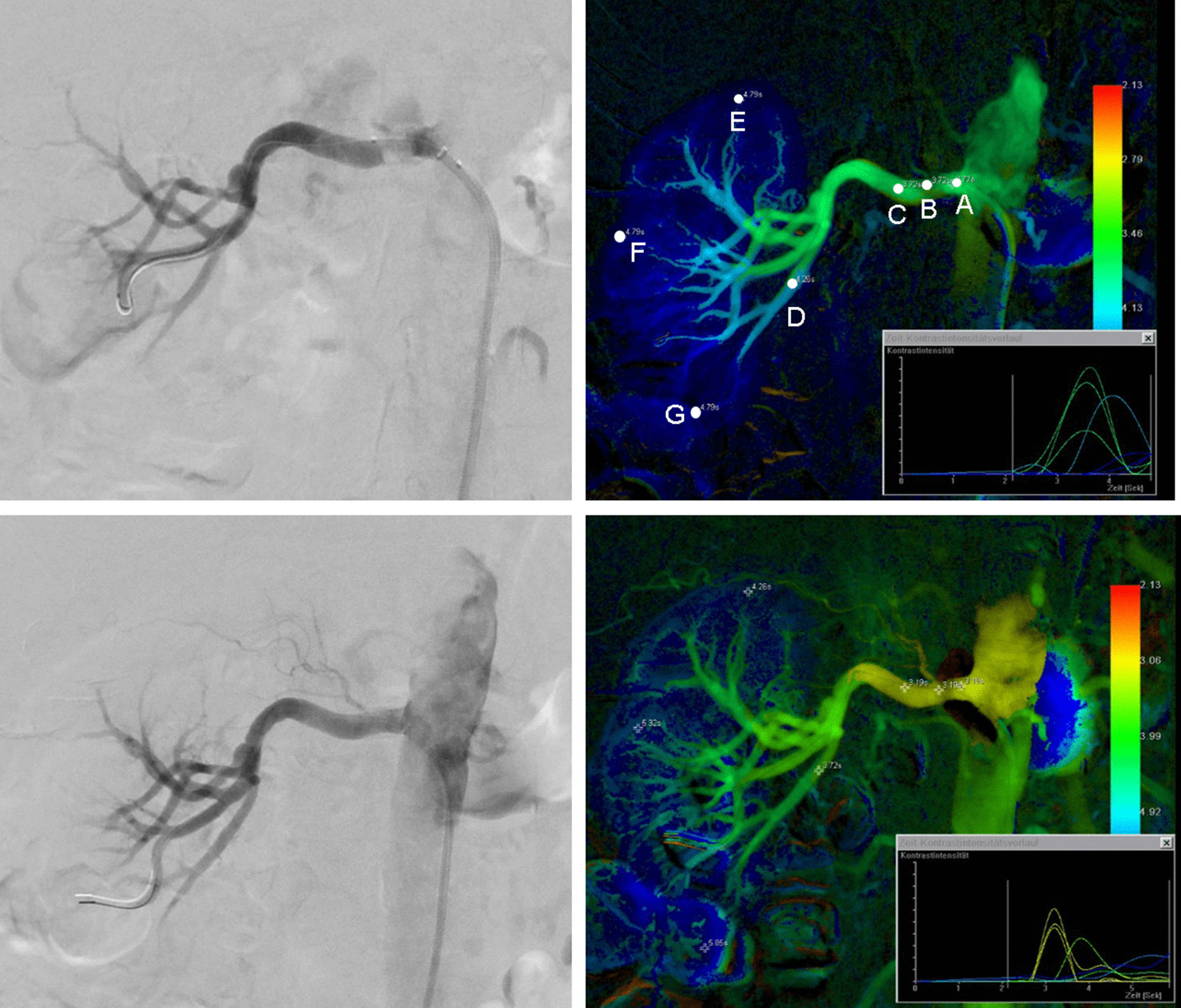
Methods: We retrospectively evaluated 32 patients who underwent endovascular renal artery revascularization and applied color-coded summation imaging on selected monochromatic DSA images. The differences in time to peak (dTTP) of contrast enhancement in predefined anatomical measuring points were analyzed. Furthermore, differences in systolic blood pressure values (SBP) and serum creatinine were obtained. The value of underlying diabetes mellitus as a predictor for clinical outcome was assessed. Correlation analysis between the patients´ gender as well as the presence of diabetes mellitus and dTTP was performed.
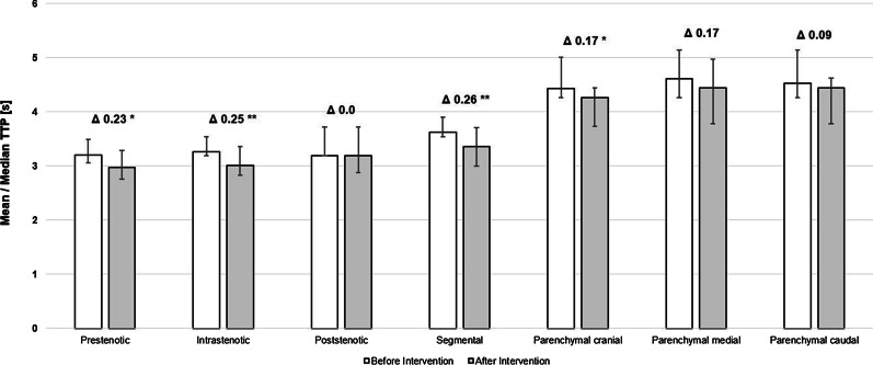
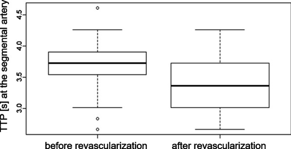
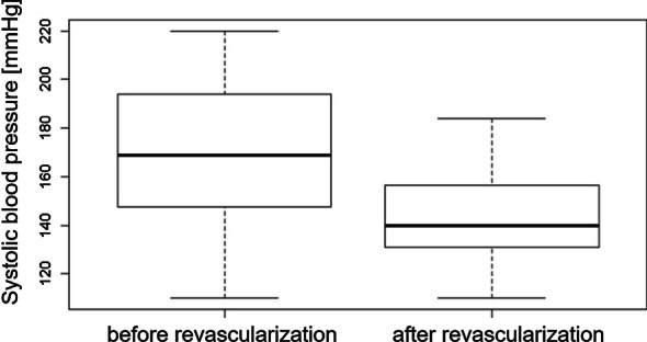
Results: Endovascular revascularization resulted in statistically significant improvement in 4/7 regions of interest. Highly significant improvement of perfusion in terms of shortened TTP values could be found at the segmental artery level and in the intrastenotical segment (p < 0.001), significant improvement prestenotical and in the apical renal parenchyma (p < 0.05). In the other anatomic regions, differences revealed not to be significant. Differences between SBP and serum creatinine levels before and after the procedure were significant (p = 0.004 and 0.0004). Patients´ gender as well as the presence of diabetes mellitus did not reveal to be predictors for the clinical success of the procedure. Furthermore, diabetes and gender did not show relevant correlation with dTTP in the parenchymal measuring points.
Conclusions: The supplementary use of color-coding DSA and the data gained from parametric images may provide helpful information in the evaluation of the procedures´ technical success. The segmental artery might be a particularly suitable vascular territory for analyzing differences in blood flow characteristics. Further studies with larger cohorts are needed to further confirm the diagnostic value of this technique.
Keywords: Color-coded; Digital subtraction angiography; Endovascular; PTA; Renal artery.
Conflict of interest statement
The authors declare that they have no competing interests.
Figures

References
-
- Turgutalp K, Kiykim A, Ozhan O, Helvaci I, Ozcan T, Yildiz A. Comparison of diagnostic accuracy of Doppler USG and contrast-enhanced magnetic resonance angiography and selective renal arteriography in patients with atherosclerotic renal artery stenosis. Med Sci Monit. 2013;19:475–482. doi: 10.12659/MSM.889035. - DOI - PMC - PubMed
Publication types
MeSH terms
LinkOut - more resources
Full Text Sources
Other Literature Sources
Medical
