Review
. 2021 Feb 10;85(1):e00021-19.
doi: 10.1128/MMBR.00021-19. Print 2021 Feb 17.

Uncovering the Hidden Credentials of Brucella Virulence

Affiliations
Review

Uncovering the Hidden Credentials of Brucella Virulence

R Martin Roop 2nd et al. Microbiol Mol Biol Rev. .

Abstract

Bacteria in the genus Brucella are important human and veterinary pathogens. The abortion and infertility they cause in food animals produce economic hardships in areas where the disease has not been controlled, and human brucellosis is one of the world's most common zoonoses. Brucella strains have also been isolated from wildlife, but we know much less about the pathobiology and epidemiology of these infections than we do about brucellosis in domestic animals. The brucellae maintain predominantly an intracellular lifestyle in their mammalian hosts, and their ability to subvert the host immune response and survive and replicate in macrophages and placental trophoblasts underlies their success as pathogens. We are just beginning to understand how these bacteria evolved from a progenitor alphaproteobacterium with an environmental niche and diverged to become highly host-adapted and host-specific pathogens. Two important virulence determinants played critical roles in this evolution: (i) a type IV secretion system that secretes effector molecules into the host cell cytoplasm that direct the intracellular trafficking of the brucellae and modulate host immune responses and (ii) a lipopolysaccharide moiety which poorly stimulates host inflammatory responses. This review highlights what we presently know about how these and other virulence determinants contribute to Brucella pathogenesis. Gaining a better understanding of how the brucellae produce disease will provide us with information that can be used to design better strategies for preventing brucellosis in animals and for preventing and treating this disease in humans.

Keywords: Brucella; pathogenesis; virulence determinants.


Figures

FIG 1
FIG 1
Natural hosts and zoonotic potential of Brucella strains. The Brucella species shown in blue are recognized zoonotic pathogens, and the thickness of the solid arrows represents the relative frequency with which these hosts serve as sources of human infection. The dashed arrows indicate that these Brucella strains have been isolated from human disease but direct transmission from the corresponding natural host to humans has not been documented. The question mark indicates that the natural host for B. inopinata is unknown.
FIG 2
FIG 2
Natural disease cycle of brucellosis in domestic animals. The blue triangles denote the oral, nasal, and venereal routes of infection.
FIG 3
FIG 3
Contributions of the T4SS effectors, the LPS O-chain, Omp22, Omp25d, and cyclic β-1,2-d-glucan (CβG) to the development of the replicative Brucella-containing vacuole in host macrophages. The empty black and orange circles represent membrane vesicles trafficking from the endolysosomal pathway, endoplasmic reticulum, and Golgi apparatus to the Brucella-containing vacuoles (BCVs). The change in the colors of the BCV membranes represents their change in composition as they transition from eBCVs to rBCVs. The outermost blue membrane of the aBCV represents engulfment of the rBCV by the host cell autophagosomal pathway. eBCV, endosomal BCV; rBCV, replicative BCV; aBCV, autophagosomal BCV.
FIG 4
FIG 4
Brucella virulence determinants that influence the capacity of macrophages to modulate the host immune response. , activation; ⊣, inhibition. The dashed arrow indicates that the Brucella LPS does not signal strongly through the TLR4 pathway and stimulates a diminished inflammatory response. The red X indicates that the Brucella flagellin is not recognized by TLR5.
FIG 5
FIG 5
Brucella virulence determinants that impact the ability of dendritic cells to modulate the host immune response. , activation; ⊣, inhibition. The dashed arrow indicates that the Brucella LPS does not signal strongly through the TLR4 pathway and stimulates a diminished inflammatory response.
FIG 6
FIG 6
Genetic regulators and the corresponding stimuli that control expression of the genes encoding the T4SS in Brucella. , activation; ⊣, repression. The question mark indicates that the environmental stimuli recognized by these regulators have not been determined. Both activation and repression of virB expression have been reported for BabR.
FIG 7
FIG 7
Virulence determinants that allow the brucellae to resist the reactive oxygen and nitrogen species they encounter during their intracellular residence in host phagocytes. Reactive oxygen and nitrogen species are shown in red. OM, outer membrane; CM, cytoplasmic membrane.
FIG 8
FIG 8
Fe, Mn, Zn, and Mg acquisition systems and defenses against metal toxicity that have been shown to play a role in the virulence of Brucella. The solid arrows represent direct regulatory links that have been documented experimentally, and the dashed arrows and lines denote suspected regulatory links. OM, outer membrane; CM, cytoplasmic membrane.
FIG 9
FIG 9
Overlapping regulation of genes encoding major virulence determinants in Brucella by BvrRS, VjbR, RpoE, CtrA, and LovhK. BvrRS and LovhK are shown twice to reflect the fact that these regulators have been shown to have both direct and indirect effects on genes encoding these virulence determinants.
FIG 10
FIG 10
Proposed role of C12-HSL signaling and VjbR in temporal regulation of virB expression during development of the Brucella-containing vacuoles in host cells. It is thought that accumulation of C12-HSL in the spatial confines of the BCVs prevents virB expression during the later stages of BCV development. The phagosome membrane is depicted in brown.
FIG 11
FIG 11
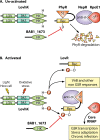
Regulation of the general stress response (GSR) and other virulence determinants in Brucella by LovhK. The yellow and red lightning bolts in panel B denote the stimuli thought to activate the histidine kinase activity of Lovhk through its LOV and PAS domains. LovR has been proposed to function as a phosphate sink and to modulate LovhK signaling. HK, histidine kinase domain; SL, sigma factor-like domain; Rec, response regulator receiver domain.
FIG 12
FIG 12
Unipolar cell division and coordination of cell division with the cell cycle in Brucella. The polar locations of the AT adhesins BtaE, BtaF, and BmaC are shown, and the red boxes indicate that Brucella cells in the G1 phase of the cell cycle are the most infectious for mammalian cells.
FIG 13
FIG 13
Regulation of CtrA and CpdR activity by PdhS and the CckA-ChpT phosphorelay in Brucella. Arrows indicate the direction of phosphate transfer. The red X denotes CtrA degradation by ClpPX. The multiple dashed arrows between PdhS and CckA indicate that the exact number and nature of the regulatory steps between these two regulators have not been determined.
FIG 14
FIG 14
Conserved roles that MucR/Ros-based transcriptional modules are proposed to play in controlling host-specific gene expression in Agrobacterium, Sinorhizobium, and Brucella.

References

    1. Moreno E. 2014. Retrospective and prospective perspectives on zoonotic brucellosis. Front Microbiol 5:213. doi:10.3389/fmicb.2014.00213. - DOI - PMC - PubMed
    1. Olsen SC, Palmer MV. 2014. Advancement of knowledge of Brucella over the past 50 years. Vet Pathol 51:1076–1089. doi:10.1177/0300985814540545. - DOI - PubMed
    1. Eisenberg T, Hamann KP, Kaim U, Schelz K, Seeger H, Seeger N, Schauerte N, Melzer F, Tomaso H, Scholz HC, Koylass MS, Whatmore AM, Zschöck M. 2012. Isolation of potentially novel Brucella spp. from frogs. Appl Environ Microbiol 78:3753–3755. doi:10.1128/AEM.07509-11. - DOI - PMC - PubMed
    1. Soler-Lloréns PF, Quance CR, Lawhon SD, Stuber TP, Edwards JF, Ficht TA, Robbe-Austerman S, O’Callaghan D, Keriel A. 2016. A Brucella spp. isolate from a Pac-Man frog (Ceratophrys ornata) reveals characteristics departing from classical brucellae. Front Cell Infect Microbiol 6:116. doi:10.3389/fcimb.2016.00116. - DOI - PMC - PubMed
    1. El-Tras WF, Tayel AA, Eltholth MM, Guitian J. 2010. Brucella infection in freshwater fish: evidence for natural infection of Nile catfish, Clarias gariepinus, with Brucella melitensis. Vet Microbiol 141:321–325. doi:10.1016/j.vetmic.2009.09.017. - DOI - PubMed

Publication types

MeSH terms

LinkOut - more resources