. 2021 Apr 12;95(9):e01953-20.
doi: 10.1128/JVI.01953-20. Print 2021 Apr 12.

HIV-Infected Macrophages Are Infected and Killed by the Interferon-Sensitive Rhabdovirus MG1

Affiliations

HIV-Infected Macrophages Are Infected and Killed by the Interferon-Sensitive Rhabdovirus MG1

Teslin S Sandstrom et al. J Virol. .

Abstract

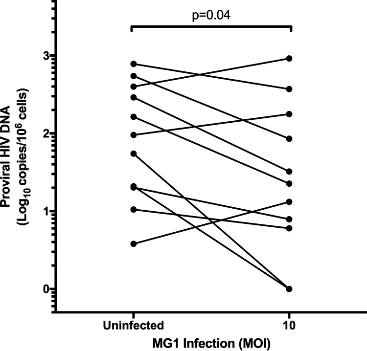
The use of unique cell surface markers to target and eradicate HIV-infected cells has been a longstanding objective of HIV-1 cure research. This approach, however, overlooks the possibility that intracellular changes present within HIV-infected cells may serve as valuable therapeutic targets. For example, the identification of dysregulated antiviral signaling in cancer has led to the characterization of oncolytic viruses capable of preferentially killing cancer cells. Since impairment of cellular antiviral machinery has been proposed as a mechanism by which HIV-1 evades immune clearance, we hypothesized that HIV-infected macrophages (an important viral reservoir in vivo) would be preferentially killed by the interferon-sensitive oncolytic Maraba virus MG1. We first showed that HIV-infected monocyte-derived macrophages (MDM) were more susceptible to MG1 infection and killing than HIV-uninfected cells. As MG1 is highly sensitive to type I interferons (IFN-I), we then investigated whether we could identify IFN-I signaling differences between HIV-infected and uninfected MDM and found evidence of impaired IFN-α responsiveness within HIV-infected cells. Finally, to assess whether MG1 could target a relevant, primary cell reservoir of HIV-1, we investigated its effects in alveolar macrophages (AM) obtained from effectively treated individuals living with HIV-1. As observed with in vitro-infected MDM, we found that HIV-infected AM were preferentially eliminated by MG1. In summary, the oncolytic rhabdovirus MG1 appears to preferentially target and kill HIV-infected cells via impairment of antiviral signaling pathways and may therefore provide a novel approach to an HIV-1 cure.IMPORTANCE Human immunodeficiency virus type 1 (HIV-1) remains a treatable, but incurable, viral infection. The establishment of viral reservoirs containing latently infected cells remains the main obstacle in the search for a cure. Cure research has also focused on only one cellular target of HIV-1 (the CD4+ T cell) while largely overlooking others (such as macrophages) that contribute to HIV-1 persistence. In this study, we address these challenges by describing a potential strategy for the eradication of HIV-infected macrophages. Specifically, we show that an engineered rhabdovirus-initially developed as a cancer therapy-is capable of preferential infection and killing of HIV-infected macrophages, possibly via the same altered antiviral signaling seen in cancer cells. As this rhabdovirus is currently being explored in phase I/II clinical trials, there is potential for this approach to be readily adapted for use within the HIV-1 cure field.

Keywords: HIV; MG1; interferon; macrophage; oncolytic virus.


Figures

FIG 1
FIG 1
Enrichment of HSA+ MDM. (A) Representative dot plots and histogram showing the frequency of HSA+ MDM presort (black), as well as within the HSA-negative (blue) and HSA-enriched (red) cell fractions. The HSA isotype control is shown in gray. Histogram peak counts (y axis) were normalized to that of the isotype control for visualization purposes. SS Lin, side scatter, linear scale. (B) Purity of HSA-enriched MDM fraction postsort (n = 6). (C) HIV-1 p24 antigen release by enriched HSA+ and HSA MDM (n = 4). Data represent mean ± standard error of the mean (SEM); n values represent separate biological replicates.
FIG 2
FIG 2
MG1 preferentially infects HSA+ MDM. At 6 days post-HIV-1 infection, MDM cultures were infected with MG1 or left uninfected. UV-inactivated MG1 was included as an additional experimental condition (UV Ctrl.). UV-inactivated viral particles were added to MDM cultures at a ratio of 10:1, as calculated from pre-UV inactivation virus titers. At 48 h post-OV infection, frequencies of HSA+ and GFP+ MDM were measured by flow cytometry. (A) Example of gating strategy employed during data analysis. Intact cells were analyzed (black gate), after which HSA (blue gate) and HSA+ (red gate) MDM were gated upon. The percentages of GFP+ cells were then measured within HSA+ and HSA populations, as shown in representative histograms. Histogram peak counts (y axis) for HSA and HSA+ populations were normalized to that of the uninfected control for visualization purposes. (B) Frequencies of GFP+ cells within HSA+ and HSA MDM populations at 48 h post-MG1 infection (n = 7; P < 0.0001 by 2-way repeated-measures ANOVA; *, P < 0.001 by Bonferroni posttest). (C) Flow cytometry gating strategy and representative histograms depicting LDLR expression on HSA (blue) and HSA+ (red) MDM. Intact cells were gated (black), after which HSA (blue) and HSA+ (red) MDM were defined. The PE FMO control is shown as a filled, gray peak. Histogram peak counts (y axis) for HSA and HSA+ populations were normalized to that of the PE FMO control for visualization purposes. (D) LDLR expression on HSA (blue) and HSA+ (red) MDM (n = 7; P = 0.003 by paired, two-tailed t test). Data represent mean ± SEM; n values represent separate biological replicates.
FIG 3
FIG 3
MG1-infected, HSA+ MDM preferentially killed by MG1. (A) Representative dot plots depicting the gating strategy used to identify HSA/GFP+/annexin V+ and HSA+/GFP+/annexin V+ MDM, as shown in panels B and C. HSA (blue) and HSA+ (red) MDM were first defined by flow cytometry. To demonstrate that the surface expression of phosphatidylserine was increased following MG1 infection, the frequency of annexin V+ cells within the HSA or HSA+ gates was measured at 24 h post-MG1 infection (top scatterplots; teal gates). To measure the amount of annexin V+ cells within the MG1-infected population, GFP+ cells were gated upon (bottom; green gate), and the frequency of annexin V+ MDM within HSA+/GFP+ and HSA/GFP+ cell gates was measured (bottom; purple gate). (B) Cumulative data showing the difference in frequency of annexin V+ cells between MG1-infected and uninfected MDM cultures (n = 4; P = 0.013 by 2-way repeated-measures ANOVA; *, P < 0.05 by Bonferroni posttest). (C) Cumulative data showing the frequency of annexin V+ cells within HSA+/GFP+ and HSA/GFP+ MDM gates. This was shown as the percentage of HSA/GFP+/annexin V+ MDM and HSA+/GFP+/annexin V+ MDM at 24 h post-MG1 infection (uninfected and at an MOI of 10, n = 5; at an MOI of 1, n = 4; P = 0.004 by 2-way repeated-measures ANOVA; **, P < 0.01 by Bonferroni posttest). (D) Proviral HIV-1 DNA, measured relative to the MG1-uninfected control, at 48 h post-MG1 infection (n = 10; P = 0.0004 by 1-way repeated-measures ANOVA; **, P < 0.01; ***, P < 0.001 by Bonferroni posttest). (E) Quantification of HIV-1 p24 antigen in culture supernatants following MG1 infection (uninfected, MOI 0.1, and MOI 1, n = 6; UV inactivated, n = 4; P < 0.0001 by 2-way ANOVA; *, P < 0.05; **, P < 0.01; ***, P < 0.001 by Bonferroni posttest; ns, not significant). Data represent mean ± SEM; n values represent separate biological replicates.
FIG 4
FIG 4
Conditioned supernatants from MG1-infected, HIV-1-negative MDM block p24 release without the preferential killing of HIV-infected MDM. (A) Supernatant transfer experimental workflow. MDM from healthy donors were plated in duplicate and either infected with MG1 (MOI of 10, UV-inactivated MG1 [UV], or uninfected [Uninf.]) or infected with HIV NL4.3 BAL-IRES-HSA. UV-inactivated viral particles were added to MDM cultures at a ratio of 10:1. Supernatants from MG1-infected MDM were collected at 2 dpi, filtered with Amicon ultra centrifugal filter units (MWCO, 100 kDa), and stored at −80°C. At 6 dpi, HIV-infected MDM cultures were infected with MG1 at an MOI of 10, left uninfected, or treated with UV-inactivated MG1. In duplicate, HIV-infected MDM were treated with filtered supernatants collected from autologous HIV-1-uninfected MDM. At 48 h post-MG1 infection, cell viability was assessed by the MTT assay, and proviral HIV-1 DNA was measured by qPCR. In certain experiments, cell supernatants were collected at 0, 2, 4, and 6 days post-MG1 infection for the measurement of HIV-1 p24 release by ELISA. (B) MDM viability, as measured by the MTT assay relative to the respective uninfected control, at 48 h post-MG1 infection or supernatant transfer. (C) HIV-1 p24 antigen in culture supernatants, relative to MG1-uninfected control, at 6 days post-MG1 infection or supernatant transfer. (D) Proviral HIV-1 DNA, measured relative to the respective uninfected control, at 48 h post-MG1 infection or supernatant transfer. n = 6; P values were calculated by a paired, two-tailed t test. Data represent mean ± SEM; n values represent separate biological replicates.
FIG 5
FIG 5
MG1 infection induces cytokine secretion by MDM. Supernatants were collected at 48 hpi from MG1-infected, HIV-uninfected MDM, and concentrations of IFN-α2, IL-10, TNF-α, IL-6, and IL-4 were measured via Luminex technology. n = 9; P values were calculated by Wilcoxon matched-pairs signed rank test (nonparametric), Data represent median with interquartile range; n values represent separate biological replicates.
FIG 6
FIG 6
IFN-α protects HIV-infected and bystander MDM from MG1 infection and killing in a dose-dependent manner. (A) To assess the effect of IFN-I on MG1 infection of HSA and HSA+ MDM, cells were pretreated with increasing doses of IFN-α 24 h prior to MG1 infection. Frequencies of GFP+ MDM within HSA and HSA+ cell populations at 48h post-MG1 infection were then measured by flow cytometry (n = 6; P < 0.0001 by 2-way repeated-measures ANOVA; *, P < 0.05, **, P < 0.01 by Bonferroni posttest.). (B) Proviral HIV-1 DNA, measured relative to MG1-uninfected control, at 48 h post-MG1 infection. MDM were pretreated with IFN-α 24 h prior to MG1 infection (n = 7; P values were calculated by paired, two-tailed t test). Data represent mean ± SEM; n values represent separate biological replicates.
FIG 7
FIG 7
Differences in basal and IFN-α-induced ISG expression exist between HIV-infected and bystander MDM. (A) Flow cytometry gating strategy. Intact cells were gated (black), after which HSA (blue) and HSA+ (red) MDM were defined. Representative histograms show PKR (top) and ISG15 (bottom) induction in IFN-α-stimulated MDM. Respective isotype controls are shown in gray; HSA MDM are shown in blue (filled, unstimulated; open, stimulated); HSA+ MDM are shown in red (filled, unstimulated; open, stimulated). Histogram peak counts (y axis) for HSA and HSA+ populations were normalized to that of the isotype control for visualization purposes. SS Lin, side scatter, linear scale; FS Lin, forward scatter, linear scale. (B) Basal PKR expression in uninfected (white), HSA (blue), and HSA+ (red) MDM (n = 6; P = 0.001 by 1-way repeated measures ANOVA; **, P < 0.01 by Bonferroni posttest). (C) Basal ISG15 expression in uninfected (white), HSA (blue), and HSA+ (red) MDM (n = 7; P = 0.0001 by 1-way repeated measures ANOVA; ***, P < 0.001 by Bonferroni posttest). (D) Relative PKR induction following 24 h of IFN-α stimulation, normalized to respective unstimulated controls (n = 6; P = 0.023 by 2-way repeated measures ANOVA; ***, P < 0.001 by Bonferroni posttest.). (E) Relative ISG15 induction following 24 h of IFN-α stimulation, normalized to respective unstimulated controls (n = 7; P < 0.0001 by 2-way repeated measures ANOVA, *, P < 0.05, ***, P < 0.001 by Bonferroni posttest; ns, not significant). (F) PKR mRNA measured in HSA+ and HSA MDM at 16 h poststimulation with 1,000 U/ml IFN-α. (G) ISG15 mRNA measured in HSA+ and HSA MDM at 16 h poststimulation with 1,000 U/ml IFN-α. ΔΔCTs used to determine fold change were calculated by normalizing threshold cycle (CT) values to those of the respective untreated control and GAPDH (n = 4). Data represent mean ± SEM; n values represent separate biological replicates.
FIG 8
FIG 8
Surface expression of IFNAR1/2 on HSA+ and HSA MDM. (A) Flow cytometry gating strategy and representative histograms (filled gray, PE FMO control; filled light blue, HSA MDM; empty red, HSA+ MDM) depicting IFNAR1 and IFNAR2 expression on HSA and HSA+ MDM. Intact cells were gated (black), after which HSA (blue) and HSA+ (red) MDM were defined. Histogram peak counts (y axis) for HSA and HSA+ populations were normalized to that of the PE FMO control for visualization purposes. (B) IFNAR1 expression on HSA (blue) and HSA+ (red) MDM, as measured by mean fluorescent intensity (n = 7). (C) IFNAR2 expression on HSA and HSA+ MDM, as measured by mean fluorescent intensity (n = 7). Data represent mean ± SEM; n values represent separate biological replicates.
FIG 9
FIG 9
MG1 infection reduces proviral HIV-1 DNA in alveolar macrophages from PLWHIV. Alveolar macrophages were allowed to adhere for 2 h at 37°C following collection by bronchoalveolar lavage. Subsequently, nonadherent cells were removed by washing, and adherent alveolar macrophages were infected with MG1 at an MOI of 10. Cell pellets were collected at 48 h postinfection, and proviral HIV DNA was measured by ddPCR. n = 11 (representative of separate biological replicates); P value calculated by paired, two-tailed t test.

Similar articles

Cited by

References

    1. Micci L, Alvarez X, Iriele RI, Ortiz AM, Ryan ES, McGary CS, Deleage C, McAtee BB, He T, Apetrei C, Easley K, Pahwa S, Collman RG, Derdeyn CA, Davenport MP, Estes JD, Silvestri G, Lackner AA, Paiardini M. 2014. CD4 depletion in SIV-infected macaques results in macrophage and microglia infection with rapid turnover of infected cells. PLoS Pathog 10:e1004467. 10.1371/journal.ppat.1004467. - DOI - PMC - PubMed
    1. Honeycutt JB, Thayer WO, Baker CE, Ribeiro RM, Lada SM, Cao Y, Cleary RA, Hudgens MG, Richman DD, Garcia JV. 2017. HIV persistence in tissue macrophages of humanized myeloid-only mice during antiretroviral therapy. Nat Med 23:638–643. 10.1038/nm.4319. - DOI - PMC - PubMed
    1. Abreu CM, Veenhuis RT, Avalos CR, Graham S, Queen SE, Shirk EN, Bullock BT, Li M, Metcalf Pate KA, Beck SE, Mangus LM, Mankowski JL, Clements JE, Gama L. 2019. Infectious virus persists in CD4+ T cells and macrophages in antiretroviral therapy-suppressed simian immunodeficiency virus-infected macaques. J Virol 93:e00065-19. 10.1128/JVI.00065-19. - DOI - PMC - PubMed
    1. Abreu CM, Veenhuis RT, Avalos CR, Graham S, Parrilla DR, Ferreira EA, Queen SE, Shirk EN, Bullock BT, Li M, Metcalf Pate KA, Beck SE, Mangus LM, Mankowski JL, Mac Gabhann F, O’Connor SL, Gama L, Clements JE. 2019. Myeloid and CD4 T cells comprise the latent reservoir in antiretroviral therapy-suppressed SIVmac251-infected macaques. mBio 10:e01659-19. 10.1128/mBio.01659-19. - DOI - PMC - PubMed
    1. Ganor Y, Real F, Sennepin A, Dutertre C-A, Prevedel L, Xu L, Tudor D, Charmeteau B, Couëdel-Courteille A, Marion S, Zenak A-R, Jourdain J-P, Zhou Z, Schmitt A, Capron C, Eugenin EA, Cheynier R, Revol M, Cristofari S, Hosmalin A, Bomsel M. 2019. HIV-1 reservoirs in urethral macrophages of patients under suppressive antiretroviral therapy. Nat Microbiol 4:633–644. 10.1038/s41564-018-0335-z. - DOI - PubMed

Publication types

Grants and funding