. 2021 Feb 10;13(580):eabd3438.
doi: 10.1126/scitranslmed.abd3438.

Engineering adeno-associated viral vectors to evade innate immune and inflammatory responses

Affiliations

Engineering adeno-associated viral vectors to evade innate immune and inflammatory responses

Ying Kai Chan et al. Sci Transl Med. .

Abstract

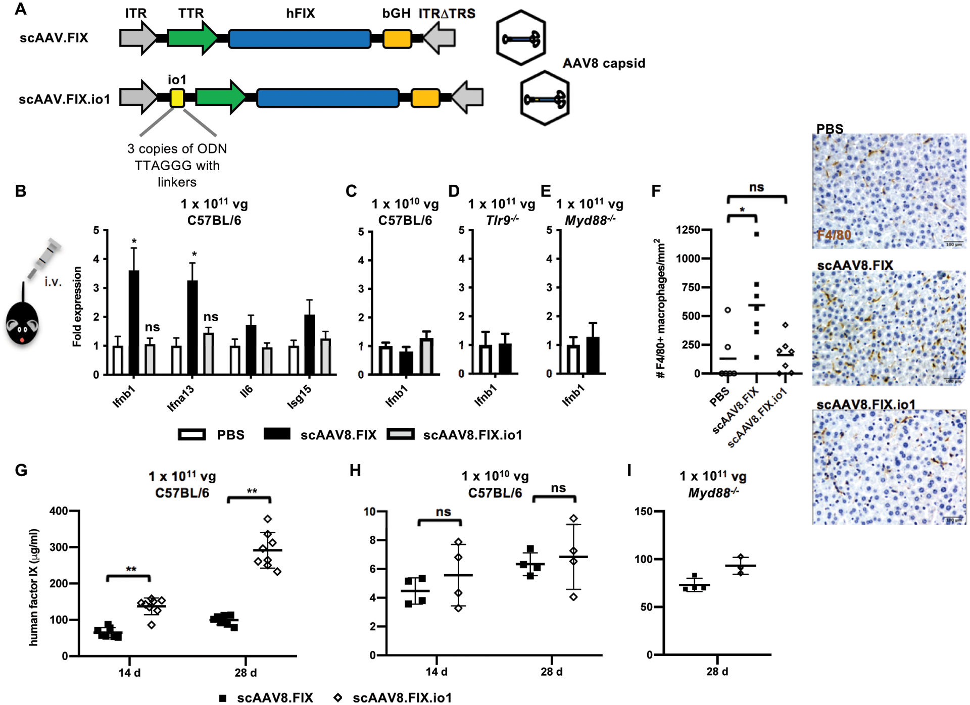
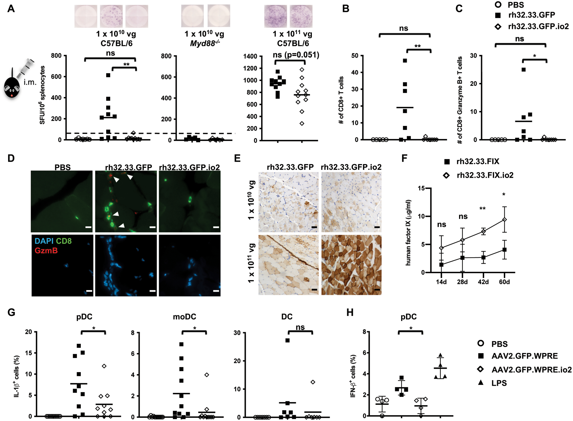
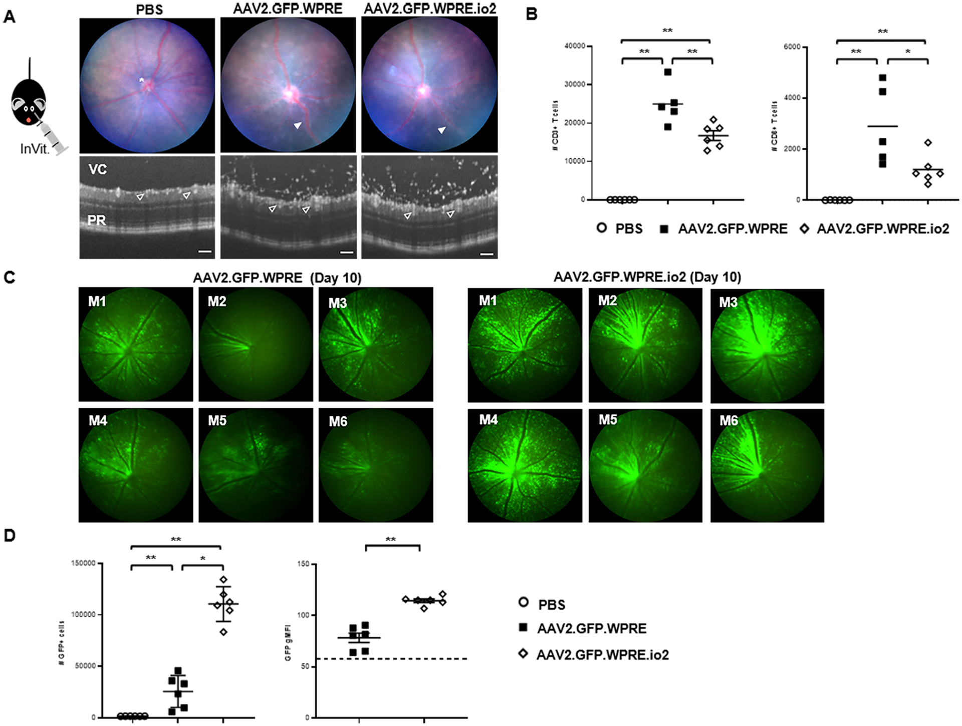
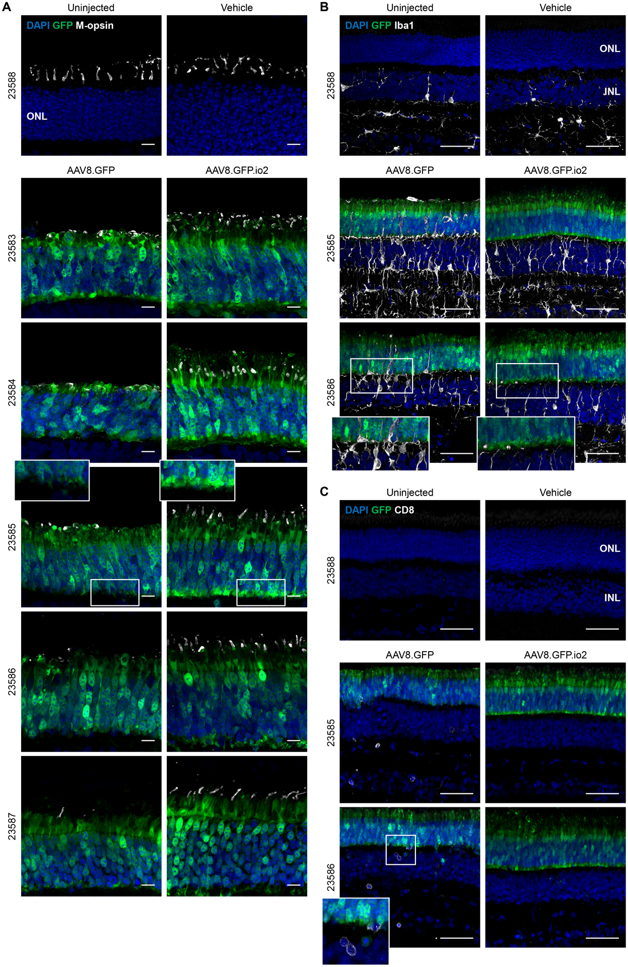
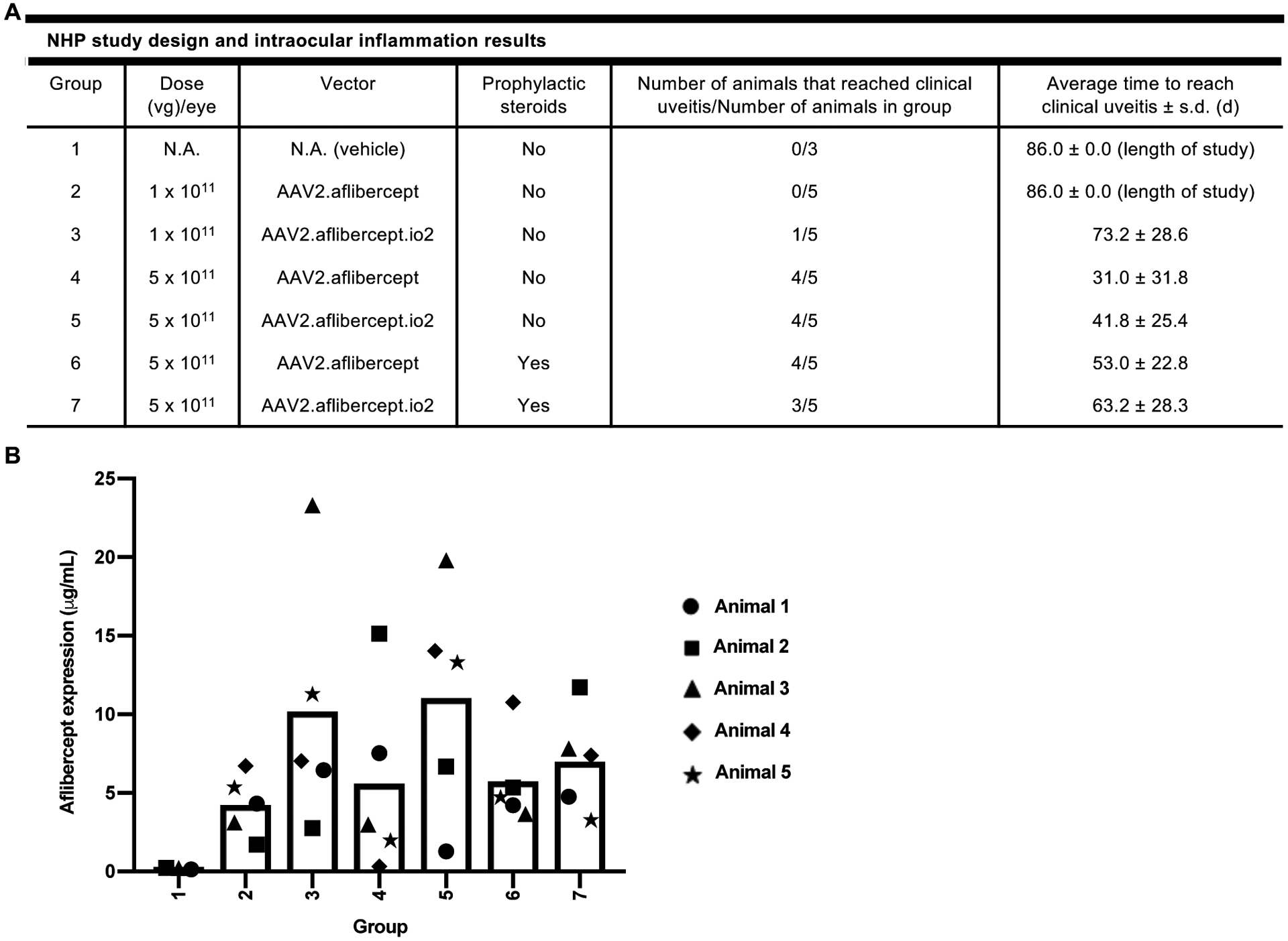
Nucleic acids are used in many therapeutic modalities, including gene therapy, but their ability to trigger host immune responses in vivo can lead to decreased safety and efficacy. In the case of adeno-associated viral (AAV) vectors, studies have shown that the genome of the vector activates Toll-like receptor 9 (TLR9), a pattern recognition receptor that senses foreign DNA. Here, we engineered AAV vectors to be intrinsically less immunogenic by incorporating short DNA oligonucleotides that antagonize TLR9 activation directly into the vector genome. The engineered vectors elicited markedly reduced innate immune and T cell responses and enhanced gene expression in clinically relevant mouse and pig models across different tissues, including liver, muscle, and retina. Subretinal administration of higher-dose AAV in pigs resulted in photoreceptor pathology with microglia and T cell infiltration. These adverse findings were avoided in the contralateral eyes of the same animals that were injected with the engineered vectors. However, intravitreal injection of higher-dose AAV in macaques, a more immunogenic route of administration, showed that the engineered vector delayed but did not prevent clinical uveitis, suggesting that other immune factors in addition to TLR9 may contribute to intraocular inflammation in this model. Our results demonstrate that linking specific immunomodulatory noncoding sequences to much longer therapeutic nucleic acids can "cloak" the vector from inducing unwanted immune responses in multiple, but not all, models. This "coupled immunomodulation" strategy may widen the therapeutic window for AAV therapies as well as other DNA-based gene transfer methods.


Conflict of interest statement

Competing interests: Y.K.C. and G.M.C. are co-inventors on patent applications filed by Harvard University related to the described work (WO2017214378A1 “Engineered viral vector reduces induction of inflammatory and immune responses” and WO2019094548A1 “Compositions and methods for inhibiting viral vector-induced inflammatory responses”). Y.K.C., J.J.C., T.L. and G.M.C. are co-founders of Ally Therapeutics. G.M.C. also is a founder of Editas Medicine and eGenesis Bio (for the full disclosure list, please see http://arep.med.harvard.edu/gmc/tech.html). Y.K.C., S.K.W., J.J.C., M.K.W. and H.J.K. have consulted for Ally Therapeutics. J.J.C., M.K.W., W.J.N. and T.L. are currently employees of Ally Therapeutics. Y.K.C., J.J.C., M.K.W., W.J.N., T.L., G.G. and G.M.C. have equity holdings or stock options in Ally Therapeutics. A.D.D. has consulted for Ally Therapeutics, Affibody, Novartis and Genentech. C.L.C. is a founder of GenSight Biologics and serves on the Scientific Advisory Boards of the Institute of Ophthalmology, Basel and Sigilon Therapeutics. F.M. is currently an employee of Spark Therapeutics. G.G. is a co-founder of Voyager Therapeutics, Adrenas Therapeutics and Aspa Therapeutics. G.G. serves as a scientific consultant for AAVAA Therapeutics, Aevitas Therapeutics, Ally Therapeutics, CanBridge Pharma., Catalent, Decibel Therapeutics, Iveric bio., Modalis Therapeutics and Saturn Therapeutics. P.W.T. serves as a scientific consultant for Biogen and Shape Therapeutics on work not related to the present study. All other authors declare no competing interests.

Figures

Figure 1.
Figure 1.. Innate immune response and human factor IX expression in mice following intravenous AAV8 administration.
(A) Schematic diagram of vector organization of scAAV.FIX and scAAV.FIX.io1. The io1 sequence is not expected to be transcribed or translated due to its placement in an untranslated region upstream of the promoter. (B) Innate immune response in liver assayed by qPCR 2 h after 1 × 10¹¹ vg AAV8 administration in C57BL/6 mice. n = 6 – 7 animals per condition. (C) Innate immune response in liver assayed by qPCR 2 h after 1 × 10¹⁰ vg AAV8 administration in C57BL/6 mice. n = 4 – 5 animals per condition. (D) Innate immune response in liver assayed by qPCR 2 h after 1 × 10¹¹ vg AAV8 administration in Tlr9−/− mice. n = 4 animals per condition. (E) Innate immune response in liver assayed by qPCR 2 h after 1 × 10¹¹ vg AAV8 administration in Myd88−/− mice. n = 4 animals per condition. PBS injection was set to 1-fold expression for each gene. Data shown are mean ± s.e.m. per condition. * p<0.05 by two-tailed Mann-Whitney test and compared against PBS condition. ns, not significant, p>0.05. (F) F4/80+ macrophage infiltration in C57BL/6 mouse liver 2 h after 1 × 10¹¹ vg AAV8 administration. Data shown are mean of n = 6 – 7 animals per condition. * p<0.005 by two-tailed Mann-Whitney test. Scale bar, 100 μm. (G) Human factor IX in plasma of C57BL/6 mice at indicated time points after 1 × 10¹¹ vg AAV8 administration. n = 7 – 8 animals per condition. (H) Human factor IX in plasma of C57BL/6 mice at indicated time points after 1 × 10¹⁰ vg AAV8 administration. n = 4 animals per condition. (I) Human factor IX in plasma of Myd88−/− mice at indicated time points after 1 × 10¹¹ vg AAV8 administration. n = 3 – 4 animals per condition. Data shown are mean ± s.d. per condition. ** p<0.005 by two-tailed Mann-Whitney test. ITR, inverted terminal repeat; TTR, transthyretin promoter; hFIX, human factor IX; bGH, bovine growth hormone poly(A) signal; TRS, terminal resolution site.
Figure 2.
Figure 2.. Immune responses to single-stranded vectors in mouse skeletal muscle in vivo and human PBMCs in vitro.
(A) CD8+ T cell responses to rh32.33 capsid 21 d after intramuscular injections. Representative images of the ELISpot well are shown. Dotted line (50 SFU/106 splenocytes) indicates cutoff for a positive T cell response. (B) Number of CD8+ T cells in the muscle sections (four fields per sample) of C57BL/6 mice for PBS and 1 × 10¹⁰ vg rh32.33 vectors. (C) Number of CD8+ Granzyme B+ T cells in the muscle sections (four fields per sample) of C57BL/6 mice for PBS and 1 × 10¹⁰ vg rh32.33 vectors. (D) Representative immunohistochemical images of CD8 and Granzyme B staining in the muscle sections of C57BL/6 mice for PBS and 1 × 10¹⁰ vg rh32.33 vectors. White arrows indicate double positive cells. Scale bar, 10 μm. (E) Representative images of GFP expression (brown) by immunohistochemistry staining in muscle sections of C57BL/6 mice. Scale bar, 50 μm. (F) Human FIX in plasma of C57BL/6 mice at indicated time points following 1 × 10¹¹ vg rh32.33 vector administration. Data shown are mean ± s.d. of n = 4 animals per condition. (G) Intracellular cytokine staining of IL-1β in specific DC populations 24 h after infection of primary human PBMCs from different donors (n = 13). (H) Intracellular cytokine staining of IFN-β 24 h after infection of primary human PBMCs from different donors (n = 7). Some donor PBMCs did not respond to AAV or LPS stimulation (no innate immune response induced over PBS-treated samples) and are not shown. Data shown are mean, with each symbol representing an animal or a donor. n = 4 – 13 animals or donors per condition as indicated. *p<0.05 and **p<0.005 by two-tailed Mann-Whitney test for (A-D), two-way ANOVA with Sidak’s post-hoc test for (F), two-tailed Wilcoxon matched-pairs signed ranked test for (G), and two-tailed Student’s t-test for (H). ns, not significant, p>0.05. SFU, spot forming units.
Figure 3.
Figure 3.. Engineered vector reduces retinal infiltration and achieves greater GFP transgene expression following intravitreal injection in mice.
(A) Representative fundal and circular OCT scans of the retina 10 days after intravitreal injection with 2μl of titer-matched (5 × 10¹² vg/mL) AAV2.GFP.WPRE, AAV2.GFP.WPRE.io2 or PBS control. Scale bars, 100 μm. Image annotations: optic disc (*), vasculitis (solid white arrow), vitreous cavity (VC), retinal vessels (open white arrow), photoreceptor layer (PR). By OCT, infiltrating cells entrapped within the optically empty vitreous gel above the retinal tissue are visualized as white dots. (B) At day 11 post-injection, eyes were dissected and processed by flow cytometry to identify and enumerate the absolute number of CD45+CD3+ and CD45+CD3+CD8+ T cell populations (n = 5–6 mice/group). Data shown are mean ± s.d., with each symbol representing a single eye. *p<0.05 and **p<0.005 by two-tailed Mann-Whitney test. (C) Standardized fluorescent in vivo retinal images captured on day 10 from AAV2.GFP.WPRE or AAV2.GFP.WPRE.io2 groups (n = 6 mice/group). (D) Flow cytometry of single retinas confirms higher numbers of GFP+ retinal cells and increased GFP geometric mean fluorescence intensity (gMFI) per individual cell. Dotted line indicates background (gMFI from normal retinal cell autofluorescence in PBS-injected eyes). Data shown are mean ± s.d., with each symbol representing a single eye. n = 6 eyes per group as indicated. *p<0.05 and **p<0.005 by two-tailed Mann-Whitney test.
Figure 4.
Figure 4.. Engineered vector evades photoreceptor pathology and microglia and CD8+ T cell infiltration in subretinal-injected pig eyes.
Immunohistochemical images of the ONL of pig retinas 6 weeks after subretinal injections. Each animal is indicated by an identification number and paired images are from the two treated eyes of each animal. (A) Outer segments of cone photoreceptors were visualized by anti-red-green (M) opsin staining. Scale bars, 10 μm. (B) Microglia proliferation and activation in the retina indicated by anti-Iba1 staining. Scale bars, 50 μm. (C) Cytotoxic T cell infiltration into the retina indicated by anti-CD8 staining. Scale bars, 50 μm. ONL, outer nuclear layer; Iba1, ionized calcium-binding adaptor protein 1.
Figure 5.
Figure 5.. Engineered vector may delay, but does not prevent, intraocular inflammation following intravitreal AAV2 administration in NHPs.
(A) Summary of NHP study design and key intraocular inflammation results. The indicated number of NHPs received bilateral intravitreal injections of AAV2.aflibercept or AAV2.aflibercept.io2 at 1 × 10¹¹ vg or 5 × 10¹¹ vg on Day 1, and intraocular inflammation was scored based on the SUN criteria system during the 12-week study. Group 6 and 7 animals received intramuscular injection of prophylactic systemic steroids on Days −1 and 6. Clinical uveitis was defined as AC or VC score of 3 or higher. A two-tailed Fisher’s Exact Test with stepdown Sidak adjustment was used to analyze number of animals that reached clinical uveitis while a two-tailed generalized linear model was used to analyze average time to reach clinical uveitis. No statistically significant differences were detected between groups. (B) Aflibercept concentrations (μg/mL) in NHP vitreous humors. Aflibercept concentrations were measured in NHP vitreous humor samples collected 12 weeks post-injection. For AAV-treated animals, animals 1 and 2 were male and animals 3 to 5 were female. For vehicle-treated animals, animal 1 was male and animals 2 and 3 were female. A univariate linear regression model with aflibercept expression as the outcome was used to compare vitreous aflibercept concentrations in vector-injected groups. No statistically significant differences were detected between groups.

References

    1. Verma IM, Somia N, Gene therapy -- promises, problems and prospects. Nature 389, 239–242 (1997); published online EpubSep 18 (10.1038/38410). - DOI - PubMed
    1. Hinderer C, Katz N, Buza EL, Dyer C, Goode T, Bell P, Richman LK, Wilson JM, Severe Toxicity in Nonhuman Primates and Piglets Following High-Dose Intravenous Administration of an Adeno-Associated Virus Vector Expressing Human SMN. Hum Gene Ther 29, 285–298 (2018); published online EpubMar (10.1089/hum.2018.015). - DOI - PMC - PubMed
    1. Hordeaux J, Wang Q, Katz N, Buza EL, Bell P, Wilson JM, The Neurotropic Properties of AAV-PHP.B Are Limited to C57BL/6J Mice. Mol Ther 26, 664–668 (2018); published online EpubMar 7 (10.1016/j.ymthe.2018.01.018). - DOI - PMC - PubMed
    1. Mendell JR, Al-Zaidy S, Shell R, Arnold WD, Rodino-Klapac LR, Prior TW, Lowes L, Alfano L, Berry K, Church K, Kissel JT, Nagendran S, L’Italien J, Sproule DM, Wells C, Cardenas JA, Heitzer MD, Kaspar A, Corcoran S, Braun L, Likhite S, Miranda C, Meyer K, Foust KD, Burghes AHM, Kaspar BK, Single-Dose Gene-Replacement Therapy for Spinal Muscular Atrophy. N Engl J Med 377, 1713–1722 (2017); published online EpubNov 2 (10.1056/NEJMoa1706198). - DOI - PubMed
    1. Cukras C, Wiley HE, Jeffrey BG, Sen HN, Turriff A, Zeng Y, Vijayasarathy C, Marangoni D, Ziccardi L, Kjellstrom S, Park TK, Hiriyanna S, Wright JF, Colosi P, Wu Z, Bush RA, Wei LL, Sieving PA, Retinal AAV8-RS1 Gene Therapy for X-Linked Retinoschisis: Initial Findings from a Phase I/IIa Trial by Intravitreal Delivery. Mol Ther 26, 2282–2294 (2018); published online EpubSep 5 (10.1016/j.ymthe.2018.05.025). - DOI - PMC - PubMed

Publication types