. 2021 Feb 6;10(2):13.
doi: 10.1038/s41389-021-00302-5.

TGF-β1-mediated transition of resident fibroblasts to cancer-associated fibroblasts promotes cancer metastasis in gastrointestinal stromal tumor

Affiliations

TGF-β1-mediated transition of resident fibroblasts to cancer-associated fibroblasts promotes cancer metastasis in gastrointestinal stromal tumor

Hyunho Yoon et al. Oncogenesis. .

Abstract

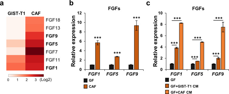
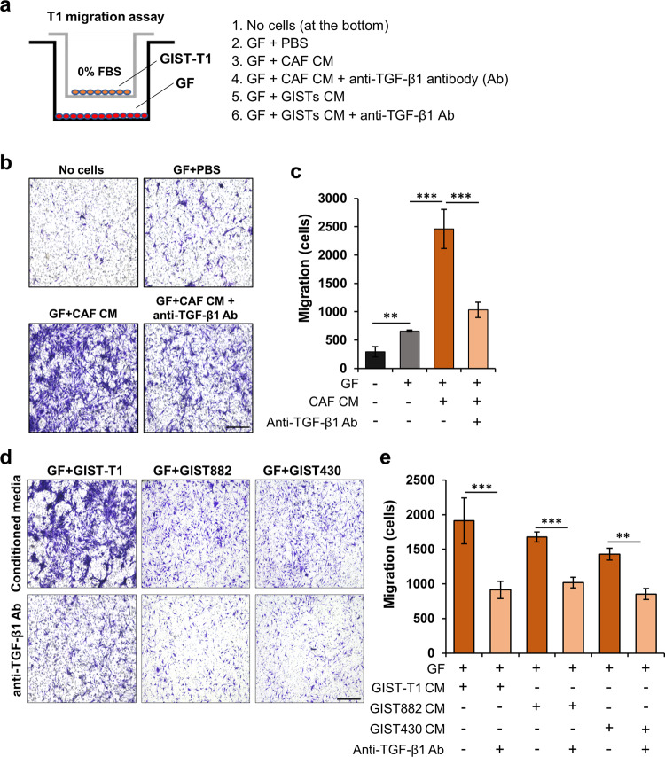
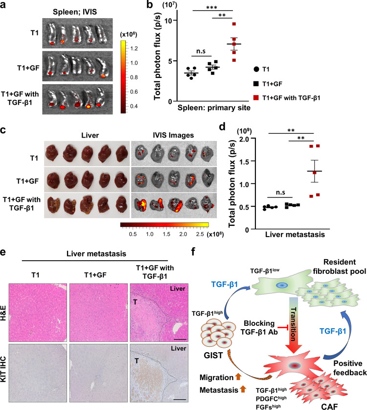
Cancer-associated fibroblasts (CAFs) are the most abundant cells in the tumor microenvironment. Crosstalk between tumor cells and CAFs contributes to tumor survival in most epithelial cancers. Recently, utilizing gastrointestinal stromal tumor (GIST) as a model for sarcomas, we identified paracrine networks by which CAFs promote tumor progression and metastasis. However, the mechanisms by which CAFs arise in sarcomas remain unclear. Here, RNA sequencing analysis revealed that transforming growth factor-β1 (TGF-β1) is highly expressed in both tumor cells and CAFs. To determine the functional role of TGF-β1, we treated normal gastric fibroblasts (GFs) with recombinant TGF-β1, which caused the GFs to adopt a more stellate morphology, as well as increased the mRNA expression of CAF-mediated genes (CCL2, RAB3B, and TNC) and genes encoding fibroblast growth factors (FGFs). Moreover, while either GIST or CAF conditioned media enhanced the transition from GFs to CAFs, a TGF-β1-blocking antibody attenuated this effect. Transwell migration assays revealed that the TGF-β1-mediated transition from GFs to CAFs enhanced tumor cell migration. This migratory effect was abrogated by an anti-TGF-β1 antibody, suggesting that TGF-β1 secreted from GIST cells or CAFs is associated with GIST migration via GF-to-CAF transition. In addition, the murine spleen-to-liver metastasis model showed that GF pre-treated with TGF-β1 promoted GIST metastasis. Collectively, these findings reveal unappreciated crosstalk among tumor cells, CAFs, and normal resident fibroblasts in the stroma of sarcomas, which enhances a GF-to-CAF transition associated with tumor migration and metastasis.


Conflict of interest statement

J.K.S. receives research funding from Novartis Pharmaceuticals, Amgen Pharmaceuticals, and Foundation Medicine, consultant fees from Grand Rounds, Loxo, and Deciphera, speaker’s fees from Roche and Deciphera, and owns stocks in Personalis.

Figures

Fig. 1
Fig. 1. GIST cells and CAFs enhance a GF-to-CAF transition.
a The images of gastric fibroblasts (GFs) and GIST-CAFs. Scale bars, 50 µm. b Expression of PDGFC mRNA in GFs and CAFs by quantitative RT-PCR (qPCR). c The effects of GIST-T1 conditioned media (CM) and CAF CM treatment on PDGFC expression in GFs. After GFs were treated with CM of GIST-T1 and CAFs for 48 h, the expression level of PDGFC was determined by qPCR. d Expression of CAF markers (CCL2, RAB3B, and TNC) in GFs and CAFs by qPCR. e The effects of GIST-T1 CM and CAF CM treatment on CAF markers expression in GFs. All graphs show mean ± SEM, and p values were represented by Student’s t test or ANOVA analysis. **p < 0.01, ***p < 0.001. f Phosphokinase array in GIST-T1 treated with CAFscr CM, CAFshPDGFC #1 CM, or CAFshPDGFC #2 CM. g The average signal (pixel density) was normalized with positive control and analyzed among samples. All graphs show mean ± SEM, and p values were represented by ANOVA analysis. *p < 0.05; **p < 0.01.
Fig. 2
Fig. 2. FGFs are overexpressed in CAFs.
a Heatmap of the expression levels of FGF genes in GIST-T1 and CAFs analyzed from RNA-seq (GSE143547). b Expression of FGF1, FGF5, and FGF9 in GFs and CAFs by qPCR. c GIST-T1 CM and CAF CM enhanced the expression of FGF1, FGF5, and FGF9 in GF. The level of expression was evaluated by qPCR. All graphs show mean ± SEM, and p values were represented by Student’s t test or ANOVA analysis. **p < 0.01, ***p < 0.001.
Fig. 3
Fig. 3. TGF-β1 is highly expressed in GIST cells and CAFs.
a Representative immunohistochemistry (IHC) images with staining for TGF-β1 in the frozen tumor sections collected from human GISTs harboring mutant KIT. Scale bars, 100 µm. b Representative immunofluorescence (IF) images of TGF-β1 (green), KIT (GIST marker; red), and DAPI (nuclei; blue) staining in the resected sections. Scale bars, 50 µm. c mRNA expression of TGFB1 in GFs, GIST-T1, GIST882, GIST430, and CAFs by qPCR. d Comparison of TGF-β1 levels measured by enzyme-linked immunosorbent assay (ELISA) in GFs, GIST-T1, GIST882, GIST430, and CAFs. After each cell line was seeded for 48 h in the absence of FBS, the supernatant was collected to perform ELISA. All graphs show mean ± SEM, and p values were represented by ANOVA analysis. **p < 0.01, ***p < 0.001.
Fig. 4
Fig. 4. TGF-β1 is associated with a GF-to-CAF transition, which increases GIST migration.
a Photographic images of GFs treated with human recombinant TGF-β1. Scale bars, 50 µm. GFs were treated with TGF-β1 (10 ng/mL) for 48 h. b Effect of TGF-β1 treatment on mRNA expression of CAF markers (CCL2, RAB3B, and TNC) in GFs by qPCR. c Effect of anti-TGF-β1 blocking antibody on the expression of FGF1, FGF5, and FGF9 in GFs treated with TGF-β1. GFs were treated with TGF-β1 (10 ng/mL) and/or anti-TGF-β1 blocking antibody (1 µg/mL) for 48 h. The expression levels of these genes were measured by qPCR. d Experimental design for Transwell migration assay of anti-TGF-β1 blocking antibody. After GFs were seeded on the bottom, TGF-β1 (10 ng/mL) and anti-TGF-β1 blocking antibody (1 µg/mL) were added for 24 h. eg Representative images (e) of the migrated GIST cells stained with 0.05% crystal violet in the indicated group and quantitative data in GIST-T1 cells (f) and GIST882 cells (g) analyzed by ImageJ software. Scale bars, 200 µm. All graphs show mean ± SEM, and p values were represented by Student’s t test or ANOVA analysis. **p < 0.01, ***p < 0.001.
Fig. 5
Fig. 5. TGF-β1 secreted from GIST-T1 cells and CAFs is involved in increased GIST migration.
a Experimental design of the Transwell migration assay. b, c Representative images (b) and quantitative data (c). GFs were treated with PBS (control), CAF CM, and/or TGF-β1 blocking antibody (1 µg/mL) for 24 h in the bottom well. The migrated GIST-T1 cells were imaged using a BZ-X800 Life Science Microscope and analyzed by ImageJ software. d Representative images showing Transwell migration assay of TGF-β1 blocking antibody in GFs treated with CM of GIST-T1, GIST882, and GIST430. e Quantitative data of the migrated GIST-T1 cells in the indicated conditions. Scale bars, 200 µm. Graphs show mean ± SEM, and p values were represented by ANOVA analysis. **p < 0.01, ***p < 0.001.
Fig. 6
Fig. 6. TGF-β1-mediated transition of GF to CAF promotes GIST metastasis.
a, b Effects of a transition from GF to CAF on the murine spleen-to-liver metastasis model. The mice were injected with GFP-labeled T1, T1 + GF, and T1 + GF pre-treated with TGF-β1. After 3 weeks, all mice were sacrificed. The harvested spleens and livers from each mouse were analyzed using the IVIS imaging system. IVIS images (a) of the spleen and quantification (b) analyzed from total photon flux (p/s) of the spleen. p Values were represented by ANOVA analysis. **p < 0.01, ***p < 0.001. n.s. nonsignificant. c Photographic images (left) and IVIS images (right) of the liver in the indicated group. d Quantitative data were analyzed by total photon flux (p/s) on the metastatic liver. p Values were represented by ANOVA analysis. **p < 0.01. e Representative H&E images (top) and IHC (bottom) stained with anti-KIT antibody in the tumor sections collected from livers. Scale bars, 100 µm. f Proposed model of TGF-β1-mediated transition of GFs to CAFs in the GIST stroma. TGF-β1 secretion from CAFs and GIST cells enhances a GF-to-CAF transition, which promotes GIST migration via paracrine signaling networks.

References

    1. Corless CL, Heinrich MC. Molecular pathobiology of gastrointestinal stromal sarcomas. Annu. Rev. Pathol. 2008;3:557–586. doi: 10.1146/annurev.pathmechdis.3.121806.151538. - DOI - PubMed
    1. Corless CL, Barnett CM, Heinrich MC. Gastrointestinal stromal tumours: origin and molecular oncology. Nat. Rev. Cancer. 2011;11:865–878. doi: 10.1038/nrc3143. - DOI - PubMed
    1. Shi E, et al. FGFR1 and NTRK3 actionable alterations in “Wild-Type” gastrointestinal stromal tumors. J. Transl. Med. 2016;14:339. doi: 10.1186/s12967-016-1075-6. - DOI - PMC - PubMed
    1. Blanke CD, et al. Phase III randomized, intergroup trial assessing imatinib mesylate at two dose levels in patients with unresectable or metastatic gastrointestinal stromal tumors expressing the kit receptor tyrosine kinase: S0033. J. Clin. Oncol. 2008;26:626–632. doi: 10.1200/JCO.2007.13.4452. - DOI - PubMed
    1. Demetri GD, et al. Efficacy and safety of regorafenib for advanced gastrointestinal stromal tumours after failure of imatinib and sunitinib (GRID): an international, multicentre, randomised, placebo-controlled, phase 3 trial. Lancet. 2013;381:295–302. doi: 10.1016/S0140-6736(12)61857-1. - DOI - PMC - PubMed

LinkOut - more resources