. 2021 Feb 10;12(1):916.
doi: 10.1038/s41467-021-21199-z.

Recent African strains of Zika virus display higher transmissibility and fetal pathogenicity than Asian strains

Affiliations

Recent African strains of Zika virus display higher transmissibility and fetal pathogenicity than Asian strains

Fabien Aubry et al. Nat Commun. .

Abstract

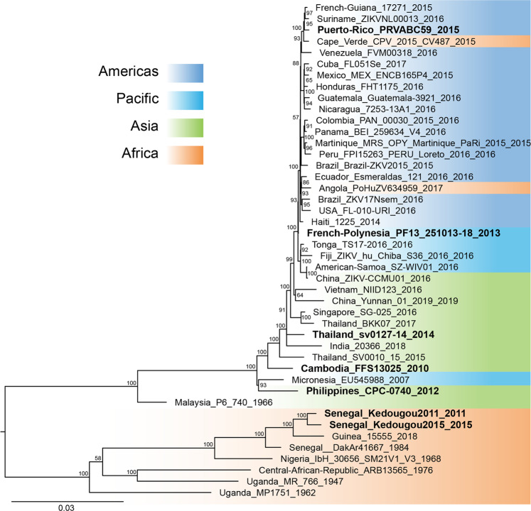
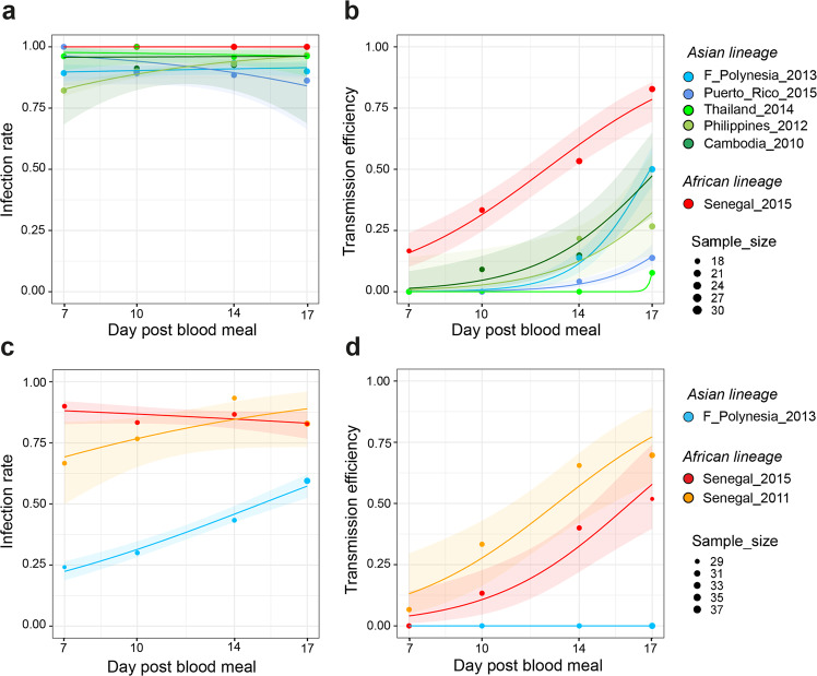
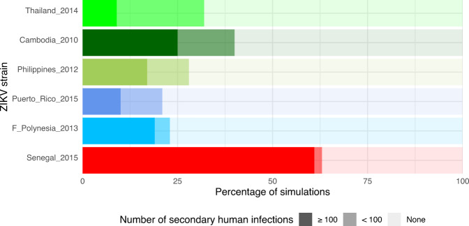
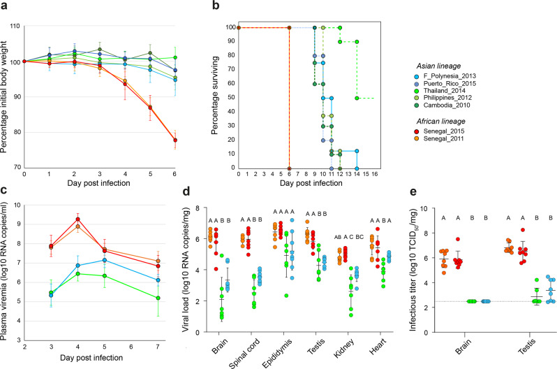
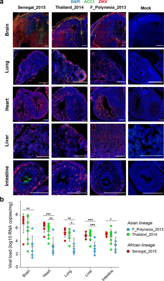
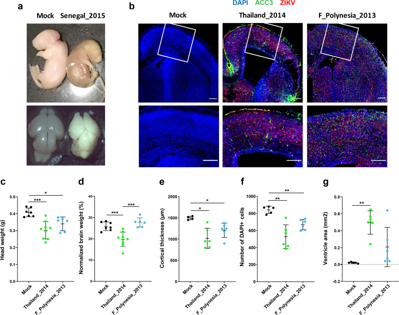
The global emergence of Zika virus (ZIKV) revealed the unprecedented ability for a mosquito-borne virus to cause congenital birth defects. A puzzling aspect of ZIKV emergence is that all human outbreaks and birth defects to date have been exclusively associated with the Asian ZIKV lineage, despite a growing body of laboratory evidence pointing towards higher transmissibility and pathogenicity of the African ZIKV lineage. Whether this apparent paradox reflects the use of relatively old African ZIKV strains in most laboratory studies is unclear. Here, we experimentally compare seven low-passage ZIKV strains representing the recently circulating viral genetic diversity. We find that recent African ZIKV strains display higher transmissibility in mosquitoes and higher lethality in both adult and fetal mice than their Asian counterparts. We emphasize the high epidemic potential of African ZIKV strains and suggest that they could more easily go unnoticed by public health surveillance systems than Asian strains due to their propensity to cause fetal loss rather than birth defects.


Conflict of interest statement

The authors declare no competing interests.

Figures

Fig. 1
Fig. 1. Phylogenetic position of ZIKV strains used in this study.
The phylogenetic tree shows the seven ZIKV strains of the panel (in bold) among a backdrop of ZIKV strains spanning the current viral genetic diversity. The colored background represents the geographic origin of ZIKV strains. The consensus tree was generated from 1000 ultrafast bootstrap replicate maximum-likelihood trees, using a GTR + F + G4 nucleotide substitution model of the full ZIKV open reading frame. The tree is midpoint rooted and the root position is verified by the Spondweni virus outgroup on amino-acid and codon-based trees. Support values next to the nodes indicate ultrafast bootstrap proportions (%) and the scale bar represents the number of nucleotide substitutions/site.
Fig. 2
Fig. 2. Mosquito infection rate and transmission efficiency of African and Asian ZIKV strains.
Wild-type Ae. aegypti mosquitoes from Colombia were orally exposed to ZIKV and collected on day 7, 10, 14, and 17 post infectious blood meal to analyze their carcasses and saliva samples collected in vitro. Infection rates and transmission efficiencies over time are shown for each ZIKV strain tested after oral exposure to a high dose (5.6–5.8 log10 FFU/ml) (a, b) or a low dose (4.7–4.8 log10 FFU/ml) (c, d) of virus. Infection rate is the proportion of ZIKV-positive carcasses among all blood-fed mosquitoes (determined by RT-PCR). Transmission efficiency is the proportion of blood-fed mosquitoes with infectious saliva (determined by FFA). The data points represent the empirically measured proportions, and their size is proportional to the sample size (high dose: n = 18–30 mosquitoes per group; low dose: n = 29–37 mosquitoes per group). The lines represent the logistic regression results and the shaded areas are the 95% confidence intervals of the logistic fits. Source data are provided as a Source Data file.
Fig. 3
Fig. 3. Simulated effect of empirical variation in ZIKV transmissibility on the risk and magnitude of human outbreaks.
A stochastic agent-based model was run 100 times based on the experimentally determined kinetics of mosquito transmission of six ZIKV strains. Other parameters of the model, such as the mosquito biting rate and infection dynamics within the human host, were shared between viruses. The figure shows the proportion of simulated outbreaks that resulted in ≥100, <100, and no secondary human infections.
Fig. 4
Fig. 4. Pathogenicity of African and Asian ZIKV strains in immunocompromised adult mice.
In a first experiment (a, b), male AG129 mice were inoculated with 103 PFU of ZIKV. Each virus strain was represented by n = 10 mice, with the exception of the F_Polynesia_2013 and Philippines_2012 strains that were represented by n = 8 mice. a Mouse weight over time is shown as the percentage of body weight prior to infection (mean ± standard error). b Mouse survival over time is shown as the percentage of mice alive. Mice were euthanized when reaching humane endpoints (weight loss >20% or/and severe symptom onset). In a second experiment (ce), male AG129 mice were inoculated with 1 PFU of ZIKV (n = 8 mice per strain). c Time course of mouse viremia is shown in log10-transformed viral genome copies per ml of plasma (mean ± standard error). Three extreme outliers were excluded for the Senegal_2015 strain. d Viral loads in organs collected on day 7 post infection are shown in log10-transformed viral genome copies per mg of tissue. Statistical significance of differences was determined by one-way ANOVA followed by Tukey’s post hoc test for brain, heart and testis, by Brown–Forsythe and Welch ANOVA followed by Games-Howell’s post hoc test (two sided) for epididymis and spinal cord, and by Kruskal–Wallis rank-sum test followed by Dunn’s post hoc test (two sided) for kidney. Viral loads were significantly higher for African than for Asian ZIKV strains in the brain (p < 0.0001), spinal cord (p < 0.0001), testis (p < 0.0001), kidney (p < 0.0001), and heart (p < 0.0001). e Infectious virus in brain and testis collected on day 7 post infection are shown in log10-transformed 50% tissue-culture infectious dose (TCID50) per mg of tissue. The horizontal dotted line indicates the lower limit of detection of the assay (310 TCID50 units per mg of tissue). Statistical significance of differences was determined by Kruskal–Wallis rank-sum test followed by Steel-Dwass’s post hoc test for brain and by one-way ANOVA followed by Tukey’s post hoc test for testis. Infectious titers were significantly higher for African than for Asian ZIKV strains in the brain (p < 0.0001) and testis (p < 0.0001). In (d, e), data are presented as mean ± standard deviation and ZIKV strains not sharing a letter above the graph are statistically significantly different (p < 0.05). Source data are provided as a Source Data file.
Fig. 5
Fig. 5. Organ tropism and viral load of African and Asian strains of ZIKV in vertically infected mouse embryos.
Immunocompetent mouse embryos were infected at E10.5 by intraplacental injection of 500–1000 PFU of ZIKV and analyzed at E14.5 by microdissection. a Immunolabeling of embryonic brain, lung, heart, liver and intestine sections representative of each ZIKV strain tested (n = 3 embryos per strain). Blue, green and red colors indicate DAPI, anti-cleaved caspase 3 (ACC3) and ZIKV stainings, respectively. The scale bars represent 200 µm. b Viral load of embryonic brain, lung, heart, liver and intestine are shown for each ZIKV strain in viral genome copies per organ. Data are presented as mean ± standard deviation and represent n = 6 mice for the Senegal_2015 and F_Polynesia_2013 strains and n = 8 mice for the Thailand_2014 strain. Statistical significance of the differences was determined by one-way ANOVA followed by Tukey’s post hoc test and is only shown when significant (***p < 0.001; **p < 0.01; *p < 0.05). Viral loads differed significantly between ZIKV strains (p < 0.0277) in all organs. Source data are provided as a Source Data file.
Fig. 6
Fig. 6. Brain phenotypes of mouse embryos vertically infected with African and Asian strains of ZIKV.
Immunocompetent mouse embryos were infected at E10.5 by intraplacental injection of 500–1000 PFU of ZIKV and analyzed at E18.5 by microdissection. a Representative view of E18.5 embryos (top) and dorsal view of E18.5 embryonic brains (bottom) after mock injection (left; n = 10) or infection by the Senegal_2015 ZIKV strain (right; n = 7). bg Analyses of in utero brain development of E18.5 mouse embryos after mock injection (n = 7 for head and brain measurements and n = 5 otherwise) or infection by the Thailand_2014 (n = 9 for head and brain measurements and n = 6 otherwise) or the F_Polynesia_2013 (n = 6) ZIKV strains. b Immunolabeling of embryonic brain sections representative of each ZIKV strain tested (top: full view; bottom: enlarged area within white frame). Blue, green and red colors indicate DAPI, anti-cleaved caspase 3 (ACC3) and ZIKV stainings, respectively. The scale bars represent 200 µm. c, d Embryonic heads and brains were examined morphologically by measuring (c) head weight and (d) brain weight normalized to head weight. e, f Microcephalic phenotypes were assessed by measuring (e) cortical thickness and (f) number of DAPI-positive cells. g Ventriculomegaly was estimated by measuring the ventricle area. In (cg) data are presented as mean ± standard deviation. Statistical significance of differences was determined by Brown–Forsythe and Welch ANOVA followed by Tamhane’s T2 multiple comparison test (two sided). Only statistically significant differences are shown (***p < 0.001; **p < 0.01; *p < 0.05). Embryos infected with the F_Polynesia_2013 and Thailand_2014 ZIKV strains had a significantly smaller head weight, cortical thickness and number of cortical cells than the mock-injected embryos (p < 0.05). Source data are provided as a Source Data file.

References

    1. Boyer S, Calvez E, Chouin-Carneiro T, Diallo D, Failloux AB. An overview of mosquito vectors of Zika virus. Microbes Infect. 2018;20:646–660. doi: 10.1016/j.micinf.2018.01.006. - DOI - PubMed
    1. Gutierrez-Bugallo G, et al. Vector-borne transmission and evolution of Zika virus. Nat. Ecol. Evol. 2019;3:561–569. doi: 10.1038/s41559-019-0836-z. - DOI - PMC - PubMed
    1. Dick GW, Kitchen SF, Haddow AJ. Zika virus. I. Isolations and serological specificity. Trans. R. Soc. Trop. Med. Hyg. 1952;46:509–520. doi: 10.1016/0035-9203(52)90042-4. - DOI - PubMed
    1. Musso D, Gubler DJ. Zika virus. Clin. Microbiol. Rev. 2016;29:487–524. doi: 10.1128/CMR.00072-15. - DOI - PMC - PubMed
    1. Kindhauser MK, Allen T, Frank V, Santhana RS, Dye C. Zika: the origin and spread of a mosquito-borne virus. Bull. World Health Organ. 2016;94:675–686C. doi: 10.2471/BLT.16.171082. - DOI - PMC - PubMed

Publication types