

Retinal Vascular Assessment in Psoriasis: A Multicenter Study
- PMID: 33568971
- PMCID: PMC7868328
- DOI: 10.3389/fnins.2021.629401
Retinal Vascular Assessment in Psoriasis: A Multicenter Study
Abstract
Purpose: To investigate the vascular status of the macula in psoriasis patients without history of ocular inflammation by Optical Coherence Tomography Angiography (OCTA).
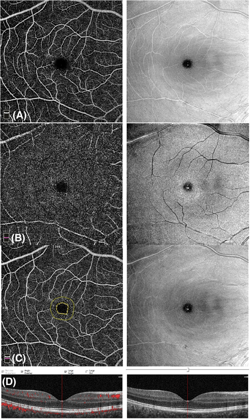
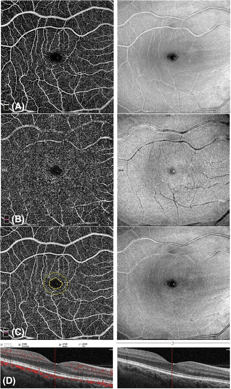
Methods: This prospective cross-sectional multicenter study included 55 psoriasis patients and 55 control healthy subjects. A complete eye examination and 6 mm × 6 mm OCTA imaging were performed. Retinal vascular status was evaluated by analyzing vascular density (VD) of superficial vascular plexus (superficial wVD) and deep vascular plexuses (deep wVD) in a 6 mm × 6 mm area and in foveal (superficial fVD and deep fVD) and parafoveal sectors (superficial pVD and deep pVD). In addition, foveal thickness (FT) and foveal avascular zone (FAZ) and clinical variables, including best corrected visual acuity (BCVA), intraocular pressure and refractive condition, were collected.
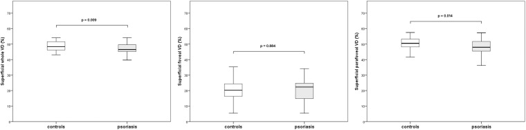
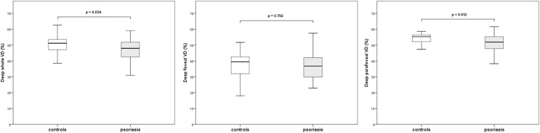
Results: BCVA, intraocular pressure and refractive condition were comparable between cases and controls. OCTA imaging showed that superficial wVD and superficial pVD were lower in the psoriasis group in comparison with controls (p = 0.009 and p = 0.01, respectively). Similarly, deep wVD and pVD were lower in the psoriasis group in comparison with control subjects (p = 0.03 and p = 0.01, respectively). In a sub-analysis of 47 patients affected by psoriasis without psoriatic arthritis, lower values of wVD and pVD in both superficial and deep capillary plexuses were registered.
Conclusion: OCTA is a useful tool which provides data on vascular status of the retina in psoriasis with no ocular involvement. VD data may suggest that vascular changes may occur earlier than clinical onset of posterior inflammation.
Keywords: macula; optical coherence tomography angiography; psoriasis; retina; vascular changes.
Copyright © 2021 Castellino, Longo, Fallico, Russo, Bonfiglio, Cennamo, Fossataro, Fabbrocini, Balato, Parisi, D’urso, Lacarrubba, Musumeci, Alosi, Petrillo, Micali, Avitabile and Reibaldi.
Conflict of interest statement
The authors declare that the research was conducted in the absence of any commercial or financial relationships that could be construed as a potential conflict of interest.
Figures
References
-
- Carnevali A., Sacconi R., Corbelli E., Tomasso L., Querques L., Zerbini G., et al. (2017). Optical coherence tomography angiography analysis of retinal vascular plexuses and choriocapillaris in patients with type 1 diabetes without diabetic retinopathy. Acta Diabetol. 54 695–702. 10.1007/s00592-017-0996-8 - DOI - PubMed
LinkOut - more resources
Full Text Sources
Other Literature Sources