

Dynamics and Concordance Abnormalities Among Indices of Intrinsic Brain Activity in Individuals With Subjective Cognitive Decline: A Temporal Dynamics Resting-State Functional Magnetic Resonance Imaging Analysis
- PMID: 33568986
- PMCID: PMC7868384
- DOI: 10.3389/fnagi.2020.584863
Dynamics and Concordance Abnormalities Among Indices of Intrinsic Brain Activity in Individuals With Subjective Cognitive Decline: A Temporal Dynamics Resting-State Functional Magnetic Resonance Imaging Analysis
Abstract
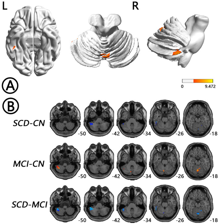
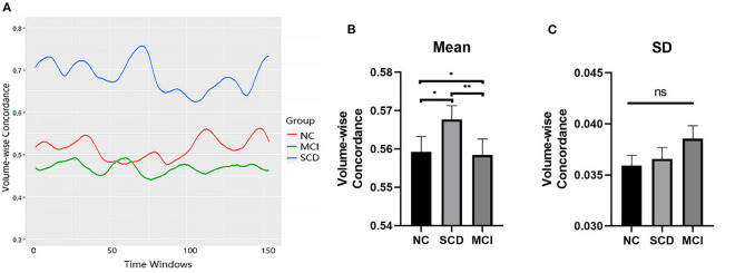
Individuals with subjective cognitive decline (SCD) are more likely to develop into Alzheimer disease (AD) in the future. Resting-state functional magnetic resonance imaging (rs-fMRI) studies have shown alterations of intrinsic brain activity (IBA) in SCD individuals. However, rs-fMRI studies to date have mainly focused on static characteristics of IBA, with few studies reporting dynamics- and concordance-related changes in IBA indices in SCD individuals. To investigate these aberrant changes, a temporal dynamic analysis of rs-fMRI data was conducted on 94 SCD individuals (71.07 ± 6.18 years, 60 female), 75 (74.36 ± 8.42 years, 35 female) mild cognitive impairment (MCI) patients, and 82 age-, gender-, and education-matched controls (NCs; 73.88 ± 7.40 years, 49 female) from the Alzheimer's Disease Neuroimaging Initiative database. The dynamics and concordance of the rs-fMRI indices were calculated. The results showed that SCD individuals had a lower amplitude of low-frequency fluctuations dynamics in bilateral hippocampus (HP)/parahippocampal gyrus (PHG)/fusiform gyrus (FG) and bilateral cerebellum, a lower fractional amplitude of low-frequency fluctuation dynamics in bilateral precuneus (PreCu) and paracentral lobule, and a lower regional homogeneity dynamics in bilateral cerebellum, vermis, and left FG compared with the other two groups, whereas those in MCI patients were higher (Gaussian random field-corrected, voxel-level P < 0.001, cluster-level P < 0.05). Furthermore, SCD individuals had higher concordance in bilateral HP/PHG/FG, temporal lobe, and left midcingulate cortex than NCs, but those in MCI were lower than those in NCs. No correlation between concordance values and neuropsychological scale scores was found. SCD individuals showed both dynamics and concordance-related alterations in IBA, which indicates a compensatory mechanism in SCD individuals. Temporal dynamics analysis offers a novel approach to capturing brain alterations in individuals with SCD.
Keywords: Alzheimer's disease; intrinsic brain activity; resting-state functional MRI; subjective cognitive decline; temporal dynamics analysis.
Copyright © 2021 Yang, Zha, Zhang, Ke, Hu, Wang, Su and Hu.
Conflict of interest statement
The authors declare that the research was conducted in the absence of any commercial or financial relationships that could be construed as a potential conflict of interest.
Figures


Similar articles
-
Intrinsic Brain Activity Alterations in Patients With Mild Cognitive Impairment-to-Normal Reversion: A Resting-State Functional Magnetic Resonance Imaging Study From Voxel to Whole-Brain Level.Front Aging Neurosci. 2022 Jan 17;13:788765. doi: 10.3389/fnagi.2021.788765. eCollection 2021. Front Aging Neurosci. 2022. PMID: 35111039 Free PMC article.
-
Dynamics and concordance alterations of regional brain function indices in vestibular migraine: a resting-state fMRI study.J Headache Pain. 2024 Jan 5;25(1):1. doi: 10.1186/s10194-023-01705-y. J Headache Pain. 2024. PMID: 38178029 Free PMC article.
-
Altered static and dynamic indices of intrinsic brain activity in patients with subcortical ischemic vascular disease: a resting-state functional magnetic resonance imaging analysis.Neuroradiology. 2023 May;65(5):923-931. doi: 10.1007/s00234-023-03135-8. Epub 2023 Mar 9. Neuroradiology. 2023. PMID: 36892613
-
Dynamics and concordance alterations of intrinsic brain activity indices in stroke-induced Broca's aphasia varies based on first language: A resting-state fMRI analysis.Brain Res Bull. 2025 May;224:111312. doi: 10.1016/j.brainresbull.2025.111312. Epub 2025 Mar 22. Brain Res Bull. 2025. PMID: 40127726
-
Functional MRI-specific alterations in frontoparietal network in mild cognitive impairment: an ALE meta-analysis.Front Aging Neurosci. 2023 Jun 28;15:1165908. doi: 10.3389/fnagi.2023.1165908. eCollection 2023. Front Aging Neurosci. 2023. PMID: 37448688 Free PMC article.
Cited by
-
Static and dynamic functional connectivity variability of the anterior-posterior hippocampus with subjective cognitive decline.Alzheimers Res Ther. 2022 Sep 3;14(1):122. doi: 10.1186/s13195-022-01066-9. Alzheimers Res Ther. 2022. PMID: 36057586 Free PMC article.
-
Functional Connectivity Dynamics Altered of the Resting Brain in Subjective Cognitive Decline.Front Aging Neurosci. 2022 Jun 24;14:817137. doi: 10.3389/fnagi.2022.817137. eCollection 2022. Front Aging Neurosci. 2022. PMID: 35813944 Free PMC article.
-
The Potential Role of Gustatory Function as an Early Diagnostic Marker for the Risk of Alzheimer's Disease in Subjective Cognitive Decline.J Alzheimers Dis Rep. 2023 Apr 3;7(1):249-262. doi: 10.3233/ADR220092. eCollection 2023. J Alzheimers Dis Rep. 2023. PMID: 37090958 Free PMC article.
-
Correlation transfer function analysis as a biomarker for Alzheimer brain plasticity using longitudinal resting-state fMRI data.Sci Rep. 2023 Dec 6;13(1):21559. doi: 10.1038/s41598-023-48693-2. Sci Rep. 2023. PMID: 38057476 Free PMC article.
-
Neuroimaging Modalities in Alzheimer's Disease: Diagnosis and Clinical Features.Int J Mol Sci. 2022 May 28;23(11):6079. doi: 10.3390/ijms23116079. Int J Mol Sci. 2022. PMID: 35682758 Free PMC article. Review.
References
-
- Brueggen K., Dyrba M., Cardenas-Blanco A., Schneider A., Fliessbach K., Buerger K., et al. . (2019). Structural integrity in subjective cognitive decline, mild cognitive impairment and Alzheimer's disease based on multicenter diffusion tensor imaging. J. Neurol. 266, 2465–2474. 10.1007/s00415-019-09429-3 - DOI - PubMed
Grants and funding
LinkOut - more resources
Full Text Sources
Other Literature Sources
Research Materials
Miscellaneous