. 2021 Jan 25:11:608871.
doi: 10.3389/fimmu.2020.608871. eCollection 2020.

Neutrophil-Derived Myeloperoxidase Facilitates Both the Induction and Elicitation Phases of Contact Hypersensitivity

Affiliations

Neutrophil-Derived Myeloperoxidase Facilitates Both the Induction and Elicitation Phases of Contact Hypersensitivity

Anna Strzepa et al. Front Immunol. .

Abstract

Background: Allergic contact dermatitis (ACD) is a common skin disorder affecting an estimated 15-20% of the general population. The mouse model of ACD is contact hypersensitivity (CHS), which consists of two phases: induction and elicitation. Although neutrophils are required for both CHS disease phases their mechanisms of action are poorly understood. Neutrophils release myeloperoxidase (MPO) that through oxidation of biomolecules leads to cellular damage.

Objectives: This study investigated mechanisms whereby MPO contributes to CHS pathogenesis.

Methods: CHS was induced in mice using oxazolone (OX) as the initiating hapten applied to the skin. After 7 days, CHS was elicited by application of OX to the ear and disease severity was measured by ear thickness and vascular permeability in the ear. The role of MPO in the two phases of CHS was determined utilizing MPO-deficient mice and a specific MPO inhibitor.

Results: During the CHS induction phase MPO-deficiency lead to a reduction in IL-1β production in the skin and a subsequent reduction in migratory dendritic cells (DC) and effector T cells in the draining lymph node. During the elicitation phase, inhibition of MPO significantly reduced both ear swelling and vascular permeability.

Conclusion: MPO plays dual roles in CHS pathogenesis. In the initiation phase MPO promotes IL-1β production in the skin and activation of migratory DC that promote effector T cell priming. In the elicitation phase MPO drives vascular permeability contributing to inflammation. These results indicate that MPO it could be a potential therapeutic target for the treatment of ACD in humans.

Keywords: contact hypersensitivity; interleukin 1β; myeloperoxidase; neutrophil; vascular permeability.


Conflict of interest statement

KP is a founder, CSO, and owner of ReNeuroGen LLC, a small pharmaceutical company whose goal is to develop KYC to treat sickle cell disease and multiple sclerosis. The remaining authors declare that the research was conducted in the absence of any commercial or financial relationships that could be construed as a potential conflict of interest.

Figures

Figure 1
Figure 1
MPO-deficiency and inhibition reduced inflammation in the OX CHS model. Mice were sham (WT, Mpo-/-) or OX (3%) sensitized and four d later were ear challenged with OX (1%). (A) Ear thickness was measured with a caliper 24 h later. (B) Mice were i.v. administered 0.03 mg/kg KYC every 12 h starting at -12 h prior to sensitization until the end of the study. Each symbol represents one ear with data from three separate experiments shown. ***p≤0.001, ns, not significant.
Figure 2
Figure 2
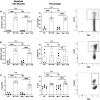
MPO-deficiency reduces the induction of the adaptive immune response. WT and Mpo-/- mice were OX (3%) or sham immunized. After four d lymph nodes cells were collected, and single cell suspensions were stained with fluorochrome-conjugated antibodies and analyzed by flow cytometry and the absolute cell number of CD4+ T cells (A), CD8+ T cells (B), CD4+CD62L- T cells (D), CD8+CD62L- T cells (E), CD4+IFN-γ+ T cells (G), and CD8+IFN-γ+ T cells (H) was determined. Representative flow cytometry gating is shown for CD3-gated CD4 and CD8 T cells (C), CD8+CD62L- T cells (F), and CD8+IFN-γ+ T cells (I). Each symbol represents data from one mouse with data from two to three independent experiments shown. *p≤0.05; **p≤0.01; ***p≤0.001; ns, not significant.
Figure 3
Figure 3
MPO-deficiency does not influence CD4 and CD8 T cell proliferation or Treg numbers in the draining lymph node during induction of CHS. WT and Mpo-/- mice were OX (3%) or sham immunized. After four d lymph nodes cells were collected, and single cell suspensions were stained with fluorochrome-conjugated antibodies and analyzed by flow cytometry. The absolute cell number and frequency of Ki-67+TCRβ+CD4+ T cells (A), Ki-67+TCRβ+CD8+ T cells (B) and TCRβ+CD4+Foxp3+ Treg (C) was determined by flow cytometry. Each symbol represents data from one mouse with data from two to three separate experiments shown. Representative flow cytometry gating for each cell type is shown in the right column (A–C). **p≤0.01; ***p≤0.001; ns, not significant.
Figure 4
Figure 4
MPO-deficiency reduces DC migration from skin to the draining lymph nodes during CHS. WT and Mpo-/- mice were OX (3%) or sham immunized. After 4 days, skin-draining auxiliary lymph nodes were collected, and single cell suspensions were stained with fluorochrome-conjugated antibodies and analyzed by flow cytometry and the absolute cell number and percentage of B220-CD11c+ MHC class II+ DC (A) was determined. WT and Mpo-/- mice were skin painted with 0.5% FITC and 6 h later the absolute number and percentage of FITC+ DC among B220-FITC+ cells was determined (B). Each symbol represents data from one mouse with data from three (A) separate experiments shown or is one representative experiment of two (B). Representative flow cytometry gating is shown on the right for each analysis. (A, B). *p≤0.05; ***p≤0.001; ns, not significant.
Figure 5
Figure 5
MPO-deficiency reduces IL-1β levels in skin biopsies. WT and Mpo-/- mice were OX (3%) or sham immunized. After 6 h skin biopsies were collected and IL-1β levels were quantitated by ELISA. Each symbol represents data from one mouse with data from two separate experiments shown. *p≤0.05; ***p≤0.001; ns, not significant.
Figure 6
Figure 6
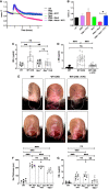
MPO supports the elicitation of CHS by increasing vascular permeability. (A, B) Confluent monolayers of bEnd3 cells were established on collagen-coated gold electrodes and impendence measurements at 4 kHz were conducted. (A) After establishment of the steady state level of impendence, impendence was measured in medium alone (control) or in the presence of unstimulated neutrophils (neut), PMA (20 μM), PMA + KYC (25 μM), PMA + neutrophils, or PMA + neutrophils + KYC. (B) At the 2.5 h time point impendence level was quantitated for each condition. The experiment shown is representative of two. C-E) Mice were sham (WT, Mpo-/-) or OX (3%) sensitized and 4 days later were ear challenged with OX (1%). Vascular permeability was measured 24 h later by Evans blue leakage from the vascular into the tissue 60 min after i.v. administration (C, D). (D, E) Mice were i.v. administered 0.03 mg/kg KYC every 12 h starting at -12 h prior to sensitization until the end of the study. Each symbol represents one ear with data from three separate experiments shown. (E) Twenty-four hours after OX challenge mouse ears were photographed using Zeiss Lumar.V12 stereoscope at 6.4x. Blood vessels are indicated by arrows and areas exhibiting inflammation are circled. The top panels show the dorsal side and the bottom panels the ventral side of the ear. (F, G) WT or Mpo-/- mice were OX (3%) immunized and four d later Teff cells were isolated and depleted of neutrophils and adoptively transferred into WT or Mpo-/- recipients that were subsequently ear challenged with OX (1%). Twenty-four h later ear thickness (F) and vascular permeability (G) was measured. Each symbol represents one ear with data from one representative experiment of two shown. *p≤0.05; **p≤0.01; ***p≤0.001; ns, not significant.
Figure 7
Figure 7
Model outlining the role of MPO in the induction and elicitation phases of CHS. (A) For induction of CHS oxazolone (OX) is applied to the skin, which results in migration of neutrophils into the skin and release of MPO and subsequent induction of IL-1β production within the skin. Skin resident dendritic cells (DC) take up Ox and become activated that is facilitated by IL-1β. The activated DC migrate to the draining lymph node. IL-1β is decreased in Mpo-/- mice resulting in a reduction in the number of activated DC. (B) Once in the draining lymph node, DC interact with naïve T cells (T) and present Ox via MHC class I and II to activate antigen-specific CD4+ and CD8+ T cells, respectively. Due to a reduction of activated DC reaching the draining lymph node in Mpo-/- mice, T cell priming is reduced. (C) For the elicitation of CHS, Ox is applied to the ears, which results in the migration of effector T cells and neutrophils into the ears. Neutrophils release MPO (C), which catalyzes the production of free radicals and oxidants that drive vascular permeability leading to swelling as indicated by an increase in the thickness of the ears (D). (D) In the absence of MPO, the production of free radicals and oxidants is reduced thereby reducing vascular permeability and ear swelling. (A–D) Treatment with KYC by inhibiting MPO activity would result in similar biological outcomes as those observed in Mpo-/- mice.

References

    1. Alinaghi F, Bennike NH, Egeberg A, Thyssen JP, Johansen JD. Prevalence of contact allergy in the general population: A systematic review and meta-analysis. Contact Dermatitis (2019) 80:77–85. 10.1111/cod.13119 - DOI - PubMed
    1. Nassau S, Fonacier L. Allergic Contact Dermatitis. Med Clin North Am (2020) 104:61–76. 10.1016/j.mcna.2019.08.012 - DOI - PubMed
    1. Strzepa A, Majewska-Szczepanik M, Lobo FM, Wen L, Szczepanik M. Broad spectrum antibiotic enrofloxacin modulates contact sensitivity through gut microbiota in a murine model. J Allergy Clin Immunol (2017) 140:121–133 e123. 10.1016/j.jaci.2016.11.052 - DOI - PubMed
    1. Gaspari AA, Katz SI, Martin SF. Contact Hypersensitivity. Curr Protoc Immunol (2016) 113(1):4.2.1–7. 10.1002/0471142735.im0402s113 - DOI - PubMed
    1. Honda T, Egawa G, Grabbe S, Kabashima K. Update of immune events in the murine contact hypersensitivity model: toward the understanding of allergic contact dermatitis. J Invest Dermatol (2013) 133:303–15. 10.1038/jid.2012.284 - DOI - PubMed

Publication types