Review
. 2020 Dec 18;12(6):1343-1359.
doi: 10.1007/s12551-020-00771-9. eCollection 2020 Dec.

Calcium signaling: breast cancer's approach to manipulation of cellular circuitry

Affiliations
Review

Calcium signaling: breast cancer's approach to manipulation of cellular circuitry

Stephen J P Pratt et al. Biophys Rev. .

Abstract

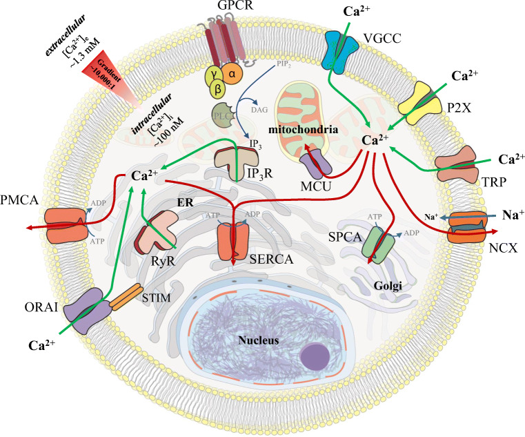
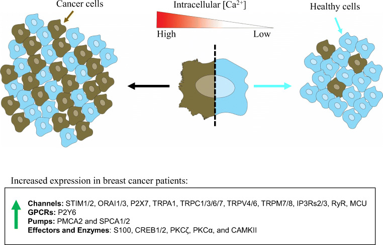
Calcium is a versatile element that participates in cell signaling for a wide range of cell processes such as death, cell cycle, division, migration, invasion, metabolism, differentiation, autophagy, transcription, and others. Specificity of calcium in each of these processes is achieved through modulation of intracellular calcium concentrations by changing the characteristics (amplitude/frequency modulation) or location (spatial modulation) of the signal. Breast cancer utilizes calcium signaling as an advantage for survival and progression. This review integrates evidence showing that increases in expression of calcium channels, GPCRs, pumps, effectors, and enzymes, as well as resulting intracellular calcium signals, lead to high calcium and/or an elevated calcium- mobilizing capacity necessary for malignant functions such as migratory, invasive, proliferative, tumorigenic, or metastatic capacities.

Keywords: Breast cancer; Calcium signaling.


Conflict of interest statement

Competing interestsThe authors declare that they have no conflicts of interest.

Figures

Fig. 1
Fig. 1
Mechanisms for cellular calcium mobilization. The plasma membrane and intracellular membrane proteins responsible for calcium mobilization are shown. Calcium ions are differentially concentrated across cell membranes between the outside of the cell, the cytoplasm, and within organelles. This separation of calcium is key to calcium signaling. In a resting cell, high free calcium concentrations are maintained outside the cell (~ 1.3 mM), while free calcium is at very low concentrations in cytoplasm (~ 0.1 mM), establishing an ~ 10:000:1 gradient across the plasma membrane. Resting free calcium concentrations within the cellular organelles vary (nucleus (~ 0.3–0.2 mM), Golgi apparatus (~ 0.3 mM), ER (~ 0.5–0.7 mM), mitochondria ~ 0.2 mM), lysosomes (~ 0.4 mM) (note that these are approximations)). Calcium can enter the cell into the cytoplasm through plasma membrane ion channels (VGCCs, P2X, TRPs, ORAI) or ER membrane channels (IP3Rs and RyRs). Alternatively, cytosolic calcium can be depleted through mitochondrial calcium uptake (via MCU channels), ATP-driven pumps (PMCA, SERCA, SPCA), or the sodium–calcium exchanger (NCX)
Fig. 2
Fig. 2
Mechanism of Fluo-4 cellular uptake. The most common method for measuring intracellular calcium is through fluorometric microscopy-based visualization of calcium using calcium-binding fluorescent indicators such as Fluo-4. Dye structures for Fluo-4 AM and Fluo-4 were derived from https://pubchem.ncbi.nlm.nih.gov/. The Fluo-4 AM molecule contains ester groups that renders the molecule uncharged and can thus freely diffuse across the cell plasma membrane into the cell (left). Once inside the cell, intracellular esterases cleave these groups from the molecule, and the molecule becomes charged and is thus impermeable to the cell membrane (right). The trapped intracellular charged Fluo-4 is also sensitive to binding calcium and will become brightly fluorescent only in the calcium-bound state
Fig. 3
Fig. 3
Calcium signaling is altered in breast cancer. The general hypothesis of abnormally elevated calcium signaling in the pathogenesis of breast cancer is illustrated. In general, human patient and cell line data suggest that breast cancer tumors and cells have high concentrations of intracellular calcium and/or an elevated capacity to mobilize calcium. This is based on the overexpression of various plasma membrane calcium channels (P2X, TRP), intracellular release mechanisms (P2Y, RYR, IP3R), intracellular calcium store re-fill proteins (STIM/ORAI), and calcium store pumps (PMCA, SPCA). This is further supported by overexpression and activation of calcium effectors (NFAT, CREB, CAMK, PKC). Moreover, experimental data using overexpression or knockdown of many calcium channels, pumps, GPCRs, and effectors in cells show that they are necessary for the migratory, invasive, proliferative, tumorigenic, or metastatic capacity of breast cancer cells. Finally, some data measuring intracellular calcium signaling directly suggest that breast cancer cells rely not only on the calcium-related proteins but also the associated intracellular calcium signals

References

    1. 2015 CRC handbook of chemistry and physics, 96th ed. CRC Press, 2015
    1. Abdoul-Azize S, Buquet C, Li H, Picquenot JM, Vannier JP. Integration of Ca(2+) signaling regulates the breast tumor cell response to simvastatin and doxorubicin. Oncogene. 2018;37:4979–4993. - PubMed
    1. Abdul M, Ramlal S, Hoosein N. Ryanodine receptor expression correlates with tumor grade in breast cancer. Pathol Oncol Res. 2008;14:157–160. - PubMed
    1. al-Mohanna FA, Caddy KW, Bolsover SR. The nucleus is insulated from large cytosolic calcium ion changes. Nature. 1994;367:745–750. - PubMed
    1. Anguita E, Villalobo A. Ca(2+) signaling and Src-kinases-controlled cellular functions. Arch Biochem Biophys. 2018;650:59–74. - PubMed

LinkOut - more resources