Review
. 2020 Apr 11;8(1):8-24.
doi: 10.1016/j.gendis.2020.04.006. eCollection 2021 Jan.

Notch signaling: Its essential roles in bone and craniofacial development

Affiliations
Review

Notch signaling: Its essential roles in bone and craniofacial development

Mikhail Pakvasa et al. Genes Dis. .

Abstract

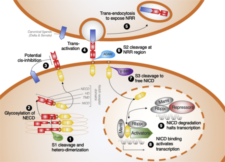
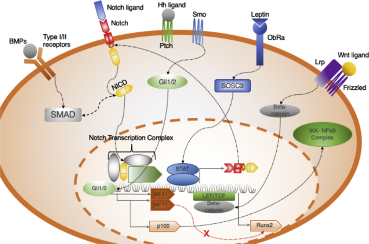
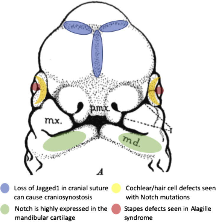
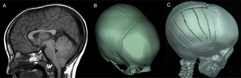
Notch is a cell-cell signaling pathway that is involved in a host of activities including development, oncogenesis, skeletal homeostasis, and much more. More specifically, recent research has demonstrated the importance of Notch signaling in osteogenic differentiation, bone healing, and in the development of the skeleton. The craniofacial skeleton is complex and understanding its development has remained an important focus in biology. In this review we briefly summarize what recent research has revealed about Notch signaling and the current understanding of how the skeleton, skull, and face develop. We then discuss the crucial role that Notch plays in both craniofacial development and the skeletal system, and what importance it may play in the future.

Keywords: Alagille syndrome; Bone; Craniofacial development; Craniosynostosis; Notch; Oncogenesis; Osteogenesis; Spondylocostal dysosotosis.


Figures

Figure 1
Figure 1
Notch signaling overview. This figure summarizes the signal transduction pathway of Notch. (1) Furin cleaves the precursor receptor in the golgi apparatus and the NECD heterodimerizes with the NICD. (2) NECD is glycosylated. (3) Notch ligands attached to the same cell are able to inhibit (cis-inhibition) Notch receptors. (4) Notch ligands on adjacent cells activate (trans-activation) the Notch heterodimer. (5) NECD is trans-endocytosed by the ligand cell. (6) S2 site exposed on the NRR region is cleaved by ADAMs proteins. (7) NICD is freed from the cell membrane through S3 cleavage by γSec. (8) NICD translocates to the nucleus where it forms a transcriptional regulatory complex with Rbpjκ and Maml. (9) NICD is ubiquinated and degraded reverting the complex to state of repression.
Figure 2
Figure 2
Summary of signaling crosstalk discussed in current review. This figure highlights the interaction between the different pathways discussed in the review. BMP activates the SMAD protein which is thought to interact with the intracellular NICD. Hh signaling activates Gli1/2 which causes transcription of Hes1, a shared target with Notch. Leptin signaling activates SOSC3 which downstream upregulates transcription of the Notch receptor. Wnt signaling causes transcription of the Runx2 transcription factor, which is inhibited by Hey1.
Figure 3
Figure 3
The role of Notch signaling in craniofacial development. This figure highlights the regions discussed in the review in which Notch has shown to be important in the developing face and skull, notably in the mandible, ear, and cranial suture.
Figure 4
Figure 4
Preoperative and post-operative 3D reconstruction of a CT scan performed on a child with Alagille syndrome who had complete fusion of all sutures except the coronal suture.(A) Sagittal T1 magnetic resonance image shows cerebellar tonsils hanging down below the foramen magnum. (B) Preoperative 3-dimensional cranial tomography reconstruction shows findings indicating that only the coronal suture is open and other sutures were fused. (C) Postoperative cranial tomography reconstruction image shows the details of craniotomy performed and the fused lambdoid suture. Reprint permission was granted by the authors and publisher (Yilmaz et al 2012, Pediatric Neurology, License number: 4775991160929).

References

    1. Morgan T.H. The theory of the gene. Am Nat. 1917;51(609):513–544.
    1. Hori K., Sen A., Artavanis-Tsakonas S. Notch signaling at a glance. J Cell Sci. 2013;126(10):2135–2140. - PMC - PubMed
    1. Kidd S., Lieber T., Young M.W. Ligand-induced cleavage and regulation of nuclear entry of Notch in Drosophila melanogaster embryos. Genes Dev. 1998;12(23):3728–3740. - PMC - PubMed
    1. Kopan R., Ilagan MaXG. The canonical notch signaling pathway: unfolding the activation mechanism. Cell. 2009;137(2):216–233. - PMC - PubMed
    1. Bray S.J. Notch signalling: a simple pathway becomes complex. Nat Rev Mol Cell Biol. 2006;7(9):678–689. - PubMed

LinkOut - more resources