. 2021 Jun;58(6):2940-2953.
doi: 10.1007/s12035-021-02296-y. Epub 2021 Feb 10.

The Coding and Small Non-coding Hippocampal Synaptic RNAome

Affiliations

The Coding and Small Non-coding Hippocampal Synaptic RNAome

Robert Epple et al. Mol Neurobiol. 2021 Jun.

Erratum in

Abstract

Neurons are highly compartmentalized cells that depend on local protein synthesis. Messenger RNAs (mRNAs) have thus been detected in neuronal dendrites, and more recently in the pre- and postsynaptic compartments as well. Other RNA species such as microRNAs have also been described at synapses where they are believed to control mRNA availability for local translation. A combined dataset analyzing the synaptic coding and non-coding RNAome via next-generation sequencing approaches is, however, still lacking. Here, we isolate synaptosomes from the hippocampus of young wild-type mice and provide the coding and non-coding synaptic RNAome. These data are complemented by a novel approach for analyzing the synaptic RNAome from primary hippocampal neurons grown in microfluidic chambers. Our data show that synaptic microRNAs control almost the entire synaptic mRNAome, and we identified several hub microRNAs. By combining the in vivo synaptosomal data with our novel microfluidic chamber system, our findings also support the hypothesis that part of the synaptic microRNAome may be supplied to neurons via astrocytes. Moreover, the microfluidic system is suitable for studying the dynamics of the synaptic RNAome in response to stimulation. In conclusion, our data provide a valuable resource and point to several important targets for further research.

Keywords: Gene expression; RNA sequencing; Synapse; Synaptosomes; lncRNA; mRNA; microRNA; snoRNA.


Conflict of interest statement

The authors declare no conflict of interest.

Figures
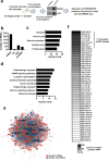
Fig. 1
Fig. 1
The coding and small non-coding RNAome of hippocampal synaptosomes. a Experimental scheme. b Bar graph showing the detected RNA species. c GO analysis showing that the identified mRNAs represent the synaptic compartment. d KEGG-pathway analysis showing that the synaptic mRNAome consists of transcripts that are essential for the function of hippocampal synapses. e microRNA-mRNA interaction network of the synaptic RNAome. Red circles represent the identified mRNAs that form a highly connected network, while blue circles indicate the detected microRNAs. Only the names of the top hub microRNAs are shown. f Heat map showing the synaptic microRNAome ranked by their confirmed mRNA targets that were found at synapses
Fig. 2
Fig. 2
Analyzing the synaptic RNAome in microfluidic chambers via RNA-sequencing. a Microfluid chambers built from PDMS. Left panel shows the scheme of the microfluidic chamber indicating the perfusion channel in which most of synapses form. The principle is based on chambers first reported by Taylor and colleagues [20] but has been substantially modified (see Fig S1 for more details). The middle panel shows the bright-field image of neurons growing in these chambers, and the right panel shows immunostaining for PSD-95 and synaptophysin within the perfusion channel (upper image) and the part of the chambers that contains the cell bodies (lower image). b Scheme and image showing our newly devised tool for cutting the perfusion channel from the microfluidic chambers, named SNIDER. c Schematic illustration of the cutting of the microfluidic chambers. d Venn diagram showing the comparison of the total RNA-seq data obtained from primary hippocampal cultures grown in normal dishes (primary neuronal culture) and corresponding data obtained from the perfusion channel isolated from microfluidic chambers in which primary hippocampal neurons were grown. e Bar chart showing the detected RNA species. f GO analysis showing that the identified mRNAs represent the synaptic compartment. g KEGG-pathway analysis showing that the synaptic mRNAome consists of transcripts that are essential for the function of hippocampal synapses. h Venn diagram showing the overlap of mRNAs detected in hippocampal synaptosomes and in microfluidic chambers. i Upper panel: GO analysis showing that the 1244 mRNAs specifically detected in microfluidic chambers represent the synaptic compartment and “cell projection.” Lower panel: KEGG-pathway analysis of the same dataset
Fig. 3
Fig. 3
A core synaptic microRNAome. a MicroRNA-mRNA interaction network of the synaptic RNAome detected in microfluidic chambers. Red circles represent the identified mRNAs that form a highly connected network, while blue circles indicate the detected microRNAs that control this network. Only the names of the top hub microRNAs are shown. b Venn diagram comparing microRNAs detected in microfluidic chambers (Chambers) and synaptosomes. c MicroRNA-mRNA interaction network of the 219 synaptic mRNAs commonly detected in synaptosomes and microfluidic chambers and the 17 commonly detected microRNAs. Only the names of the top hub microRNAs are shown
Fig. 4
Fig. 4
Comparing microRNAs from astrocytic exosomes to the synaptic RNAome. a Venn diagram comparing the 48 microRNAs exclusively detected in synaptosomes to the list of microRNAs found in astrocytic exosomes. b MicroRNA-mRNA interaction network showing that 203 of the commonly detected 219 mRNAs and 21 of the 23 microRNAs found in synaptosomes and astrocytic exosomes form an interaction network. Only the names of the top hub microRNAs are shown. c KEGG-pathway analysis of the 203 mRNAs within the network. d KEGG pathway analysis of the 16 common synaptic mRNAs that are not targeted by the overlapping microRNAs shown in a
Fig. 5
Fig. 5
The synaptic mRNAome upon stimulation. a Experimental scheme. b Volcano plot showing a substantial upregulation of synaptic RNAs upon KCL treatment. c GO analysis showing that the identified mRNAs represent the synaptic compartment. d KEGG pathway analysis showing that the changes of the synaptic mRNAome upon KCL treatment represent transcripts mainly linked to ribosomal function. e Upper panel shows images of the KEGG pathway for “ribosome.” Colored subunits represent transcripts significantly increased. Lower panel: bar chart showing that 50% of the genes that comprise the ribosome KEGG pathway are increased at the synaptic compartment upon KCL treatment

Similar articles

Cited by

References

    1. Biever A, Glock C, Tushev G, Ciirdaeva E, Dalmay T, Langer JD, Schuman EM. Monosomes actively translate synaptic mRNAs in neuronal processes. Science (New York, NY) 2020;367(6477):eaay4991. - PubMed
    1. Doyle M, Kiebler MA. Mechanisms of dendritic mRNA transport and its role in synaptic tagging. EMBO J. 2011;30:3540–3552. - PMC - PubMed
    1. Fonkeu Y, Kraynyukova N, Hafner AS, Kochen L, Sartori F, Schuman EM, Tchumatchenko T. How mRNA localization and protein synthesis sites influence dendritic protein distribution and dynamics. Neuron. 2019;103:1109–1122. - PubMed
    1. Holt CE, Martin KC, Schuman EM. Local translation in neurons: visualization and function. Nat Struct Mol Biol. 2019;26(7):557–566. - PubMed
    1. Kosik KS. Low Copy Number: How Dendrites Manage with So Few mRNAs. Neuron. 2016;92:1168–1180. - PubMed