. 2021 Apr;11(4):1209-1222.
doi: 10.1002/2211-5463.13114. Epub 2021 Mar 11.

Circular RNA profiles and the potential involvement of down-expression of hsa_circ_0001360 in cutaneous squamous cell carcinogenesis

Affiliations

Circular RNA profiles and the potential involvement of down-expression of hsa_circ_0001360 in cutaneous squamous cell carcinogenesis

Pingjiao Chen et al. FEBS Open Bio. 2021 Apr.

Abstract

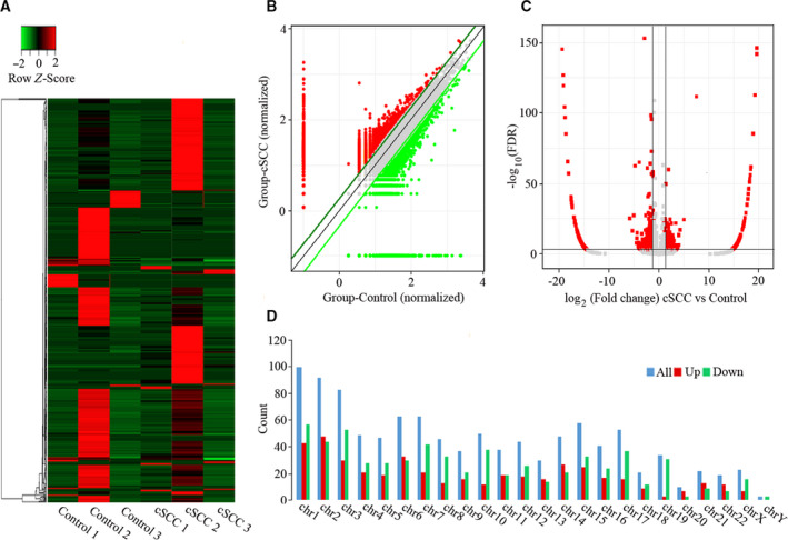
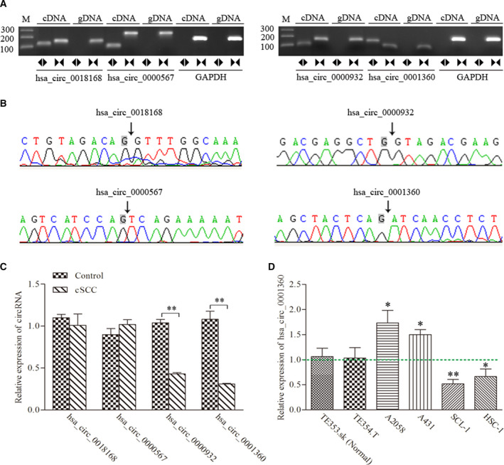
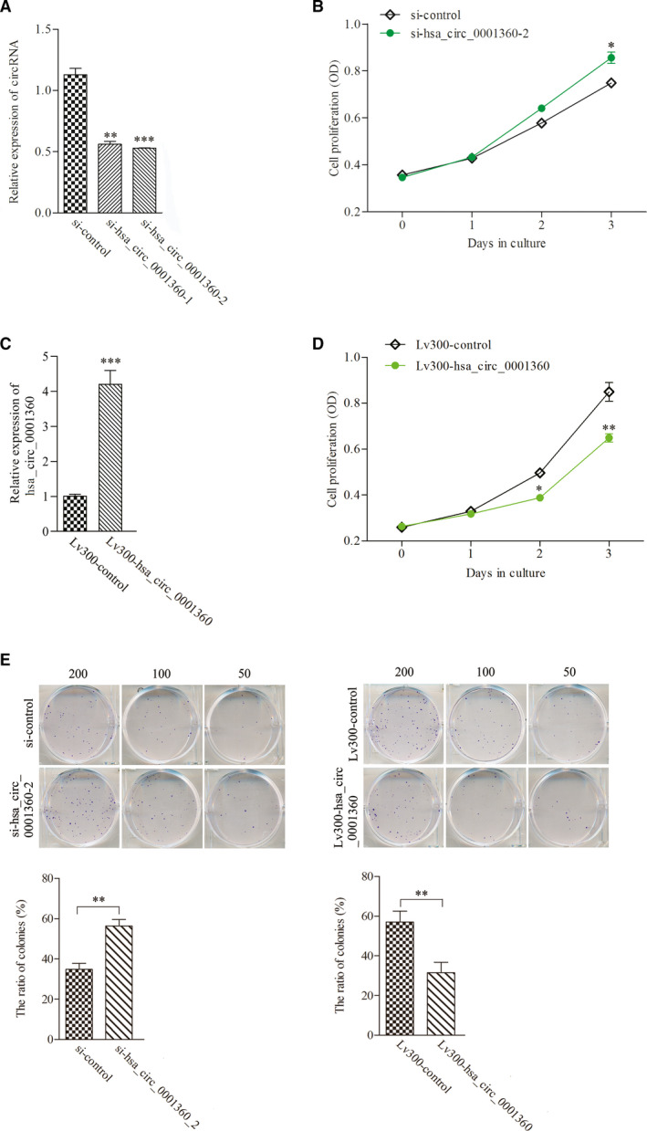
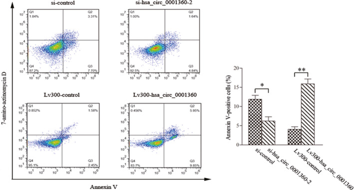
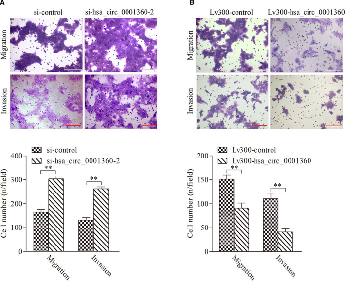
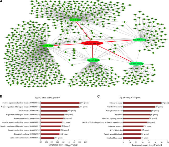
Circular RNAs (circRNAs) act as sponges of noncoding RNAs and have been implicated in many pathophysiological processes, including tumor development and progression. However, their roles in cutaneous squamous cell carcinoma (cSCC) are not yet well understood. This study aimed to identify differentially expressed circRNAs and their potential functions in cutaneous squamous cell carcinogenesis. The expression profiles of circRNAs in three paired cSCC and adjacent nontumorous tissues were detected with RNA sequencing and bioinformatics analysis. The candidate circRNAs were validated by PCR, Sanger sequencing and quantitative RT-PCR in another five matched samples. The biological functions of circRNAs in SCL-1 cells were assessed using circRNA silencing and overexpression, 3-(4,5-dimethylthiazol-2-yl)-5-(3-carboxymethoxyphenyl)-2-(4-sulfophenyl)-2H-tetrazolium inner salt (MTS), flow cytometry, transwell and colony formation assays. In addition, the circRNA-miRNA-mRNA interaction networks were predicted by bioinformatics. In summary, 1115 circRNAs, including 457 up-regulated and 658 down-regulated circRNAs (fold change ≥ 2 and P < 0.05), were differentially expressed in cSCC compared with adjacent nontumorous tissues. Of four selected circRNAs, two circRNAs (hsa_circ_0000932 and hsa_circ_0001360) were confirmed to be significantly decreased in cSCC using PCR, Sanger sequencing and quantitative RT-PCR. Furthermore, hsa_circ_0001360 silencing was found to result in a significant increase of the proliferation, migration and invasion but a significant decrease of apoptosis in SCL-1 cells in vitro, whereas hsa_circ_0001360 overexpression showed the opposite regulatory effects. hsa_circ_0001360 was predicted to interact with five miRNAs and their corresponding genes. In conclusion, circRNA dysregulation may play a critical role in carcinogenesis of cSCC, and hsa_circ_0001360 may have potential as a biomarker for cSCC.

Keywords: circular RNA; cutaneous squamous cell carcinoma; expression profile; hsa_circ_0001360.


Conflict of interest statement

The authors declare no conflict of interest.

Figures

Fig. 1
Fig. 1
Differences and characterizations in circRNA expression profiles between cSCC and adjacent nontumorous tissues. (A) Hierarchically clustered heatmap analyzed circRNA expression profiles of the six samples, with columns representing samples and rows representing circRNAs. Up‐regulation was shown in red, and down‐regulation was in green. (B) Scatterplots displayed the differential circRNA expression between cSCC (y axis) and adjacent nontumorous tissues (x axis). The circRNAs above the top green line and below the bottom green line indicated more than 2‐fold change of circRNAs between the two groups of compared samples. (C) Volcano plots represented the differential circRNA expression between cSCC and adjacent nontumorous tissues. The red points in the plot represented the differentially expressed circRNAs with statistical significance. (D) Chromosomal distributions of differentially expressed circRNAs.
Fig. 2
Fig. 2
Identification of four candidate circRNAs. (A) RT‐PCR with divergent (circular) and convergent (line) primers was used to confirm the candidate circRNAs in samples of cSCC tissues. Divergent (circular) primers (◄►) can amplify a single fragment at the expected sizes in cDNA, but not in gDNA. Convergent (line) primers (►◄) can amplify a single fragment at the expected sizes in cDNA and gDNA. (B) Sanger sequencing of the candidate circRNAs showed the backsplice junction. (C) qRT‐PCR showed the expression levels of the four candidate circRNAs between cSCC and adjacent nontumorous tissues. (D) qRT‐PCR showed the expression levels of hsa_circ_0001360 in TE353.sk, TE354.T, A2058, A431, SCL‐1 and HSC‐1 cell lines. Student's t‐test and one‐way ANOVA were used. The data were expressed as mean ± standard deviation (n = 3). *P < 0.05, **P < 0.01 represented statistical difference.
Fig. 3
Fig. 3
hsa_circ_0001360 regulated proliferation and colony formation activity of SCL‐1 cells. (A) The expression levels of hsa_circ_0001360 were significantly decreased after transfection with specifically synthesized siRNA in SCL‐1 cells. (B) The proliferation activity of SCL‐1 cells was significantly increased after transfection with si‐hsa_circ_0001360‐2. (C) The expression levels of hsa_circ_0001360 were significantly up‐regulated after transfection with Lv300‐hsa_circ_0001360‐2. (D) The proliferation activity of SCL‐1 cells was decreased after transfection with Lv300‐hsa_circ_0001360‐2. (E) The colony formation activity of SCL‐1 cells was increased after transfection with si‐hsa_circ_0001360‐2 and was decreased after transfection with Lv300‐hsa_circ_0001360‐2. Student's t‐test and one‐way ANOVA were used. The data were expressed as mean ± standard deviation (n = 5). *P < 0.05, **P < 0.01, ***P < 0.001 represented statistical difference.
Fig. 4
Fig. 4
Flow cytometry assays showed that hsa_circ_0001360 regulated apoptosis in SCL‐1 cells. Student's t‐test was used. The data were expressed as mean ± standard deviation (n = 3). *P < 0.05, **P < 0.01 represented statistical difference.
Fig. 5
Fig. 5
hsa_circ_0001360 regulated the migration and invasion of SCL‐1 cells. (A) Both the migration and invasion of SCL‐1 cells were significantly increased after transfection with si‐hsa_circ_0001360‐2. (B) Both the migration and invasion of SCL‐1 cells were significantly decreased after transfection with Lv300‐hsa_circ_0001360‐2. Scale bars represent 20 μm. Student's t‐test was used. The data were expressed as mean ± standard deviation (n = 5). **P < 0.01 represented statistical difference.
Fig. 6
Fig. 6
circRNA–miRNA–mRNA network, GO, and KEGG pathway analysis for hsa_circ_0001360. (A) The predicted hsa_circ_0001360 targeted the circRNA–miRNA–mRNA gene coexpression network. The five miRNAs (hsa‐miR‐8055, hsa‐miR‐8063, hsa‐miR‐4494, hsa‐miR‐888‐3p, and hsa‐miR‐6824‐5p) and their mRNA target genes were displayed based on sequence‐pairing prediction. (B) GO enrichment analysis for hsa_circ_0001360 in terms of biological processes (BP). The top 10 significantly enriched target genes and their scores (negative logarithm of P value) were listed as the x axis and the y axis, respectively. (C) KEGG pathway analysis for hsa_circ_0001360. The top 10 significantly enriched pathways and their scores (negative logarithm of P value) were listed as the x axis and the y axis, respectively.

Similar articles

Cited by

References

    1. Que SKT, Zwald FO and Schmults CD (2018) Cutaneous squamous cell carcinoma: incidence, risk factors, diagnosis, and staging. J Am Acad Dermatol 78, 237–247. - PubMed
    1. Work G, Invited R, Kim JYS, Kozlow JH, Mittal B, Moyer J, Olenecki T and Rodgers P (2018) Guidelines of care for the management of cutaneous squamous cell carcinoma. J Am Acad Dermatol 78, 560–578. - PMC - PubMed
    1. Yu X and Li Z (2016) The role of miRNAs in cutaneous squamous cell carcinoma. J Cell Mol Med 20, 3–9. - PMC - PubMed
    1. Zhou M, Zhou L, Zheng L, Guo L, Wang Y, Liu H, Ou C and Ding Z (2014) miR‐365 promotes cutaneous squamous cell carcinoma (CSCC) through targeting nuclear factor I/B (NFIB). PLoS One 9, e100620. - PMC - PubMed
    1. Karia PS, Han J and Schmults CD (2013) Cutaneous squamous cell carcinoma: estimated incidence of disease, nodal metastasis, and deaths from disease in the United States, 2012. J Am Acad Dermatol 68, 957–966. - PubMed

Publication types