. 2021 Jan 12;5(1):143-155.
doi: 10.1182/bloodadvances.2020002732.

Immune reconstitution and infectious complications following axicabtagene ciloleucel therapy for large B-cell lymphoma

Affiliations

Immune reconstitution and infectious complications following axicabtagene ciloleucel therapy for large B-cell lymphoma

John H Baird et al. Blood Adv. .

Abstract

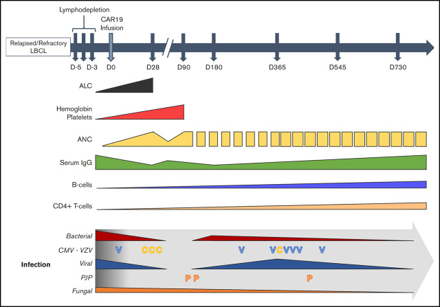
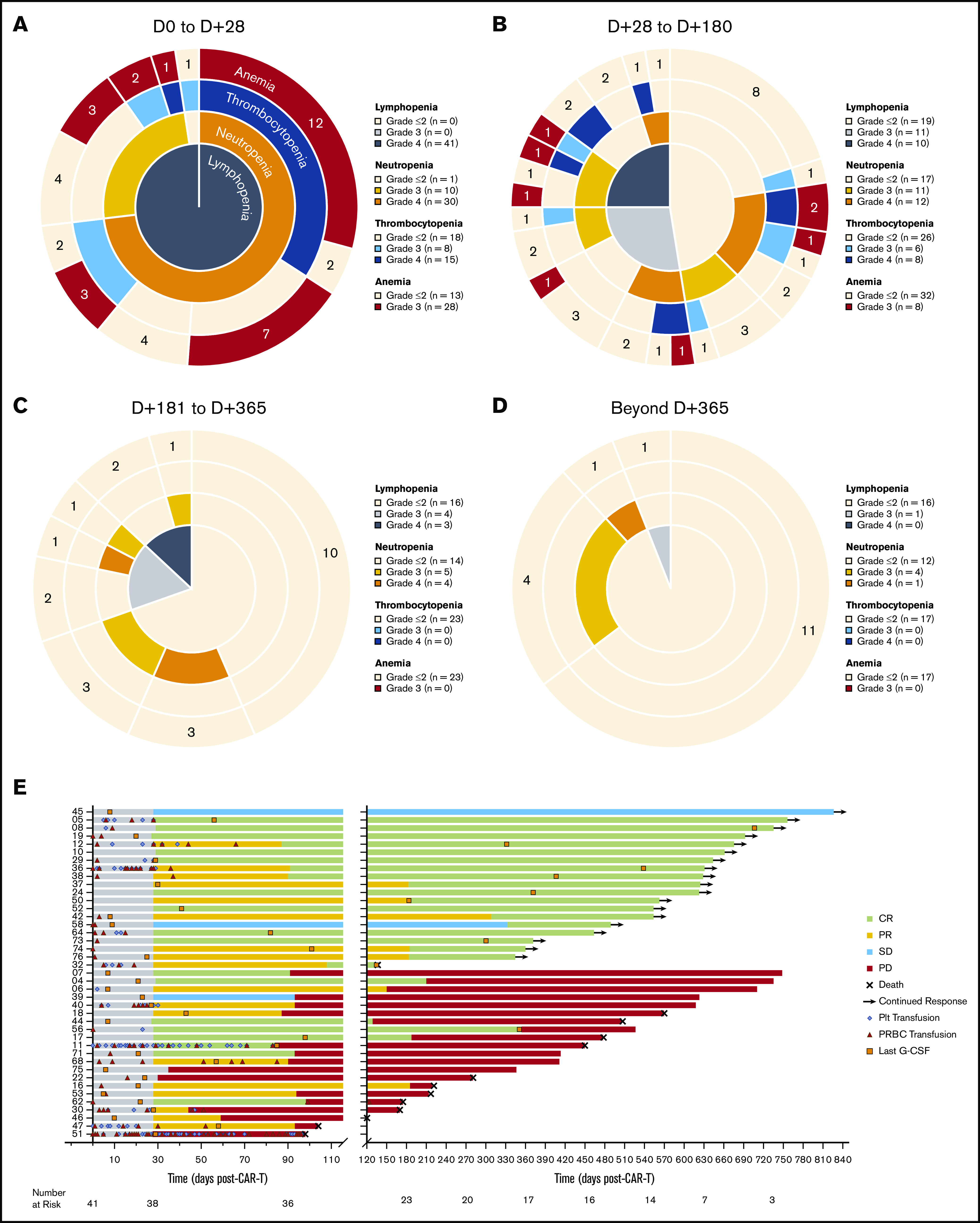
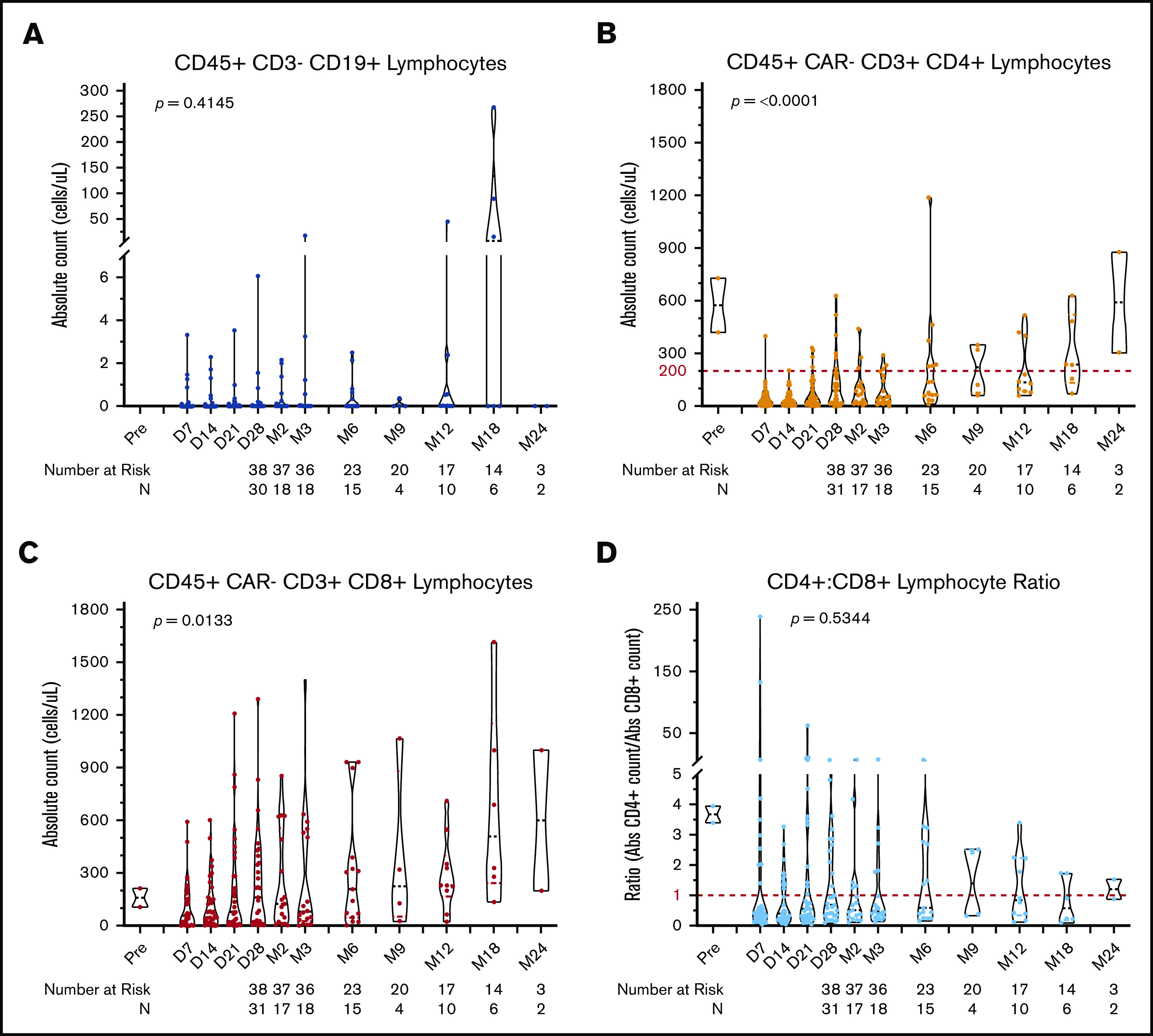
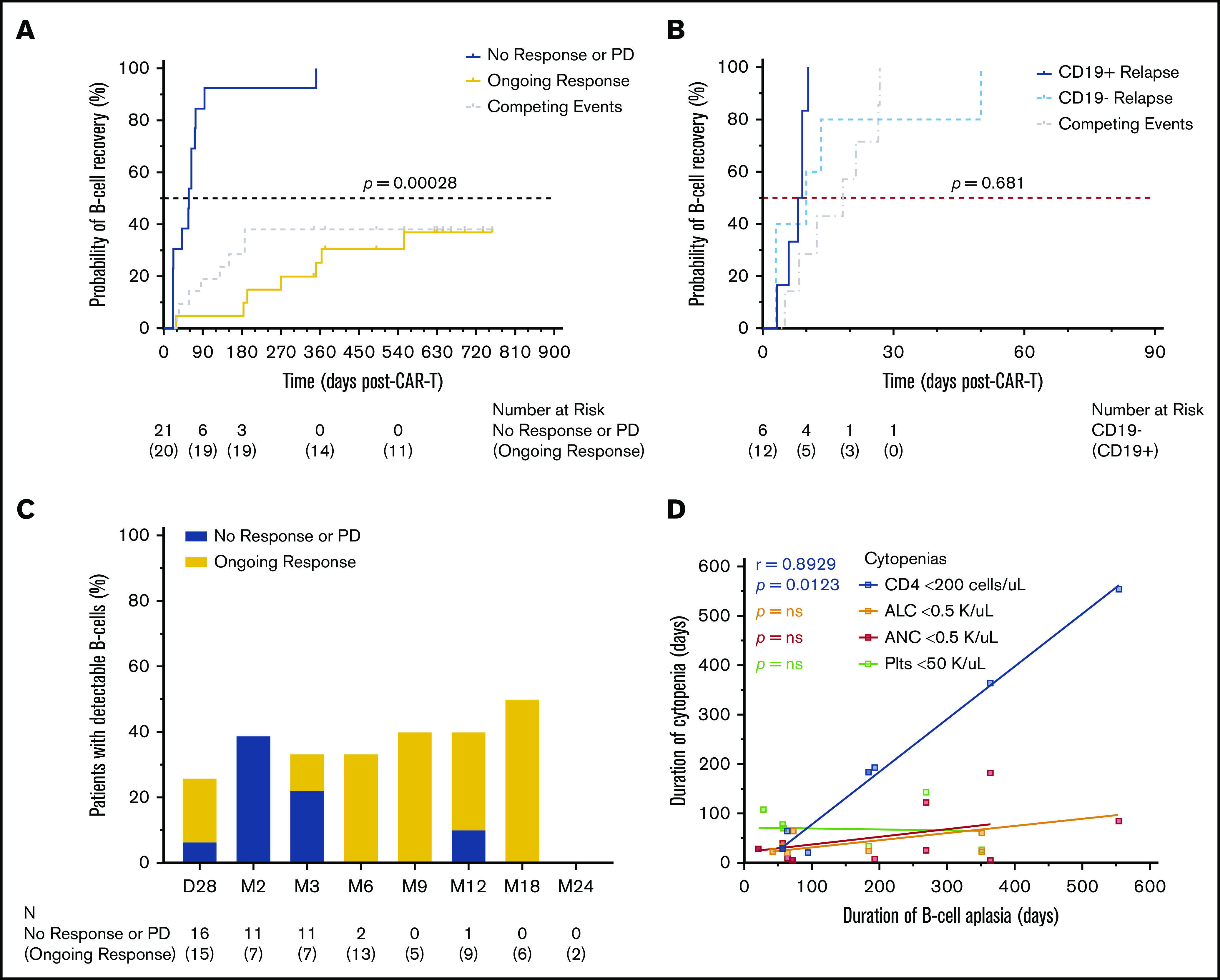
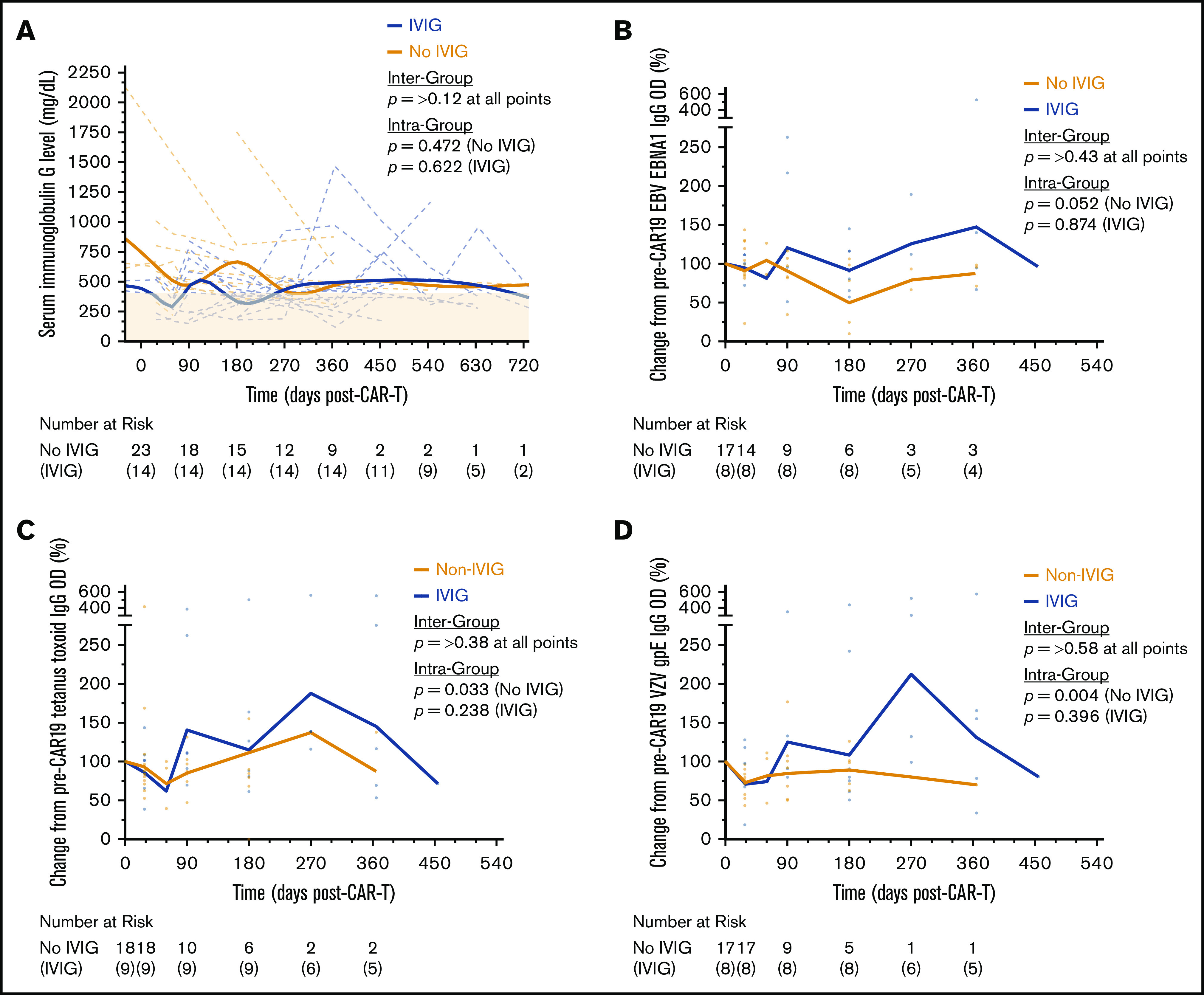
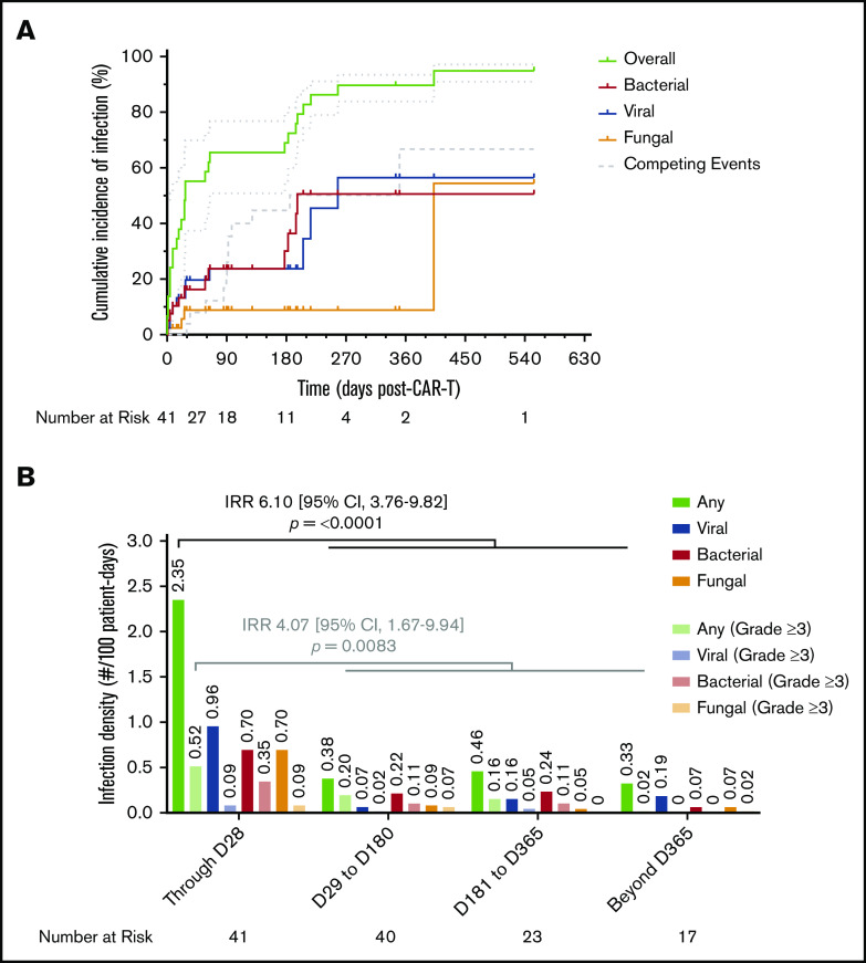
Chimeric antigen receptor (CAR) T-cell therapy targeting CD19 has significantly improved outcomes in the treatment of refractory or relapsed large B-cell lymphoma (LBCL). We evaluated the long-term course of hematologic recovery, immune reconstitution, and infectious complications in 41 patients with LBCL treated with axicabtagene ciloleucel (axi-cel) at a single center. Grade 3+ cytopenias occurred in 97.6% of patients within the first 28 days postinfusion, with most resolved by 6 months. Overall, 63.4% of patients received a red blood cell transfusion, 34.1% of patients received a platelet transfusion, 36.6% of patients received IV immunoglobulin, and 51.2% of patients received growth factor (granulocyte colony-stimulating factor) injections beyond the first 28 days postinfusion. Only 40% of patients had recovered detectable CD19+ B cells by 1 year, and 50% of patients had a CD4+ T-cell count <200 cells per μL by 18 months postinfusion. Patients with durable responses to axi-cel had significantly longer durations of B-cell aplasia, and this duration correlated strongly with the recovery of CD4+ T-cell counts. There were significantly more infections within the first 28 days compared with any other period of follow-up, with the majority being mild-moderate in severity. Receipt of corticosteroids was the only factor that predicted risk of infection in a multivariate analysis (hazard ratio, 3.69; 95% confidence interval, 1.18-16.5). Opportunistic infections due to Pneumocystis jirovecii and varicella-zoster virus occurred up to 18 months postinfusion in patients who prematurely discontinued prophylaxis. These results support the use of comprehensive supportive care, including long-term monitoring and antimicrobial prophylaxis, beyond 12 months after axi-cel treatment.


Conflict of interest statement

Conflict-of-interest disclosure: P.S. has received research support from Kite Pharma-Gilead. A.R.R. has received research support from Pharmacyclics/AbbVie; has served on ad hoc scientific advisory boards for Nohla Therapeutics and Kaleido; has served as an expert witness for US Department of Justice; and his brother works for Johnson & Johnson. T.L. has been a member of the speaker’s bureau for Kite Pharma-Gilead. C.L.M. has acted as a consultant for Lyell, NeoimmuneTech, Nektar Therapeutics, and Apricity Therapeutics; has received royalties from the National Institutes of Heath and Juno Therapeutics for CD22-CARl and has equity in Lyell and Allogene Therapeutics. D.B.M. has acted as a consulting for Kite Pharma-Gilead, Juno Therapeutics-Celgene, Novartis, Janssen Pharmaceuticals, and Pharmacyclics and has received research support from Kite Pharma-Gilead, Allogene Therapeutics, Pharmacyclics, Miltenyi Biotec, and Adaptive Biotechnologies. S.S. has acted as a consultant for Janssen Pharmaceuticals. The remaining authors declare no competing financial interests.

Figures

None
Graphical abstract
Figure 1.
Figure 1.
Hematologic recovery and transfusion utilization following CAR19. (A-D) Sunburst charts showing the overlapping prevalence of lymphopenia, neutropenia, thrombocytopenia, and anemia across 4 time periods post–axi-cel infusion. Concentric rings are organized in a hierarchical structure outward from the origin, with overlapping segments representing shared cytopenias within the same patient(s); the numbers shown in each outer ring segment represent patient(s) with that unique combination of cytopenias. The prevalence of individual cytopenias are shown in the accompanying keys. (E) Swimmer plot showing utilization of blood and platelet transfusions and time of last growth factor (G-CSF) administration for severe neutropenia. Patients are identified by study number on the left, and clinical response categorization is indicated by the bar color.
Figure 2.
Figure 2.
Lymphocyte subset recovery following CAR19. Violin plots showing the distribution of absolute cell counts for CD19+ B cells (A), CD4+ T cells (B), CD8+ T cells (C), and CD4/CD8 ratio measured by flow cytometry (D) performed on peripheral blood samples over the total duration of follow-up post–axi-cel infusion. The dashed red lines in B and D denote clinically actionable thresholds below which antimicrobial prophylaxis is used at the study center. The number of patients at risk, as well as the number of patients who underwent testing (N), is shown by time point below each plot. P values were obtained using the Kruskal-Wallis test; each time point was treated as an independent group.
Figure 3.
Figure 3.
B-cell aplasia and clinical response following CAR19. (A) Time-to-event analysis for recovery of CD19+ B cells detectable by flow cytometry performed on peripheral blood showed a significantly longer duration of B-cell aplasia in those patients who maintained a durable response to axi-cel. Patients were censored at the time of last follow-up; disease relapse or progression and death were considered competing events. Analysis was performed using the K-M method. (B) When patients who developed disease relapse or progression were stratified as CD19+ or CD19 status at relapse by immunohistochemistry or flow cytometry performed on lymphoma cells, there was no significant difference between the duration of B-cell aplasia observed. CD19 status at the time of relapse was available for 18 of 21 (85.7%) patients. (C) The proportion of patients with detectable B cells at each assessment time point, stratified by clinical response. The number of patients who underwent testing (N) is shown by time point below the plot. (D) Correlations between the duration of B-cell aplasia and the durations of other concurrent severe cytopenias; only noncensored durations from time-to-event analysis were included. Lines represent linear regression using least-squares fitting. P values shown were obtained by the likelihood ratio test computed using Cox proportional-hazards regression of the time-to-event variable vs the factor defining the 2 groups (A-B) and Spearman’s correlation analysis (D).
Figure 4.
Figure 4.
Serum immunoglobulin and antigen-specific antibody titers following CAR19. (A) Individual patient time courses showing recovery of serum IgG levels post–axi-cel infusion, stratified by whether patients received IVIG. The bold (Non-IVIG and IVIG) trend lines represents locally weighted scatterplot smoothing. Enzyme-linked immunosorbent assays were performed to detect antigen-specific IgG antibodies against Epstein-Barr virus (EBV) nuclear antigen 1 protein (EBNA1) (B), tetanus toxoid protein (C), and varicella-zoster virus (VZV) glycoprotein E (gpE) (D). Individual patient values relative to their pretreatment baseline are shown by the markers at each time point, with the bold (Non-IVIG and IVIG) trend lines connecting the geometric mean, stratified by whether patients received IVIG. P values shown were obtained by unpaired Student t tests between groups and Kruskal-Wallis tests within groups where each time point was treated as an independent group; both were adjusted for multiple comparisons using the Holm-Šídák method.
Figure 5.
Figure 5.
Cumulative incidence of infection and infection density following CAR19. (A) Cumulative incidence time-to-event analysis among the total cohort (N = 41) showing overall time to first infection of any type, as well as subsets for bacterial, viral, and fungal infections, over the total duration of follow-up after axi-cel infusion. Patients were censored at the time of last follow-up; disease relapse or progression and death were defined as competing events. Dotted lines represent 95% CI. (B) Infection densities are shown for any type and severity, as well as subsets for bacterial, viral, and fungal infections of any severity, occurring during 4 time periods following axi-cel infusion. The same groupings are interleaved for severe (CTCAE grade ≥3) infections alone. Calculated infection densities are shown above each bar. P values shown were obtained by calculating an IRR between the first 28 days of follow-up and the remainder of follow-up using the mid-P method and comparing using Fisher’s exact test.

References

    1. Locke FL, Ghobadi A, Jacobson CA, et al. . Long-term safety and activity of axicabtagene ciloleucel in refractory large B-cell lymphoma (ZUMA-1): a single-arm, multicentre, phase 1-2 trial. Lancet Oncol. 2019;20(1):31-42. - PMC - PubMed
    1. Neelapu SS, Rossi JM, Jacobson CA, et al. . CD19-loss with preservation of other B cell lineage features in patients with large B cell lymphoma who relapsed post-axi-cel. Blood. 2019;134(suppl 1):203.
    1. Lee DW, Santomasso BD, Locke FL, et al. . ASTCT consensus grading for cytokine release syndrome and neurologic toxicity associated with immune effector cells. Biol Blood Marrow Transplant. 2019;25(4):625-638. - PubMed
    1. Neelapu SS, Locke FL, Bartlett NL, et al. . Axicabtagene ciloleucel CAR T-cell therapy in refractory large B-cell lymphoma. N Engl J Med. 2017;377(26):2531-2544. - PMC - PubMed
    1. Strati P, Varma A, Adkins S, et al. . Hematopoietic recovery and immune reconstitution after axicabtagene ciloleucel in patients with large B-cell lymphoma [published online ahead of print 30 Jul 2020]. Haematologica. - PMC - PubMed

Publication types