. 2021 Feb 11;38(3):24.
doi: 10.1007/s12032-021-01464-3.

Dual inhibition of TGFβ and AXL as a novel therapy for human colorectal adenocarcinoma with mesenchymal phenotype

Affiliations

Dual inhibition of TGFβ and AXL as a novel therapy for human colorectal adenocarcinoma with mesenchymal phenotype

Davide Ciardiello et al. Med Oncol. .

Abstract

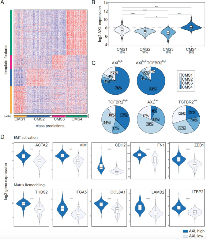
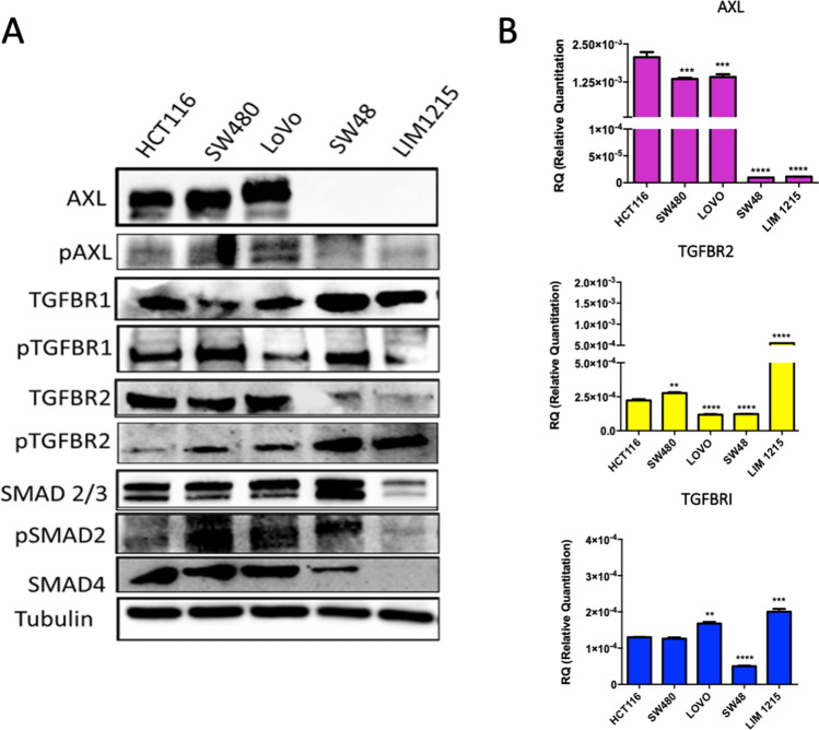
A subset of colorectal cancer (CRC) with a mesenchymal phenotype (CMS4) displays an aggressive disease, with an increased risk of recurrence after surgery, reduced survival, and resistance to standard treatments. It has been shown that the AXL and TGFβ signaling pathways are involved in epithelial-to-mesenchymal transition, migration, metastatic spread, and unresponsiveness to targeted therapies. However, the prognostic role of the combination of these biomarkers and the anti-tumor effect of AXL and TGFβ inhibition in CRC still has to be assessed. To evaluate the role of AXL and TGFβ as negative biomarker in CRC, we conducted an in-depth in silico analysis of CRC samples derived from the Gene Expression Omnibus. We found that AXL and TGFβ receptors are upregulated in CMS4 tumors and are correlated with an increased risk of recurrence after surgery in stage II/III CRC and a reduced overall survival. Moreover, we showed that AXL receptor is differently expressed in human CRC cell lines. Dual treatment with the TGFβ galunisertib and the AXL inhibitor, bemcentinib, significantly reduced colony formation and migration capabilities of tumor cells and displayed a strong anti-tumor activity in 3D spheroid cultures derived from patients with advanced CRC. Our work shows that AXL and TGFβ receptors identify a subgroup of CRC with a mesenchymal phenotype and correlate with poor prognosis. Dual inhibition of AXL and TGFβ could represent a novel therapeutic strategy for patients with this aggressive disease.

Keywords: AXL; Colorectal cancer; EMT; TGFβ.


Conflict of interest statement

DC reports travel grant from Sanofi; FM: Advisory Boards: MSD, Lilly; Institutional Research Grants: AstraZeneca; TT reports advisory board for Amgen, Bayer, Merck, Novartis, Roche, Sanofi; FC reports advisory board for Merck, Roche, Amgen, Bayer, Servier, Symphogen, Pfizer and research funding from Roche, Merck, Amgen, Bayer, Ipsen; EM advisory board for Amgen, Bayer, Merck, Roche, Sanofi, Servier and expert opinion for ESMO (European Society of Medical Oncology); all the other authors reports no conflict of interest.

Figures

Fig. 1
Fig. 1
AXL and TGFBR2 are associated with CRC progression. a Heatmap of assigned CMS subtypes to tumor samples based on template features by the CMS caller algorithm. The subtypes are highlighted in yellow (CMS1), blue (CMS2), pink (CMS3), and green (CMS4). The bars are not filled due to non-matched samples. The p-value is given from 0 to 1. b Violin and box plot of AXL expression in the CMS subtypes. The percentages below indicate the relative number of samples that were assigned to the individual CMS subtypes, respectively. c Pie charts of the CMS subtype distributions within the gene/s high versus gene/s low group. The cut-off of AXL and TGFBR2 was analyzed with maximum log-rank statistics. Rounding errors may cause little deviations from 100%. d Violin and box plots of several genes associated with epithelial-to-mesenchymal transition and matrix remodeling in AXL high compared to AXL low. Significance: p-value > 0.05 ns, < 0.05*, < 0.01**, < 0.001***, < 0.0001****
Fig. 2
Fig. 2
Kaplan–Meier survival analysis in CRC based on AXL and TGFBR2 expression. a The relapse-free survival (RFS) of CRC patients with low and high AXL and/or TGFBR2 expression in tumor tissue. b The overall survival (OS) of CRC patients with low and high AXL and/or TGFBR2 expression in tumor tissue. Variables: Time in days (survival or censoring time), number at risk, gene (gene low or highly expressed). Single-gene survival plots: low (black), high (red); gene signature plot: low/low (blue), low/high (shades of gray), high/high (red). Significance: p-value > 0.05 ns, < 0.05*, < 0.01**, < 0.001***, < 0.0001****
Fig. 3
Fig. 3
AXL and TGF-β expression and functional inhibition in a panel of CRC cell lines. a Western blot analysis of AXL and TGFβ receptor expression in human CRC cell lines. AXL is expressed in HCT116, SW480, and LoVo cells. b Relative gene expression of AXL, TGFBR2 and TGFBR1 in a panel of CRC cell lines
Fig. 4
Fig. 4
Colony-forming and migration assay in HCT116 and LoVo cells. a Colony-forming assay of HCT116 and LoVo cells treated with galunisertib 10 µM and/or bemcentinib 1 µM. Error bars indicate the standard deviation compared to single treatment. T-student test was used for statistical analysis, p-value < 0.05*, < 0.01**, < 0.001***, < 0.0001****. b Transwell migration assay at 48 h of LoVo cells treated with galunisertib 10 µM and/or bemcentinib 1 µM. Each assay was performed in duplicate. T-student test was used for statistical analysis, p-value < 0.05*, < 0.01**, < 0.001***, < 0.0001****
Fig. 5
Fig. 5
Anti-tumor activity of TGFBR1 and AXL is blocked in 3D patient-derived spheroid culture. MTT proliferation assay for colorectal cancer spheroids of case 1–7 treated with 10 µM galunisertib and 1 µM bemcentinib. Graphical representation of representative images with a magnification of 40×. T-student test was used for statistical analysis for comparing the treatments with the untreated control, p-value < 0.05*, < 0.01**, < 0.001***, < 0.0001****

References

    1. Howlader N, Noone AM, Krapcho M, et al (eds). SEER Cancer Statistics Review, 1975–2016, National Cancer Institute, Bethesda, MD, https://seer.cancer.gov/csr/1975_2016/, based on November 2018 SEER data submission, posted to the SEER website, April 2019.
    1. Van der Jeught K, Xu HC, Li YJ, Lu XB, Ji G. Drug resistance and new therapies in colorectal cancer. World J Gastroenterol. 2018;24(34):3834–3848. doi: 10.3748/wjg.v24.i34.3834. - DOI - PMC - PubMed
    1. Yaeger R, Chatila WK, Lipsyc MD, et al. Clinical sequencing defines the genomic landscape of metastatic colorectal cancer. Cancer Cell. 2018;33(1):125–136.e3. doi: 10.1016/j.ccell.2017.12.004. - DOI - PMC - PubMed
    1. Guinney J, Dienstmann R, Wang X, et al. The consensus molecular subtypes of colorectal cancer. Nat Med. 2015;21(11):1350–1356. doi: 10.1038/nm.3967. - DOI - PMC - PubMed
    1. Massagué J. TGFbeta in cancer. Cell. 2008;134(2):215–230. doi: 10.1016/j.cell.2008.07.001. - DOI - PMC - PubMed

MeSH terms