. 2021 Jan 27;7(5):eabe1235.
doi: 10.1126/sciadv.abe1235. Print 2021 Jan.

EphrinB2 clustering by Nipah virus G is required to activate and trap F intermediates at supported lipid bilayer-cell interfaces

Affiliations

EphrinB2 clustering by Nipah virus G is required to activate and trap F intermediates at supported lipid bilayer-cell interfaces

Joyce J Wong et al. Sci Adv. .

Abstract

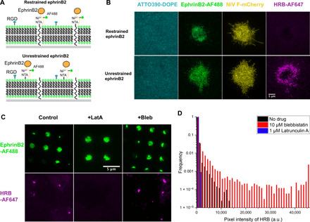
Paramyxovirus membrane fusion requires an attachment protein that binds to a host cell receptor and a fusion protein that merges the viral and host membranes. For Nipah virus (NiV), the G attachment protein binds ephrinB2/B3 receptors and activates F-mediated fusion. To visualize dynamic events of these proteins at the membrane interface, we reconstituted NiV fusion activation by overlaying F- and G-expressing cells onto ephrinB2-functionalized supported lipid bilayers and used TIRF microscopy to follow F, G, and ephrinB2. We found that G and ephrinB2 form clusters and that oligomerization of ephrinB2 is necessary for F activation. Single-molecule tracking of F particles revealed accumulation of an immobilized intermediate upon activation. We found no evidence for stable F-G protein complexes before or after activation. These observations lead to a revised model for NiV fusion activation and provide a foundation for investigating other multicomponent viral fusion systems.


Figures

Fig. 1
Fig. 1. Interactions of the NiV G ectodomain on SLBs.
(A) Membrane fusion schematic for a single viral glycoprotein, as in HIV and influenza virus. (B) Schematic for two-component membrane fusion machinery, such as NiV. (C) Schematic of ephrinB2 binding to DIBO–Alexa Fluor 647 (AF647)–labeled NiV Gecto bound to Ni–nitrilotriacetic acid (NTA) on supported lipid bilayers (SLBs). (D) EphrinB2 monomer binding to isolated receptor tetramers. (E) EphrinB2-Fc dimers could lead to larger cross-linked oligomers. (F) Experimental setup for NiV F–NiV G ectodomain complex formation. NiV Gecto Wt 40 Å–p-azido-phenylalanine (pAzF) was coincubated with increasing concentrations of NiV Fecto. (G) Schematic of potential assemblies that could form with NiV F trimers (blue triangle) and G tetramers (tetrameric rectangle), looking down on the SLB plane. Discrete assemblies of 2:1 F:G complexes could form or progress to larger arrays of F:G complexes. (H) Step size distribution of NiV Gecto Wt 40 Å-pAzF displacements following titration with ephrinB2-Fc-mNG. (I) Step size distribution of NiV Gecto Wt 40 Å-pAzF displacements following titration of monomeric ephrinB2-167-mNG. (J) Step size distribution of NiV Gecto Wt 40 Å-pAzF, NiV G L181Aecto 40 Å-pAzF, and NiV G V182Aecto 40 Å-pAzF displacements. (K) Step size distribution of NiV Gecto Wt 40 Å-pAzF displacements following titration of NiV Fecto.
Fig. 2
Fig. 2. Investigation of NiV F–NiV G interaction.
(A) Experimental setup for overlay of full-length NiV G and NiV F cotransfected Chinese hamster ovary cells on patterned substrates. EphrinB2 ectodomain-functionalized SLB circles are arrayed between RGD-ligand functionalized PLL-PEG. (B) Schematic of patterned bilayer substrate. (C) Cellular distribution of NiV G–enhanced green fluorescent protein (eGFP) and NiV F Wt-mSci in cells overlaid on patterned ephrinB2-AF488–functionalized SLBs. (D) Cellular distribution of NiV G–eGFP and NiV F N100C-A119C-mSci in cells overlaid on patterned ephrinB2-AF488–functionalized SLBs. (E) Cellular distribution of NiV G–eGFP and NiV F Wt-mSci in cells overlaid on patterned scFv5B3–functionalized SLBs.
Fig. 3
Fig. 3. NiV F is activated at the live cell–SLB interface.
(A) Schematic of the F conformation from pre- to postfusion conformation showing HRB peptide trapping at the prehairpin intermediate. (B) Binding of HRB to regions of NiV G–ephrinB2 cluster-mediated NiV F activation. NiV Gfull Wt cells cotransfected with pCAGGS, NiV Ffull Wt, or NiV F N100C-A119C were overlaid on SLB functionalized with ephrinB2-AF488 in the presence of 5 μM HRB-AF647. (C) Pixel intensity profile of HRB-AF647 binding fluorescence averaged over three cells. (D) The F membrane–spanning prehairin intermediate is restricted from diffusing out of bilayer areas. (E) Schematic of the confocal scanning microscope illumination planes for observation of lipid mixing. (F) Reconstitution of lipid mixing on a live cell–continuous SLB setup. Confocal microscopy images in the ephrinB2-AF488 channel were taken at the plane of the bilayer to image the SLB and in TR-DHPE channel at 5 μm above plane of the bilayer to selectively image fluorescent dye transfer into the cell membrane. a.u., arbitrary units.
Fig. 4
Fig. 4. NiV F is immobilized upon activation by NiV G and ephrinB2.
(A) Overlay of F-Halo-JF646 tracks, spots, and bilayer areas. Red tracks = mobile; blue tracks = immobile. (B) Displacement distributions of F-Halo-JF646 in F-Halo cells with ephrinB2-functionalized bilayers. (C) Displacement distributions of F-Halo-JF646 in F-Halo/Gfull-Wt cells with nonfunctionalized bilayers. (D) Displacement distributions of F-Halo-JF646 in F-Halo/Gfull-Wt cells with ephrinB2-functionalized bilayers. (E) Difference of average displacement fractions (inside versus outside) from F-Halo-JF646 tracking. (F) Averaged sum of squared differences of F-Halo-JF646 or G-Halo-JF646 step size distributions. n = 6 for −G +E, +G −E, and +G +E. n = 3 for +E +G +HRB. n = 5 for G-Halo. P values from one-way analysis of variance (ANOVA) followed by Dunnett’s T3 test, compared to G-Wt/F-Wt control. (G) Displacement distributions of F-Halo in F-Halo/Gfull-Wt cells with ephrinB2-functionalized bilayers and HRB. (H) Fraction of immobilized particles inside and outside bilayer regions for (A) to (D). (I) Displacement distributions of G-Halo in G-Halo/NiV Ffull-Wt cells with ephrinB2-functionalized bilayers. (J) Difference of average displacement fractions (inside versus outside) from G Halo-JF646 tracking. (K) Fraction of immobilized G-Halo-JF646 particles inside and outside bilayer regions functionalized with scFv5B3. For (H) and (K), P values were determined by paired-value t test of the immobilized fraction of particles within the same cell. n.s., not significant, **P < 0.01, ***P < 0.001.
Fig. 5
Fig. 5. Clustering of ephrinB2 and the actin cytoskeleton is necessary for F activation.
(A) Schematic of restrained (top) and unrestrained ephrinB2 (bottom) on the SLB. Restrained ephrinB2 is anchored through Ni-NTA-PEG linkers attached to the glass slide, preventing lateral diffusion. Unrestrained ephrinB2 is captured through Ni-NTA lipid that surrounds uncharged Ni-free NTA-PEG. (B) Restrained ephrinB2 does not allow for F activation, as monitored by HRB-AF647 binding (top). In contrast, unrestrained ephrinB2 captured at comparable overall densities (as measured by AF488 fluorescence) leads to activation of F (bottom). (C) Binding of HRB-AF647 to NiV Ffull Wt in bilayer areas with ephrinB2-AF488 bound to 4% Ni-NTA-DOGS in the presence of Latrunculin A or blebbistatin. (D) Pixel intensity profile of HRB-AF647 binding fluorescence averaged over nine cells in the presence or absence of blebbistatin.
Fig. 6
Fig. 6. Models for F immobilization and membrane fusion activation.
(A) Following activation, F forms a fast-diffusing prehairpin intermediate when trapped by HRB peptide binding. Without HRB peptide inhibition, F forms a slow-diffusing state that could be due to higher-order F oligomerization via HRA domains or trapping at hemifusion stalks. (B) Schematic for an ephrinB2 tetramerization model for NiV G and F activation. (C) Schematic for an oligomerization model requiring clustering of ephrinB2:G complexes to activate F.

References

    1. Kielian M., Mechanisms of virus membrane fusion proteins. Annu. Rev. Virol. 1, 171–189 (2014). - PubMed
    1. Rey F. A., Lok S.-M., Common features of enveloped viruses and implications for immunogen design for next-generation vaccines. Cell 172, 1319–1334 (2018). - PMC - PubMed
    1. Bose S., Jardetzky T. S., Lamb R. A., Timing is everything: Fine-tuned molecular machines orchestrate paramyxovirus entry. Virology 479-480, 518–531 (2015). - PMC - PubMed
    1. Sathiyamoorthy K., Chen J., Longnecker R., Jardetzky T. S., The COMPLEXity in herpesvirus entry. Curr. Opin. Virol. 24, 97–104 (2017). - PMC - PubMed
    1. Yin H.-S., Wen X., Paterson R. G., Lamb R. A., Jardetzky T. S., Structure of the parainfluenza virus 5 F protein in its metastable, prefusion conformation. Nature 439, 38–44 (2006). - PMC - PubMed

Publication types