. 2021 May;64(5):569-578.
doi: 10.1165/rcmb.2020-0166OC.

IL-33-mediated Eosinophilia Protects against Acute Lung Injury

Affiliations

IL-33-mediated Eosinophilia Protects against Acute Lung Injury

Paulette A Krishack et al. Am J Respir Cell Mol Biol. 2021 May.

Abstract

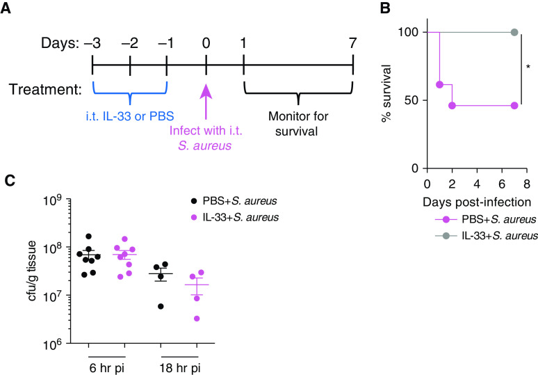
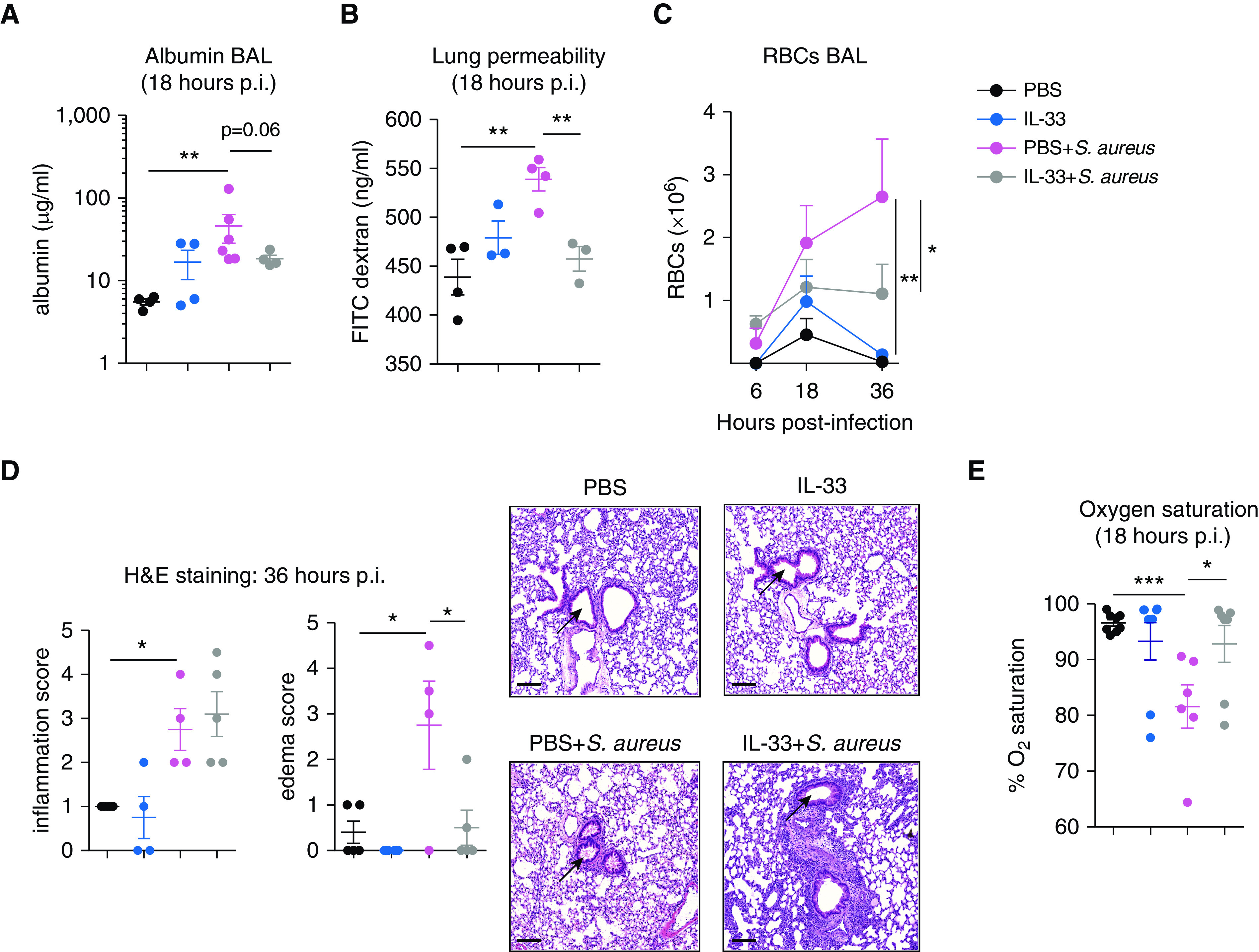
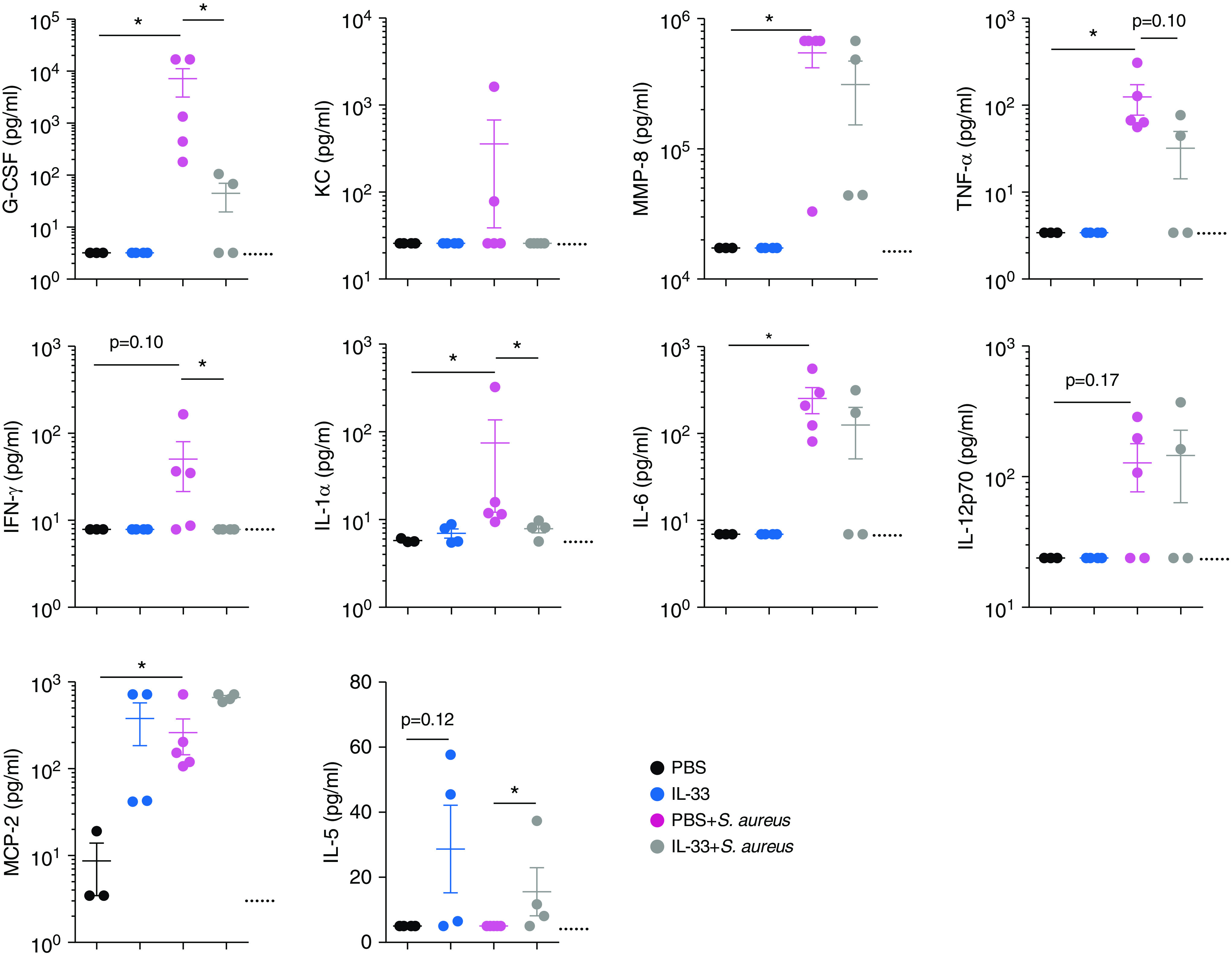
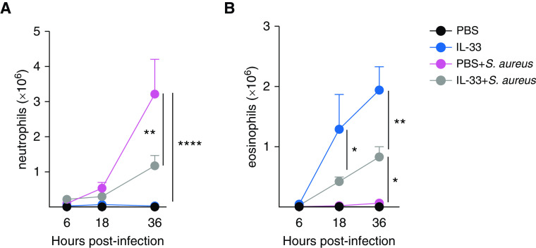
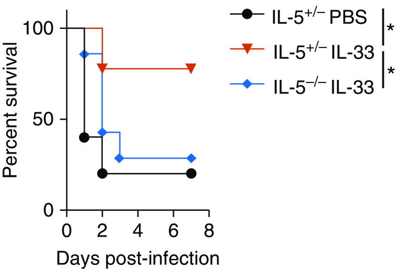
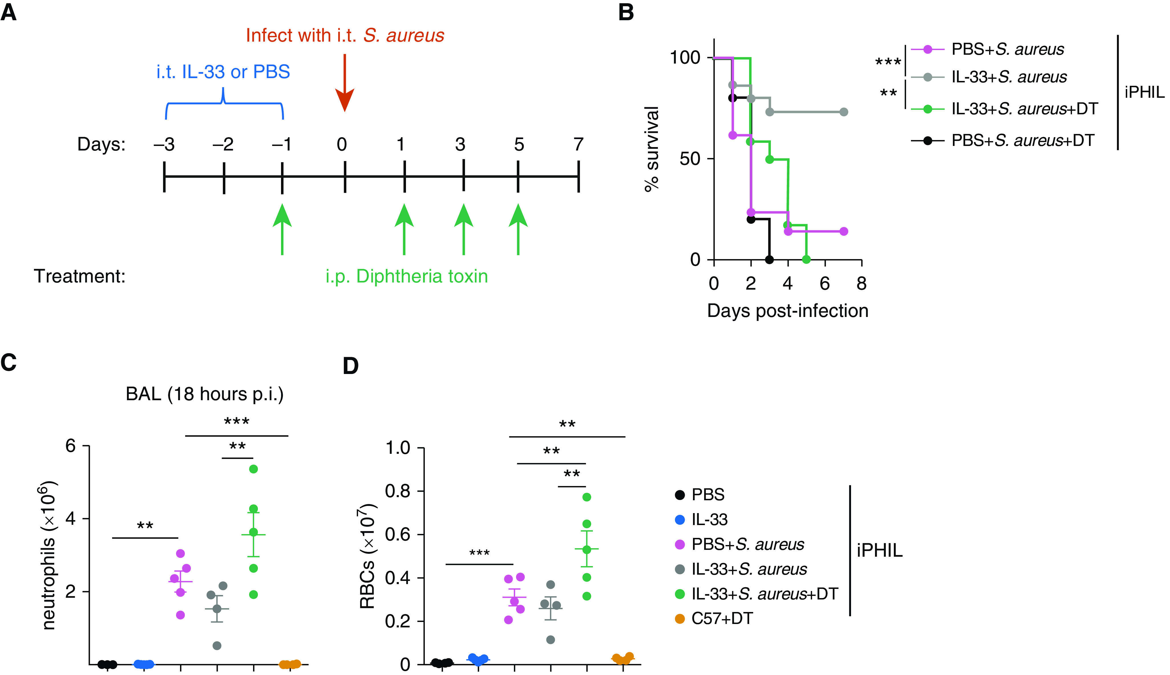
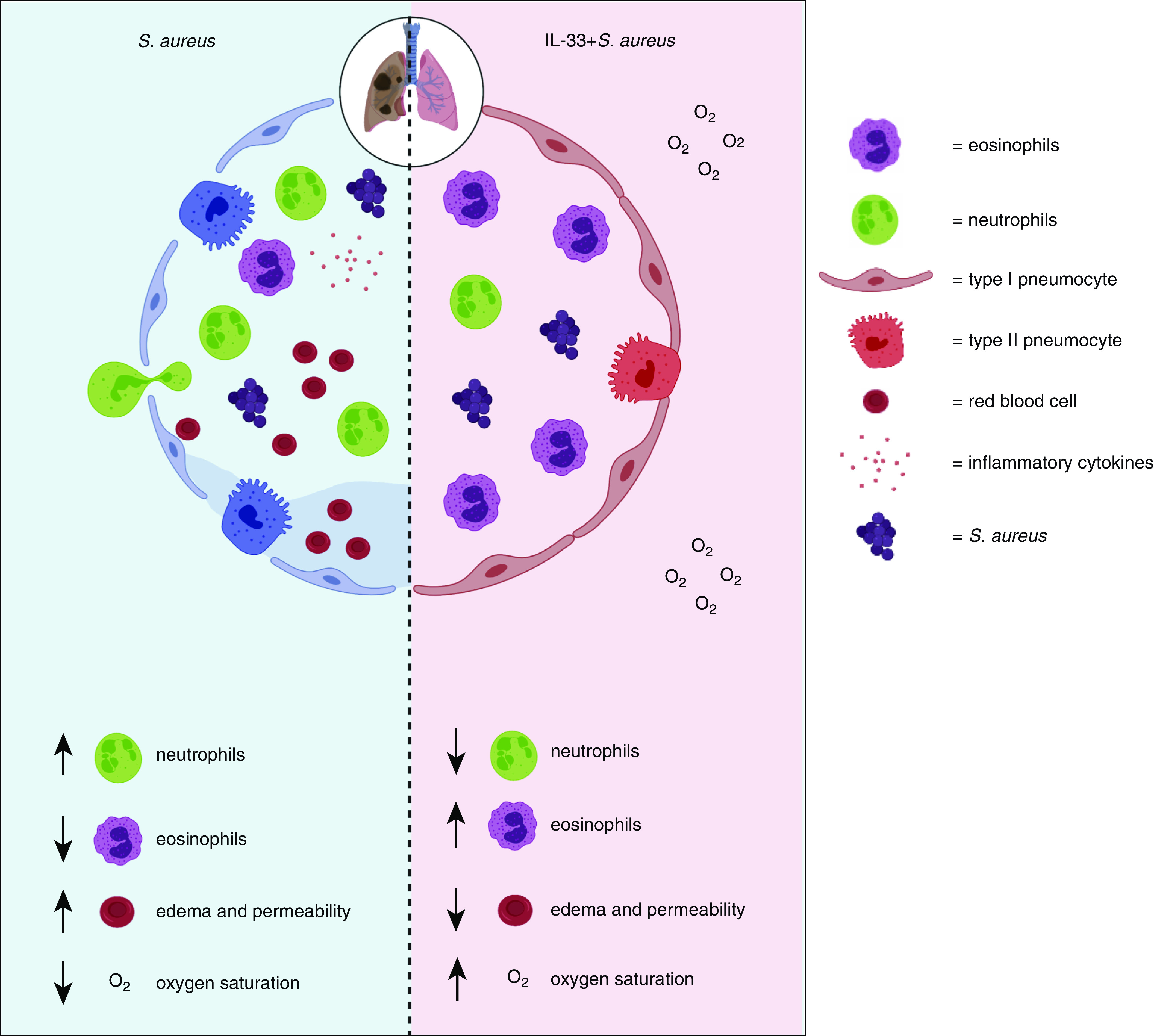
Pneumonia-induced lung injury and acute respiratory distress syndrome can develop because of an inappropriate inflammatory response to acute infections, leading to a compromised alveolar barrier. Recent work suggests that hospitalized patients with allergies/asthma are less likely to die of pulmonary infections and that there is a correlation between survival from acute respiratory distress syndrome and higher eosinophil counts; thus, we hypothesized that eosinophils associated with a type 2 immune response may protect against pneumonia-induced acute lung injury. To test this hypothesis, mice were treated with the type 2-initiating cytokine IL-33 intratracheally 3 days before induction of pneumonia with airway administration of a lethal dose of Staphylococcus aureus. Interestingly, IL-33 pretreatment promoted survival by inhibiting acute lung injury: amount of BAL fluid proinflammatory cytokines and pulmonary edema were both reduced, with an associated increase in oxygen saturation. Pulmonary neutrophilia was also reduced, whereas eosinophilia was strongly increased. This eosinophilia was key to protection; eosinophil reduction eliminated both IL-33-mediated protection against mortality and inhibition of neutrophilia and pulmonary edema. Together, these data reveal a novel role for eosinophils in protection against lung injury and suggest that modulation of pulmonary type 2 immunity may represent a novel therapeutic strategy.

Keywords: IL-33; Staphylococcus aureus; eosinophilia; lung injury; neutrophils.


Figures

Figure 1.
Figure 1.
IL-33 pretreatment protects against Staphylococcus aureus–induced pneumonia. C57BL/6 mice were treated intratracheal (i.t.) for 3 days with IL-33 or PBS. On Day 0, both groups were infected i.t. with 2 × 108 S. aureus cfu. Mice were monitored for survival. (A) Diagram demonstrating the experimental setup. (B) Kaplan-Meier survival curve. Statistical significance was determined using a log-rank (Mantel-Cox) test (n = 7–13 mice per group). (C) Bacterial burden in the lung at 6 and 18 hours post-infection (p.i.). The bacterial burden was measured in C57BL/6 mice treated with PBS or IL-33. Bacteria was quantified using serial dilution of colony-forming units (n = 4–9 mice per group). Significance was determined using an unpaired t test. *P < 0.05. cfu = colony-forming unit; S. aureus = Staphylococcus aureus.
Figure 2.
Figure 2.
IL-33 pretreatment reduces S. aureus–induced lung injury. C57BL/6 mice were treated i.t. with PBS, IL-33, PBS + S. aureus, or IL-33 + S. aureus. (A) BAL fluid albumin was measured at 18 hours p.i. (B) Quantification of lung permeability using FITC–dextran leakage from alveoli into serum at 18 hours p.i. Pulmonary edema was quantified using (C) flow cytometry measurement of BAL fluid red blood cell (RBC) counts or (D) H&E staining of lung sections with arrows indicating the area assessed for edema within the airways. RBCs were measured in the BAL fluid at 6, 18 and 36 hours p.i. and defined as single cells, live CD45Ter119+FSClo. (E) Oxygen saturation was measured in live, anesthetized mice at 18 hours p.i. for 10 minutes using the MouseOx Plus system. For AC, representative graphs of two experiments with sample sizes of three to six per group are shown. For E, two experiments were pooled, with total sample sizes between six and eight per group. For C, statistical significance was determined using a two-way ANOVA with a Tukey post hoc test, whereas for A, B, D, and E, significance was determined using an unpaired t test. Scale bars, 100 μm. *P < 0.05, **P < 0.01, and ***P < 0.001. H&E = hematoxylin and eosin.
Figure 3.
Figure 3.
IL-33 pretreatment inhibits induction of early inflammatory factors after S. aureus–induced pneumonia. C57BL/6 mice were treated i.t. with PBS, IL-33, PBS + S. aureus, or IL-33 + S. aureus. All groups were killed at 18 hours p.i. BAL fluid cytokine and chemokine levels were quantified with a multiplex assay. Dotted lines indicate the limit of detection. Statistical significance was determined using a Mann-Whitney test. Sample sizes were three to five per group. *P < 0.05. G-CSF = granulocyte colony-stimulating factor; KC = keratinocyte-derived chemokine; MCP-2 = monocyte chemotactic protein-2; MMP-8 = matrix metalloproteinase-8.
Figure 4.
Figure 4.
S. aureus–induced neutrophilia during pneumonia is suppressed by IL-33 treatment in association with an increase in eosinophils. C57BL/6 mice were treated i.t. with PBS, IL-33, PBS + S. aureus, or IL-33 + S. aureus. All groups were killed, and flow cytometry was performed on BAL fluid. Shown are graphs depicting (A) neutrophil counts or (B) eosinophil counts at 6, 18, or 36 hours p.i. Neutrophils were defined as singlet, live, CD11b+Ly6Ghi F4/80. Eosinophils were defined as singlet, live, CD45+Ly6GloCD11cSiglecF+SSChi. Significance was determined using a two-way ANOVA with a Tukey post hoc test. Each time point is a representative experiment of two independent experiments, with sample sizes of three to six per group. *P < 0.05, **P < 0.001, and ****P < 0.0001.
Figure 5.
Figure 5.
IL-5 expression is required for IL-33–mediated survival of S. aureus–induced pneumonia. Shown is a Kaplan-Meier survival curve for IL-5+/− or IL-5−/− mice. Mice were treated i.t. with PBS or IL-33 for 3 days, which was followed by i.t. infection with S. aureus on Day 0. All groups were monitored for survival for 7 days. Statistical significance was determined using a log-rank (Mantel-Cox) test. The graph is pooled from two experiments (n = 5–9 mice per group). *P < 0.05.
Figure 6.
Figure 6.
Eosinophils are required for survival and inhibition of S. aureus–induced neutrophilia and RBC infiltration during pneumonia. (A) Diagram of the experimental setup for the survival curve. On Days −3 to −1, iPHIL mice were treated i.t. with PBS or IL-33. S. aureus + diphtheria toxin (DT) mice received DT intraperitoneally on Day −1 and every other day through Day 5. All groups were infected i.t. with S. aureus on Day 0. All groups were monitored for survival for 7 days. (B) Kaplan-Meier survival curve. Statistical significance was determined using a log-rank (Mantel-Cox) test. The graph is pooled from a sample size of four experiments, with n = 10–21 per group. (C) BAL fluid neutrophil and (D) RBC counts were measured in iPHIL mice depleted of eosinophils. The treatment schedule was similar to the setup in A, with the exception of all groups being killed at 18 hours p.i. for flow cytometry analysis of BAL fluid. As a negative control, C57BL/6 mice were treated with DT alone. Statistical analysis was performed with a one-way ANOVA. Graphs are representative of two independent experiments, with n = 3–5 per group. **P < 0.01 and ***P < 0.001. iPHIL = inducible eosinophil-deficient.
Figure 7.
Figure 7.
IL-33–mediated eosinophilia protects against lethal S. aureus pneumonia by inhibiting lung injury, with up arrows indicating an increase and down arrows indicating a decrease of the indicated feature. During S. aureus–induced pneumonia, neutrophils extravasate from the blood and interstitium into alveoli early during the immune response to promote bacterial clearance. However, neutrophils can also cause tissue damage, resulting in destruction of epithelial cells lining the alveoli. This causes infiltration of inflammatory cytokines, increased pulmonary edema, and finally hypoxemia. Our study demonstrates that IL-33–mediated eosinophilia inhibited induction of airway inflammatory cytokines, neutrophilia, pulmonary edema, lung permeability, and hypoxemia.

Comment in

  • Can Eosinophils Prevent Lung Injury? Ask PHIL.
    Evans CM, McCubbrey AL. Evans CM, et al. Am J Respir Cell Mol Biol. 2021 May;64(5):523-524. doi: 10.1165/rcmb.2021-0083ED. Am J Respir Cell Mol Biol. 2021. PMID: 33651669 Free PMC article. No abstract available.

References

    1. Englert JA, Bobba C, Baron RM. Integrating molecular pathogenesis and clinical translation in sepsis-induced acute respiratory distress syndrome. JCI Insight. 2019;4:e124061. - PMC - PubMed
    1. Rhee C, Dantes R, Epstein L, Murphy DJ, Seymour CW, Iwashyna TJ, et al. CDC Prevention Epicenter Program. Incidence and trends of sepsis in US hospitals using clinical vs claims data, 2009-2014. JAMA. 2017;318:1241–1249. - PMC - PubMed
    1. Matthay MA, Zemans RL, Zimmerman GA, Arabi YM, Beitler JR, Mercat A, et al. Acute respiratory distress syndrome. Nat Rev Dis Primers. 2019;5:18. - PMC - PubMed
    1. Wuerth BA, Bonnewell JP, Wiemken TL, Arnold FW. Trends in pneumonia mortality rates and hospitalizations by organism, United States, 2002-2011. Emerg Infect Dis. 2016;22:1624–1627. - PMC - PubMed
    1. Thomer L, Schneewind O, Missiakas D. Pathogenesis of Staphylococcus aureus bloodstream infections. Annu Rev Pathol. 2016;11:343–364. - PMC - PubMed

Publication types

MeSH terms

LinkOut - more resources