

Different Roles of Mitochondria in Cell Death and Inflammation: Focusing on Mitochondrial Quality Control in Ischemic Stroke and Reperfusion
- PMID: 33572080
- PMCID: PMC7914955
- DOI: 10.3390/biomedicines9020169
Different Roles of Mitochondria in Cell Death and Inflammation: Focusing on Mitochondrial Quality Control in Ischemic Stroke and Reperfusion
Abstract
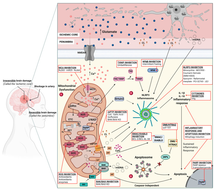
Mitochondrial dysfunctions are among the main hallmarks of several brain diseases, including ischemic stroke. An insufficient supply of oxygen and glucose in brain cells, primarily neurons, triggers a cascade of events in which mitochondria are the leading characters. Mitochondrial calcium overload, reactive oxygen species (ROS) overproduction, mitochondrial permeability transition pore (mPTP) opening, and damage-associated molecular pattern (DAMP) release place mitochondria in the center of an intricate series of chance interactions. Depending on the degree to which mitochondria are affected, they promote different pathways, ranging from inflammatory response pathways to cell death pathways. In this review, we will explore the principal mitochondrial molecular mechanisms compromised during ischemic and reperfusion injury, and we will delineate potential neuroprotective strategies targeting mitochondrial dysfunction and mitochondrial homeostasis.
Keywords: cell death; inflammation; ischemic reperfusion; ischemic stroke; mitochondrial fission; mitochondrial fusion; mitochondrial transfer; mitophagy.
Conflict of interest statement
The authors declare no conflict of interest.
Figures

Similar articles
-
Mitochondria: a target for neuroprotective interventions in cerebral ischemia-reperfusion.Curr Pharm Des. 2006;12(6):739-57. doi: 10.2174/138161206775474242. Curr Pharm Des. 2006. PMID: 16472163 Review.
-
Ginkgolide K attenuates neuronal injury after ischemic stroke by inhibiting mitochondrial fission and GSK-3β-dependent increases in mitochondrial membrane permeability.Oncotarget. 2017 Jul 4;8(27):44682-44693. doi: 10.18632/oncotarget.17967. Oncotarget. 2017. PMID: 28591721 Free PMC article.
-
The path from mitochondrial ROS to aging runs through the mitochondrial permeability transition pore.Aging Cell. 2017 Oct;16(5):943-955. doi: 10.1111/acel.12650. Epub 2017 Jul 31. Aging Cell. 2017. PMID: 28758328 Free PMC article. Review.
-
Edaravone and cyclosporine A as neuroprotective agents for acute ischemic stroke.Acute Med Surg. 2018 May 17;5(3):213-221. doi: 10.1002/ams2.343. eCollection 2018 Jul. Acute Med Surg. 2018. PMID: 29988669 Free PMC article. Review.
-
Inhibition of Bcl-2 sensitizes mitochondrial permeability transition pore (MPTP) opening in ischemia-damaged mitochondria.PLoS One. 2015 Mar 10;10(3):e0118834. doi: 10.1371/journal.pone.0118834. eCollection 2015. PLoS One. 2015. PMID: 25756500 Free PMC article.
Cited by
-
Mitochondria: Insights into Crucial Features to Overcome Cancer Chemoresistance.Int J Mol Sci. 2021 Apr 30;22(9):4770. doi: 10.3390/ijms22094770. Int J Mol Sci. 2021. PMID: 33946271 Free PMC article. Review.
-
Spreading depolarization causes reversible neuronal mitochondria fragmentation and swelling in healthy, normally perfused neocortex.J Cereb Blood Flow Metab. 2024 Dec;44(12):1561-1579. doi: 10.1177/0271678X241257887. Epub 2024 Jul 25. J Cereb Blood Flow Metab. 2024. PMID: 39053498 Free PMC article.
-
Role of Mitochondrial Dysfunctions in Neurodegenerative Disorders: Advances in Mitochondrial Biology.Mol Neurobiol. 2025 Jun;62(6):6827-6855. doi: 10.1007/s12035-024-04469-x. Epub 2024 Sep 13. Mol Neurobiol. 2025. PMID: 39269547 Review.
-
P‑hydroxybenzyl alcohol ameliorates neuronal cerebral ischemia‑reperfusion injury by activating mitochondrial autophagy through SIRT1.Mol Med Rep. 2023 Mar;27(3):68. doi: 10.3892/mmr.2023.12955. Epub 2023 Feb 17. Mol Med Rep. 2023. PMID: 36799156 Free PMC article.
-
Special Issue "Mitochondria and Brain Disease".Biomedicines. 2022 Aug 1;10(8):1854. doi: 10.3390/biomedicines10081854. Biomedicines. 2022. PMID: 36009401 Free PMC article.
References
-
- Adams H.P., Bendixen B.H., Kappelle L.J., Biller J., Love B.B., Gordon D.L., Marsh E.E. Classification of subtype of acute ischemic stroke. Definitions for use in a multicenter clinical trial. TOAST. Trial of Org 10172 in Acute Stroke Treatment. Stroke. 1993;24:35–41. doi: 10.1161/01.STR.24.1.35. - DOI - PubMed
-
- Lee T.-Y., Murphy B.D., Aviv R.I., Fox A.J., Black S.E., Sahlas D.J., Symons S., Lee D.H., Pelz D., Gulka I.B., et al. Cerebral Blood Flow Threshold of Ischemic Penumbra and Infarct Core in Acute Ischemic Stroke: A Systematic Review. Stroke. 2006;37:2201. doi: 10.1161/01.STR.0000237068.25105.aa. - DOI - PubMed
-
- Murphy B.D., Fox A.J., Lee D.H., Sahlas D.J., Black S.E., Hogan M.J., Coutts S.B., Demchuk A., Goyal M., Aviv R., et al. White Matter Thresholds for Ischemic Penumbra and Infarct Core in Patients with Acute Stroke: CT Perfusion Study 1. Radiology. 2008;247:818–825. doi: 10.1148/radiol.2473070551. - DOI - PubMed
Publication types
Grants and funding
LinkOut - more resources
Full Text Sources
Other Literature Sources
