Observational Study
. 2021 Feb 9;22(4):1725.
doi: 10.3390/ijms22041725.

Effects of Platelet-Rich Plasma on Cellular Populations of the Central Nervous System: The Influence of Donor Age

Affiliations
Observational Study

Effects of Platelet-Rich Plasma on Cellular Populations of the Central Nervous System: The Influence of Donor Age

Diego Delgado et al. Int J Mol Sci. .

Abstract

Platelet-rich plasma (PRP) is a biologic therapy that promotes healing responses across multiple medical fields, including the central nervous system (CNS). The efficacy of this therapy depends on several factors such as the donor's health status and age. This work aims to prove the effect of PRP on cellular models of the CNS, considering the differences between PRP from young and elderly donors. Two different PRP pools were prepared from donors 65‒85 and 20‒25 years old. The cellular and molecular composition of both PRPs were analyzed. Subsequently, the cellular response was evaluated in CNS in vitro models, studying proliferation, neurogenesis, synaptogenesis, and inflammation. While no differences in the cellular composition of PRPs were found, the molecular composition of the Young PRP showed lower levels of inflammatory molecules such as CCL-11, as well as the presence of other factors not found in Aged PRP (GDF-11). Although both PRPs had effects in terms of reducing neural progenitor cell apoptosis, stabilizing neuronal synapses, and decreasing inflammation in the microglia, the effect of the Young PRP was more pronounced. In conclusion, the molecular composition of the PRP, conditioned by the age of the donors, affects the magnitude of the biological response.

Keywords: aging; central nervous system; growth factors; microglia; neurons; platelet-rich plasma.


Conflict of interest statement

The authors declare no conflict of interest. The funders had no role in the design of the study; in the collection, analyses, or interpretation of data; in the writing of the manuscript; or in the decision to publish the results.

Figures

Figure A1
Figure A1
Representative image of stained AraC-treated NT2 cells used for morphometric analysis. Double immunofluorescence labeling using antibodies against the neuronal and postmitotic neuron markers β-III tubulin (green) and NeuN/Fox-3 (red), respectively. Cell nuclei were counterstained with the chromatin marker Hoechst 33342 (blue) (A). Staining corresponds to the NeuN/Fox-3 (B) and Hoechst (C) channels. Filled and empty arrowheads depict examples of neuronal and non-neuronal cells, respectively. Scale bar: 50 µm.
Figure A2
Figure A2
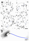
Grayscale image of the signal corresponding to the β-III tubulin channel of β-III tubulin and NeuN/Fox-3 doubly immunolabeled cells. Circles depict sampling areas (ROI). Traced neurites located with the NeuronJ plugin of ImageJ software are depicted in blue (A). (B) Detail of the framed area in (A). Scale bars: 200 µm in (A); 20 µm in (B).
Figure 1
Figure 1
Platelet concentration of the two platelet-rich plasma (PRP) groups. Error bars = standard deviation.
Figure 2
Figure 2
Molecular analysis of Young and Aged PRPs. Cytokine array of Aged PRP (A). Cytokine array of Young PRP (B). Molecular levels in the Aged PRP with respect to the Young PRP (C). The numbers and letter codes of panels A and B make reference to the coordinates of Table 4. The numbers of the X axis in panel C make reference to the number of each molecule in Table 4. * p < 0.05. Error bars = standard deviation.
Figure 3
Figure 3
Epifluorescence microscopy images of NT2 cells showing the effect of replacement of FBS by PRP on BrdU incorporation. (AE). Epifluorescence microscopy images of NT2 cells subjected to a 4-h BrdU pulse after growing for 24 h in either serum-free DMEM medium (A) or in 10% FBS (B), 2% FBS (C), 2% Aged-PRP supplemented DMEM (D), or 2% Young-PRP supplemented DMEM (E). Cells were processed for double immunofluorescence against BrdU (green), combined with Hoechst’s chromatin staining (pseudocolored red). Scale bar: 50 μm (F) Table showing results of BrdU incorporation analysis.
Figure 4
Figure 4
Analysis of phenotype density and area of cell nuclei in cultures of AraC-treated NT2 cells subjected to fetal bovine serum (FBS), Aged PRP, or Young PRP treatment. Percentage of cell of neuronal phenotype (A). Density of neuronal and non-neuronal cells (B). Area of cell nuclei in the whole cell population (C). Area of nuclei of neuronal and non-neuronal cells (D). ** p < 0.01, *** p < 0.001. Error bars = standard deviation. A-PRP: Aged PRP; Y-PRP: Young PRP.
Figure 5
Figure 5
Evaluation of PRP toxicity with calcein assay in primary cultures of neurons from rat E16-18 cortices, with different plasma concentrations (dilutions): 1:20, 1:30, 1:40, or 1:50. * p < 0.05. Error bars = standard deviation.
Figure 6
Figure 6
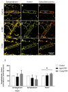
Synaptic protein expression. Protein expression of presynaptic (Synaptophysin) and postsynaptic (PSD-95) markers in neuronal cells treated in triplicate with control, Aged PRP, or Young PRP (A) and its quantification relative to the control treatment (B). (CH) Immunohistochemical analysis of presynaptic (synaptophysin) and postsynaptic (Homer) markers in neuronal cells and its quantification by the integrated density in ROI relative to the control treatment (I). A-PRP: Aged PRP; Y-PRP: Young PRP. Error bars = standard deviation.
Figure 7
Figure 7
Young PRP increases Homer postsynaptic puncta. Representative images of individual Synaptophysin and Homer puncta and colocalized puncta in neuronal primary cultures treated with either Aged PRP or Young PRP (AI). Quantification of individual Synaptophysin and Homer puncta and the colocalized Synaptophysin and Homer puncta (J). A-PRP: Aged PRP; Y-PRP: Young PRP. * p < 0.05. Error bars = standard deviation.
Figure 8
Figure 8
Expression of enzyme-inducible nitric oxide synthase (iNOS), a pro-inflammatory marker indicative of the M1 inflammatory phenotype (A) and mannose receptor (MNR), an anti-inflammatory marker indicative of the M2 restorative phenotype (B) in microglia of rats treated with different plasma concentrations. * p < 0.05; *** p < 0.001. Error bars = standard deviation.

References

    1. Le A.D.K., Enweze L., DeBaun M.R., Dragoo J.L. Platelet-rich plasma. Clin. Sports Med. 2019;38:17–44. doi: 10.1016/j.csm.2018.08.001. - DOI - PubMed
    1. Sánchez M., Anitua E., Delgado D., Sanchez P., Prado R., Orive G., Padilla S. Platelet-rich plasma, a source of autologous growth factors and biomimetic scaffold for peripheral nerve regeneration. Expert Opin. Biol. Ther. 2017;17:197–212. doi: 10.1080/14712598.2017.1259409. - DOI - PubMed
    1. Shen Y.-X., Fan Z.-H., Zhao J.-G., Zhang P. The Application of platelet-rich plasma may be a novel treatment for central nervous system diseases. Med. Hypotheses. 2009;73:1038–1040. doi: 10.1016/j.mehy.2009.05.021. - DOI - PubMed
    1. Anitua E., Pascual C., Pérez-Gonzalez R., Antequera D., Padilla S., Orive G., Carro E. Intranasal delivery of plasma and platelet growth factors using prgf-endoret system enhances neurogenesis in a mouse model of Alzheimer’s disease. PLoS ONE. 2013;8:e73118. doi: 10.1371/journal.pone.0073118. - DOI - PMC - PubMed
    1. Anitua E., Pascual C., Pérez-Gonzalez R., Orive G., Carro E. Intranasal PRGF-endoret enhances neuronal survival and attenuates NF-ΚB-dependent inflammation process in a mouse model of Parkinson’s disease. J. Control. Release. 2015;203:170–180. doi: 10.1016/j.jconrel.2015.02.030. - DOI - PubMed

Publication types

MeSH terms

LinkOut - more resources