

Pseudorabies Virus Infection Causes Downregulation of Ligands for the Activating NK Cell Receptor NKG2D
- PMID: 33572245
- PMCID: PMC7915010
- DOI: 10.3390/v13020266
Pseudorabies Virus Infection Causes Downregulation of Ligands for the Activating NK Cell Receptor NKG2D
Abstract
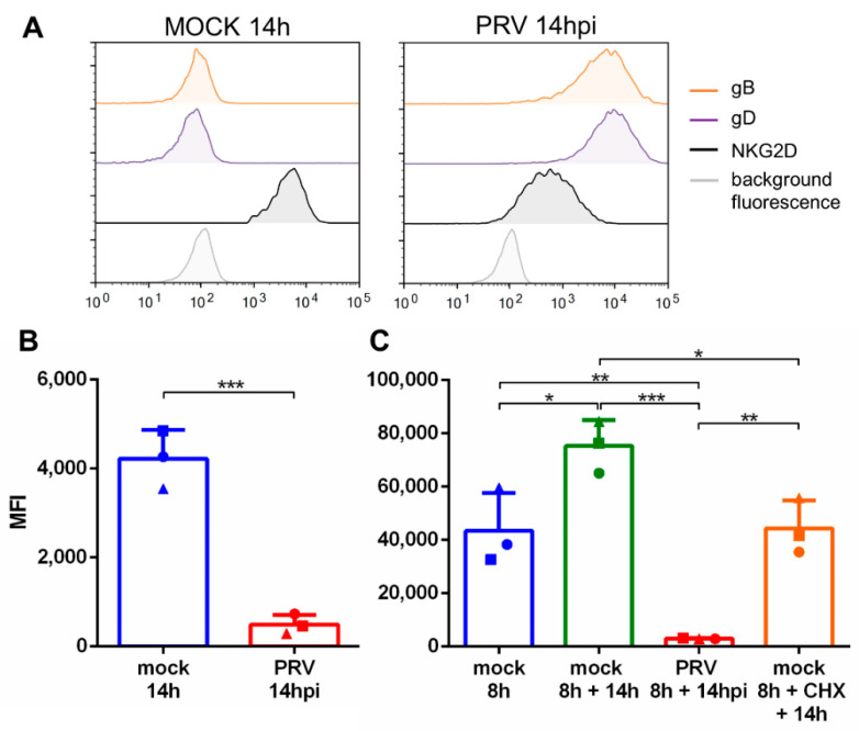
Herpesviruses display a complex and carefully balanced interaction with important players in the antiviral immune response of immunocompetent natural hosts, including natural killer (NK) cells. With regard to NK cells, this delicate balance is illustrated on the one hand by severe herpesvirus disease reported in individuals with NK cell deficiencies and on the other hand by several NK cell evasion strategies described for herpesviruses. In the current study, we report that porcine cells infected with the porcine alphaherpesvirus pseudorabies virus (PRV) display a rapid and progressive downregulation of ligands for the major activating NK cell receptor NKG2D. This downregulation consists both of a downregulation of NKG2D ligands that are already expressed on the cell surface of an infected cell and an inhibition of cell surface expression of newly expressed NKG2D ligands. Flow cytometry and RT-qPCR assays showed that PRV infection results in downregulation of the porcine NKG2D ligand pULBP1 from the cell surface and a very substantial suppression of mRNA expression of pULBP1 and of another potential NKG2D ligand, pMIC2. Furthermore, PRV-induced NKG2D ligand downregulation was found to be independent of late viral gene expression. In conclusion, we report that PRV infection of host cells results in a very pronounced downregulation of ligands for the activating NK cell receptor NKG2D, representing an additional NK evasion strategy of PRV.
Keywords: NKG2D; ligands; pMIC2; pULBP1; pig; pseudorabies virus.
Conflict of interest statement
The authors declare no conflict of interest.
Figures


Similar articles
-
Porcine UL16-binding protein 1 expressed on the surface of endothelial cells triggers human NK cytotoxicity through NKG2D.J Immunol. 2006 Aug 15;177(4):2146-52. doi: 10.4049/jimmunol.177.4.2146. J Immunol. 2006. PMID: 16887974
-
Expression of the Pseudorabies Virus gB Glycoprotein Triggers NK Cell Cytotoxicity and Increases Binding of the Activating NK Cell Receptor PILRβ.J Virol. 2019 Mar 21;93(7):e02107-18. doi: 10.1128/JVI.02107-18. Print 2019 Apr 1. J Virol. 2019. PMID: 30700600 Free PMC article.
-
The HHV-6A Proteins U20 and U21 Target NKG2D Ligands to Escape Immune Recognition.Front Immunol. 2021 Oct 15;12:714799. doi: 10.3389/fimmu.2021.714799. eCollection 2021. Front Immunol. 2021. PMID: 34721381 Free PMC article.
-
Release of Soluble Ligands for the Activating NKG2D Receptor: One More Immune Evasion Strategy Evolved by HIV-1 ?Curr Drug Targets. 2016;17(1):54-64. doi: 10.2174/1389450116666150630110329. Curr Drug Targets. 2016. PMID: 26122035 Review.
-
Leveraging NKG2D Ligands in Immuno-Oncology.Front Immunol. 2021 Jul 29;12:713158. doi: 10.3389/fimmu.2021.713158. eCollection 2021. Front Immunol. 2021. PMID: 34394116 Free PMC article. Review.
Cited by
-
M1-type polarized macrophage contributes to brain damage through CXCR3.2/CXCL11 pathways after RGNNV infection in grouper.Virulence. 2024 Dec;15(1):2355971. doi: 10.1080/21505594.2024.2355971. Epub 2024 May 23. Virulence. 2024. PMID: 38745468 Free PMC article.
-
Cellular Therapy: The Hope for Covid-19.Avicenna J Med Biotechnol. 2022 Apr-Jun;14(2):104-113. doi: 10.18502/ajmb.v14i2.8883. Avicenna J Med Biotechnol. 2022. PMID: 35633981 Free PMC article. Review.
-
Reovirus infection of tumor cells reduces the expression of NKG2D ligands, leading to impaired NK-cell cytotoxicity and functionality.Front Immunol. 2023 Sep 11;14:1231782. doi: 10.3389/fimmu.2023.1231782. eCollection 2023. Front Immunol. 2023. PMID: 37753084 Free PMC article.
-
Strategies to induce natural killer cell tolerance in xenotransplantation.Front Immunol. 2022 Aug 22;13:941880. doi: 10.3389/fimmu.2022.941880. eCollection 2022. Front Immunol. 2022. PMID: 36072599 Free PMC article. Review.
References
Publication types
MeSH terms
Substances
LinkOut - more resources
Full Text Sources
Other Literature Sources
