Review
. 2021 Feb 9;9(2):171.
doi: 10.3390/biomedicines9020171.

Cutaneous Squamous Cell Carcinoma: From Pathophysiology to Novel Therapeutic Approaches

Affiliations
Review

Cutaneous Squamous Cell Carcinoma: From Pathophysiology to Novel Therapeutic Approaches

Luca Fania et al. Biomedicines. .

Abstract

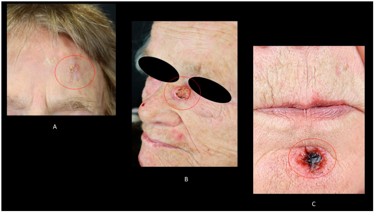
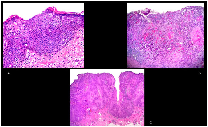
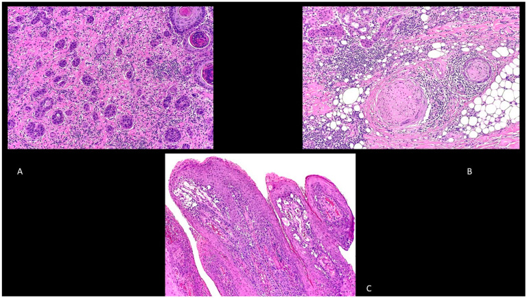
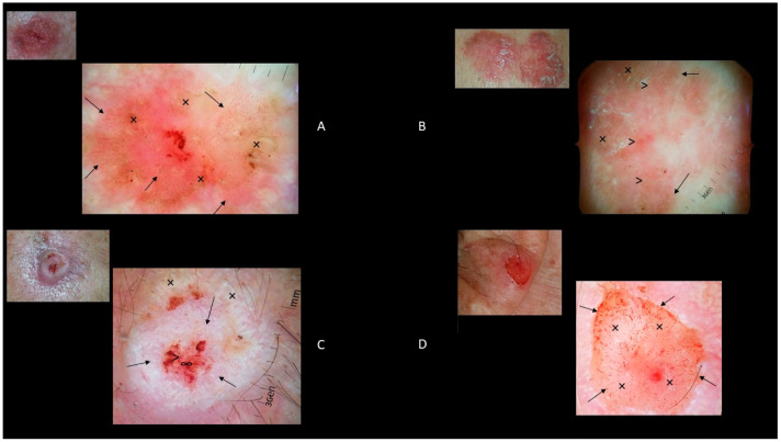
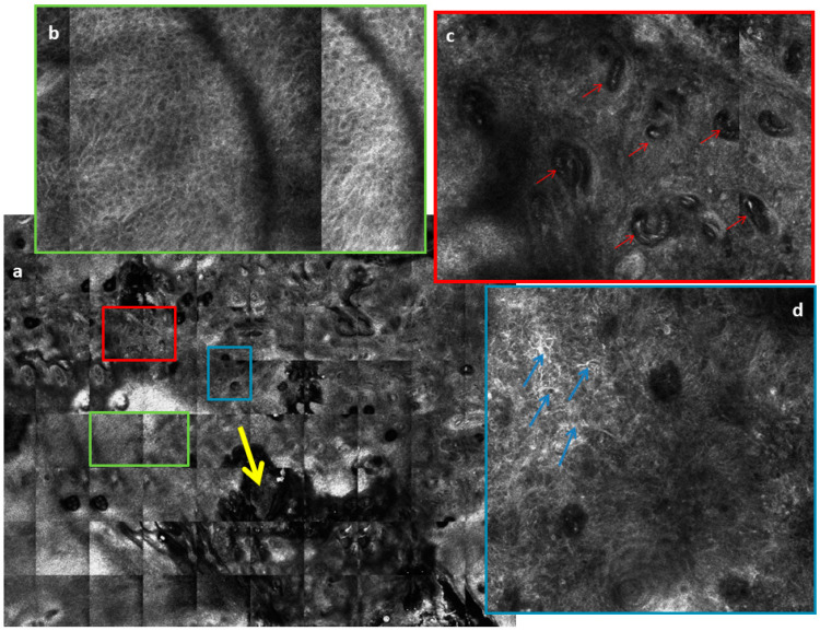
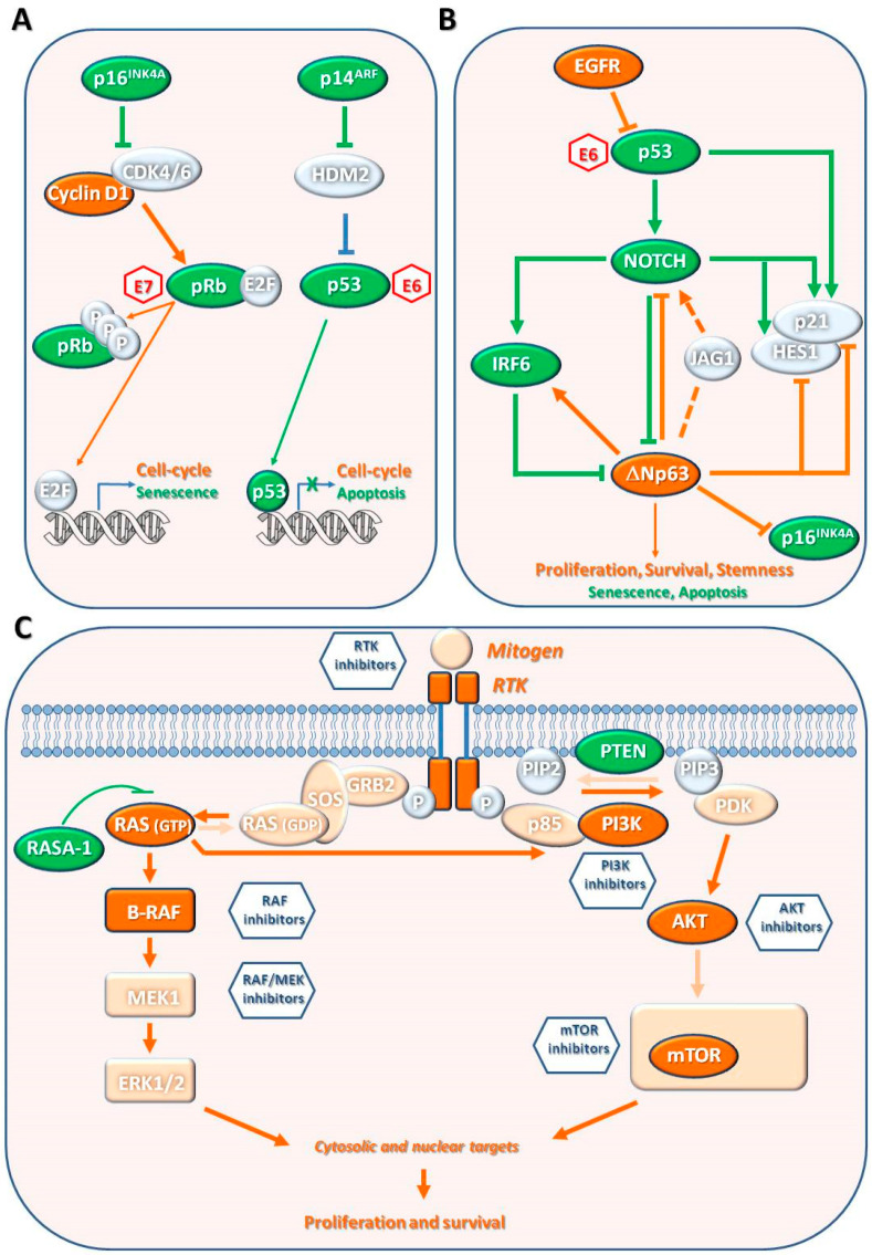
Cutaneous squamous cell carcinoma (cSCC), a non-melanoma skin cancer, is a keratinocyte carcinoma representing one of the most common cancers with an increasing incidence. cSCC could be in situ (e.g., Bowen's disease) or an invasive form. A significant cSCC risk factor is advanced age, together with cumulative sun exposure, fair skin, prolonged immunosuppression, and previous skin cancer diagnoses. Although most cSCCs can be treated by surgery, a fraction of them recur and metastasize, leading to death. cSCC could arise de novo or be the result of a progression of the actinic keratosis, an in situ carcinoma. The multistage process of cSCC development and progression is characterized by mutations in the genes involved in epidermal homeostasis and by several alterations, such as epigenetic modifications, viral infections, or microenvironmental changes. Thus, cSCC development is a gradual process with several histological- and pathological-defined stages. Dermoscopy and reflectance confocal microscopy enhanced the diagnostic accuracy of cSCC. Surgical excision is the first-line treatment for invasive cSCC. Moreover, radiotherapy may be considered as a primary treatment in patients not candidates for surgery. Extensive studies of cSCC pathogenic mechanisms identified several pharmaceutical targets and allowed the development of new systemic therapies, including immunotherapy with immune checkpoint inhibitors, such as Cemiplimab, and epidermal growth factor receptor inhibitors for metastatic and locally advanced cSCC. Furthermore, the implementation of prevention measures has been useful in patient management.

Keywords: Bowen’s disease; cemiplimab; dermoscopy; immunotherapy; keratinocyte carcinoma; non-melanoma skin cancer; radiotherapy; squamous cell carcinoma; therapy.


Conflict of interest statement

The authors declare no conflict of interest.

Figures

Figure 1
Figure 1
(A): Bowen’s disease of the forehead; (B): keratoacanthoma of the nose; (C): ulcerated squamous cell carcinoma on the chin.
Figure 2
Figure 2
(A): Bowen’s disease. Hematoxylin and Eosin, 100× (B): Well-differentiated, invasive squamous cell carcinoma, hematoxylin and eosin, 20×. (C): Verrucous squamous cell carcinoma, hematoxylin and eosin, 20×.
Figure 3
Figure 3
(A): Adenosquamous squamous cell carcinoma. Hematoxylin and eosin, 200×. (B): Perineural squamous cell carcinoma. Hematoxylin and eosin, 100×. (C): Papillary squamous cell carcinoma. Hematoxylin and eosin, 40×.
Figure 4
Figure 4
(A): Clinical image of a 12 × 8 mm nonpigmented Bowen’s disease of the chest with dermoscopic features of dotted and glomerular vessels (↑) and scaly white-to-yellow surfaces (×). (B): Clinical image of a 30 × 12 mm pigmented Bowen’s disease of the leg with dermoscopic features of brown to gray globules/dots (↑) and structureless pigmentation (×) and dotted and glomerular vessels (>). (C): Clinical image of a 14 × 11 mm well differentiated cSCC of the scalp with dermoscopic features of keratin/scales (↑), blood spots (>), white structureless areas (×), and ulcerations (∞). (D) Clinical image of a 7 × 6 mm poorly differentiated cSCC of the ear with dermoscopic features of hairpin and linear-irregular vessels (↑) and ulcerations (×).
Figure 5
Figure 5
(a) Reflectance confocal microscopy (RCM) mosaic of a well differentiated cSCC (8 × 8mm). Yellow arrow shows a scale crust and ulceration in the lower part of the figure. (b) Green rectangle: a magnification of an RCM image (1 × 1.5 mm) at the epidermal layer shows atypical honeycomb pattern, which is characterized by thickened and broadened keratinocytes outlines of varying size and shape. (c) Red rectangle: a magnification of an RCM mosaic (1 × 1.5 mm) at the dermoepidermal junction layer shows round or coiled vessels (red arrows) that run through the dermal papillae perpendicularly to the lesion surface. (d) Blue rectangle: a magnification of an RCM mosaic (1 × 1 mm) at the spinous-granular layer shows a disarranged pattern characterized by architectural disarray and the presence of dendritic (blue arrows) and plump bright cells which represent pigmented keratinocytes and melanocytes infiltrating the epidermis.
Figure 6
Figure 6
Pathways involved in cutaneous squamous cell carcinoma (cSCC) pathogenesis. Molecular alterations, which drive cSCC development, have been identified in pathways involved in cell cycle regulation, apoptosis, senescence, differentiation, and mitogenic/survival. (A) The tumor suppressor genes p16INK4A and p14ARF control retinoblastoma (pRb) and p53 pathways, respectively. Their loss of function promotes cell cycle counteracting senescence or apoptosis. Aberrant activation of E2F transcription can also be due to cyclin D activation or pRb expression loss. pRb and p53 inactivation is also mediated by E6 and E7 (red hexagons) human papilloma virus (HPV) proteins. (B) EGF-R aberrant activation, p53 inactivation, or NOTCH gene mutations inactivate the NOTCH pathway. Inactivation of NOTCH abolishes the direct or IRF6-mediated suppression of ΔNp63, favoring proliferation, survival, and stemness. NOTCH inactivation also counteracts senescence and apoptosis mediated by its targets (HES1 and p21). Moreover, ΔNp63 upregulation represses the expression of HES1, p21, and p16INK4A. (C) RAS-RAF-MEK-ERK and PI3K/AKT/mTOR pathways share the up-stream proteins, such as tyrosine kinase receptors (RTK) and RAS. Activating mutations in RTK, RAS or inactivation of negative regulator RASA1 promotes cell proliferation and survival through constitutive activation of both pathways. Aberrant activation of these pathways can also derive by B-RAF or PI3K/AKT activation, or Phosphatase and tensin homolog (PTEN) inactivation. The RTKs and the downstream pathways can be targeted with several drugs (blue hexagons) to inhibit cSCC progression. However, both pathways can be activated by RAS mutations, present in photodamaged skin, as part of a compensatory mechanism that could drive resistance to therapeutic targeting strategies. Proteins with commonly accepted tumor promoting and suppressing functions are highlighted in orange and green, respectively. Activated or downregulated processes (circles, squares and arrows) are highlighted in dark orange and green, respectively. Block and dash arrows indicate direct or indirect interactions, respectively.

References

    1. Que S.K.T., Zwald F.O., Schmults C.D. Cutaneous squamous cell carcinoma: Incidence, risk factors, diagnosis, and staging. J. Am. Acad. Dermatol. 2018;78:237–247. doi: 10.1016/j.jaad.2017.08.059. - DOI - PubMed
    1. Martincorena I., Roshan A., Gerstung M., Ellis P., van Loo P., McLaren S., Wedge D.C., Fullam A., Alexandrov L.B., Tubio J.M., et al. Tumor evolution. High burden and pervasive positive selection of somatic mutations in normal human skin. Science. 2015;348:880–886. doi: 10.1126/science.aaa6806. - DOI - PMC - PubMed
    1. Skulsky S.L., O’Sullivan B., McArdle O., Leader M., Roche M., Conlon P.J., O’Neill J.P. Review of high-risk features of cutaneous squamous cell carcinoma and discrepancies between the American Joint Committee on Cancer and NCCN Clinical Practice Guidelines In Oncology. Head Neck. 2017;39:578–594. doi: 10.1002/hed.24580. - DOI - PubMed
    1. Kim J.Y.S., Kozlow J.H., Mittal B., Moyer J., Olenecki T., Rodgers P. Guidelines of care for the management of cutaneous squamous cell carcinoma. J. Am. Acad. Dermatol. 2018;78:560–578. doi: 10.1016/j.jaad.2017.10.007. - DOI - PMC - PubMed
    1. Xiang F., Lucas R., Hales S., Neale R. Incidence of nonmelanoma skin cancer in relation to ambient UV radiation in white populations, 1978-2012: Empirical relationships. JAMA Dermatol. 2014;150:1063–1071. doi: 10.1001/jamadermatol.2014.762. - DOI - PubMed