. 2021 Jan 31;22(3):1417.
doi: 10.3390/ijms22031417.

A Cellular Assay for the Identification and Characterization of Connexin Gap Junction Modulators

Affiliations

A Cellular Assay for the Identification and Characterization of Connexin Gap Junction Modulators

Azeem Danish et al. Int J Mol Sci. .

Abstract

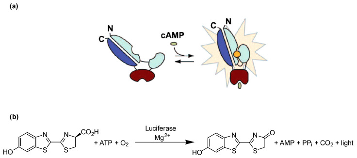
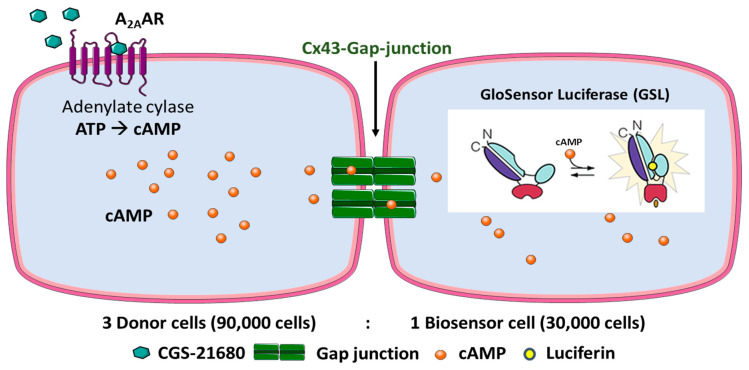
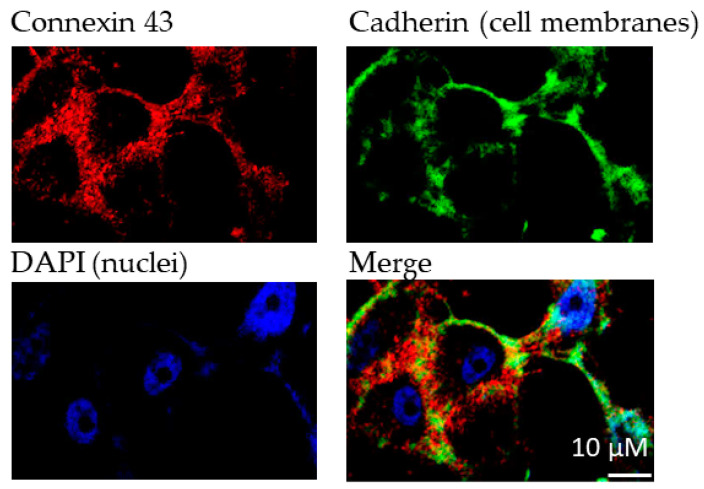
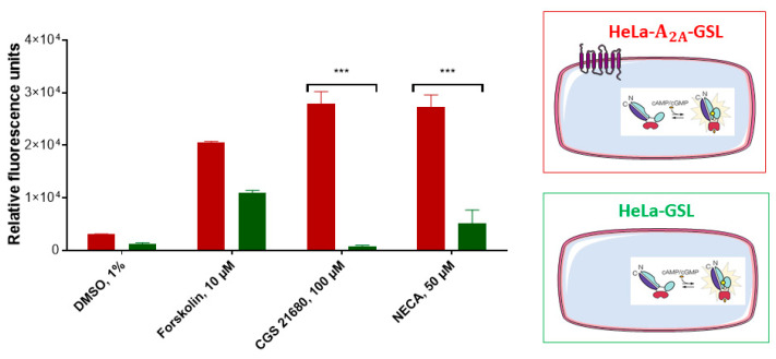
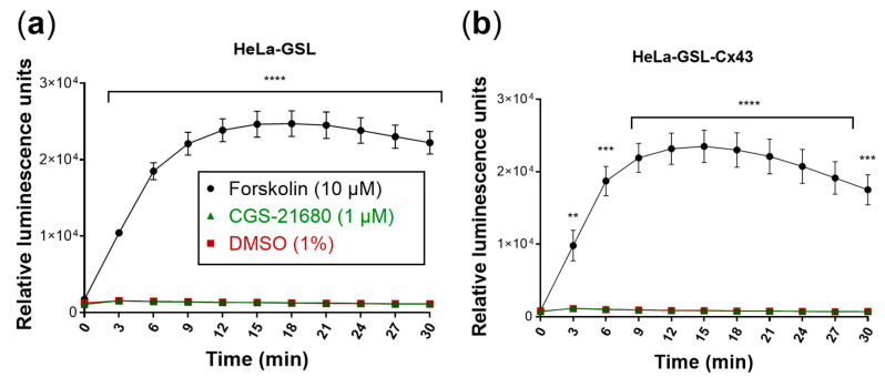
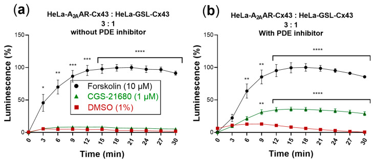
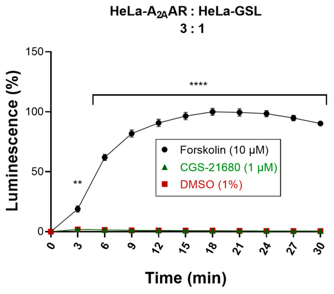
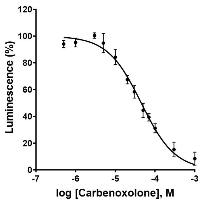
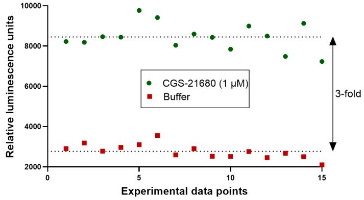
Connexin gap junctions (Cx GJs) enable the passage of small molecules and ions between cells and are therefore important for cell-to-cell communication. Their dysfunction is associated with diseases, and small molecules acting as modulators of GJs may therefore be useful as therapeutic drugs. To identify GJ modulators, suitable assays are needed that allow compound screening. In the present study, we established a novel assay utilizing HeLa cells recombinantly expressing Cx43. Donor cells additionally expressing the Gs protein-coupled adenosine A2A receptor, and biosensor cells expressing a cAMP-sensitive GloSensor luciferase were established. Adenosine A2A receptor activation in the donor cells using a selective agonist results in intracellular cAMP production. The negatively charged cAMP migrates via the Cx43 gap junctions to the biosensor cells and can there be measured by the cAMP-dependent luminescence signal. Cx43 GJ modulators can be expected to impact the transfer of cAMP from the donor to the biosensor cells, since cAMP transit is only possible via GJs. The new assay was validated by testing the standard GJ inhibitor carbenoxolon, which showed a concentration-dependent inhibition of the signal and an IC50 value that was consistent with previously reported values. The assay was demonstrated to be suitable for high-throughput screening.

Keywords: GloSensor luciferase; HeLa cells; compound library; connexin-43; gap junctions; screening.


Conflict of interest statement

The authors declare no conflict of interest.

Figures

Figure 1
Figure 1
Principle of cAMP detection using the engineered GloSensor luciferase. (a) GloSensor luciferase in the open conformation shows negligible activity resulting in a low luminescence background, whereas binding of cAMP to the cAMP binding site favors the closed conformation and hence activates the luciferase, which metabolizes luciferin to oxyluciferin, producing luminescence. (b) Luciferase catalyzes the oxidation of luciferin using molecular oxygen and ATP in the presence of Mg2+ to produce oxyluciferin, which is highly unstable in an electronically excited state and produces light upon returning to its electronical ground state. Modified based on published figure [25].
Figure 2
Figure 2
Design of the Cx43 GJ assay. HeLa cells expressing A2AAR and Cx43 are denoted as donor cells and HeLa cells expressing GloSensor luciferase and Cx43 as biosensor cells. The cell lines were cocultured in a ratio of 3:1 (donor: biosensor cells) for 4 h to allow the formation of Cx43 GJs. The cells were then equilibrated with buffer containing the substrate (luciferin) of the engineered cAMP-dependent luciferase. Upon activation of the Gs protein-coupled A2AARs, cAMP levels in the donor cells are increased. cAMP can then migrate via the Cx43 GJs to the biosensor cells. There, cAMP binds to the GloSensor luciferase which results in a conformational change that leads to an activation of the GloSensor luciferase, creating a luminescence signal by oxidation of luciferin. Depiction of GloSensor luciferase is based on reference [25].
Figure 3
Figure 3
Immunofluorescence analyses of Cx43 expression in transfected HeLa cells. Cadherin was used to stain cell membranes. Primary antibodies (1:1000): antipan cadherin (mouse), anti Cx43 (rabbit); secondary antibodies (1:500): Alexa 488 (antimouse), Alexa 594 (antirabbit). DAPI (1:10,000) was used to stain cell nuclei.
Figure 4
Figure 4
Assessment of adenosine receptor-mediated cAMP production in A2AAR-transfected (red) and nontransfected (green) HeLa- cells. The cAMP-activated GloSensor luciferase was cotransfected resulting in cAMP-dependent luminescence signals. Cells (60,000/well) were incubated with medium supplemented with 2% GloSensor luciferase reagent for 2 h at 37 °C. After addition of DMSO or compound dissolved in DMSO (forskolin, 10 µM, CGS-21680, 100 µM, or NECA, 50 µM) the cells were incubated at 37 °C for 15 min. Signals induced by adenosine receptor agonists CGS-21680 or NECA were significantly different between both cell lines (*** p < 0.001, 2-way ANOVA). For details see Section 4.
Figure 5
Figure 5
Evaluation of the biosensor cells. Forskolin (10 µM) was used as a positive control and DMSO (1%) as a negative control. Means ± SEM of three individual experiments performed in duplicates are given. (a) Biosensor cells produced luminescence only in response to forskolin (10 µM). Luminescence response to CGS-21680 (1 µM) was not different from control (DMSO). (b) Cx43 transfection of biosensor (HeLa-GSL) cells did not affect luminescence responses. Statistical significance calculated with repeated measures 2-way ANOVA and Dunnet’s multiple comparisons test comparing treatments to control (DMSO, 1%). ** p < 0.01, *** p < 0.001, **** p < 0.0001.
Figure 6
Figure 6
cAMP-dependent luminescence signal due to activation of cocultures of donor and biosensor cells (3:1) by the selective A2AAR agonist CGS-21680 (1 µM). Forskolin (10 µM) was used as positive control and DMSO (1%) served as negative control. Data are normalized to the maximal effect of forskolin (100%). Data represent means ± SEM of three individual experiments performed in duplicates. (a) Activation without PDE inhibitor. Only forskolin displayed a significantly increased luminescence signal compared to control. (b) Activation in the presence of the PDE inhibitor IBMX (200 µM). Statistical significance calculated with repeated measures 2-way ANOVA and Dunnet’s multiple comparisons test comparing treatments to control (DMSO, 1%). * p < 0.05, ** p < 0.01, *** p < 0.001, **** p < 0.0001.
Figure 7
Figure 7
Evaluation of cocultured donor and biosensor cells lacking Cx43 GJs. Activation of cocultures of Cx43-deficient donor and biosensor cells by the selective A2AAR agonist CGS-21680. Forskolin (10 µM) was used as a positive control and DMSO (1%) served as a negative control. Data are normalized to the maximal effect of forskolin (100%). Data represent means ± SEM of three individual experiments performed in duplicates. Statistical significance calculated with repeated measures 2-way ANOVA and Dunnet’s multiple comparisons test comparing treatments to control (DMSO, 1%). ** p < 0.01, **** p < 0.0001.
Figure 8
Figure 8
Concentration-dependent inhibition of Cx43 gap junctions by the blocker carbenoxolone as determined in the newly developed assay. Data points represent means ± SEM from 3 separate experiments. IC50 = 44.5 ± 4.8 µM.
Figure 9
Figure 9
Evaluation of suitability of the newly developed Cx43 GJ assay for high-throughput screening (HTS). The quality of the assay was assessed by calculating the screening window coefficient (Z´-Factor) as previously described [31]. A coculture of donor cells (90,000 cells/well) and biosensor cells (30,000 cells/well) at a ratio of 3:1 was incubated in assay buffer for 45 min. Prior to the addition of the A2AAR agonist CGS-21680 (1 µM) to the coculture, basal luminescence was measured (red data points, negative control). The coculture was then activated by the addition of CGS-21680 (1 µM). The luminescence signal had reached a stable plateau 19 min after stimulation, and data points were measured as positive controls (green data points). We measured 15 separate data points for positive and negative controls, indicating reproducibility and an assay window of about 3-fold.

Similar articles

Cited by

References

    1. Giaume C., Naus C.C., Sáez J.C., Leybaert L. Glial connexins and pannexins in the healthy and diseased brain. Physiol. Rev. 2021;101:93–145. doi: 10.1152/physrev.00043.2018. - DOI - PubMed
    1. Vinken M., Vanhaecke T., Papeleu P., Snykers S., Henkens T., Rogiers V. Connexins and their channels in cell growth and cell death. Cell. Signal. 2006;18:592–600. doi: 10.1016/j.cellsig.2005.08.012. - DOI - PubMed
    1. Bedner P., Steinhäuser C., Theis M. Functional redundancy and compensation among members of gap junction protein families? Biochim. Biophys. Acta Biomembr. 2012;1818:1971–1984. doi: 10.1016/j.bbamem.2011.10.016. - DOI - PubMed
    1. Salameh A. Life cycle of connexins: Regulation of connexin synthesis and degradation. Adv. Cardiol. 2006;42:57–70. doi: 10.1159/000092562. - DOI - PubMed
    1. Kar R., Batra N., Riquelme M.A., Jiang J.X. Biological role of connexin intercellular channels and hemichannels. Arch. Biochem. Biophys. 2012;524:2–15. doi: 10.1016/j.abb.2012.03.008. - DOI - PMC - PubMed

Publication types

LinkOut - more resources