Review
. 2021 Jan 29;11(2):100.
doi: 10.3390/life11020100.

Structure, Function, and Interactions of the HIV-1 Capsid Protein

Affiliations
Review

Structure, Function, and Interactions of the HIV-1 Capsid Protein

Eric Rossi et al. Life (Basel). .

Abstract

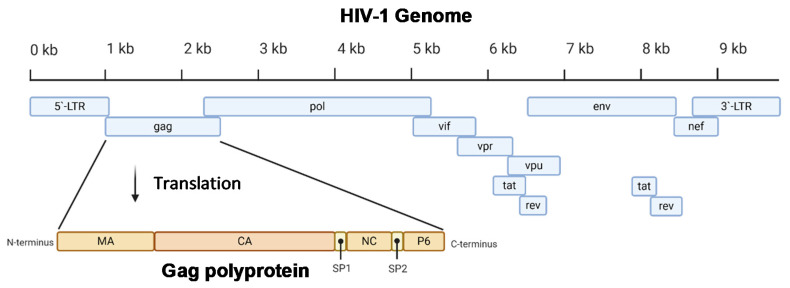
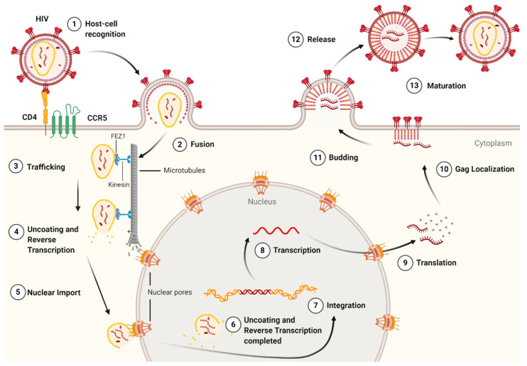
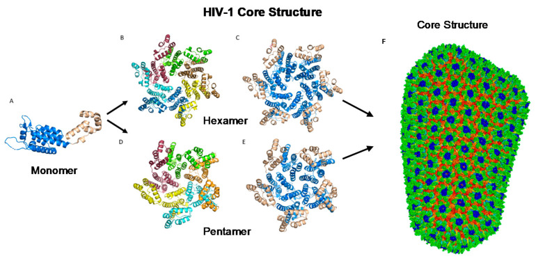
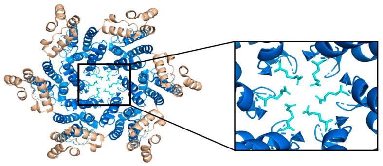
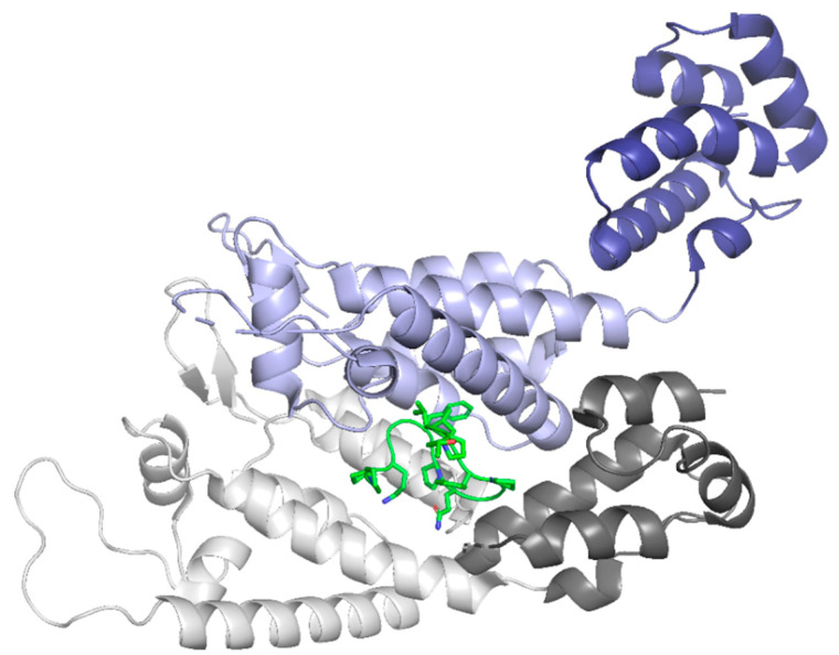
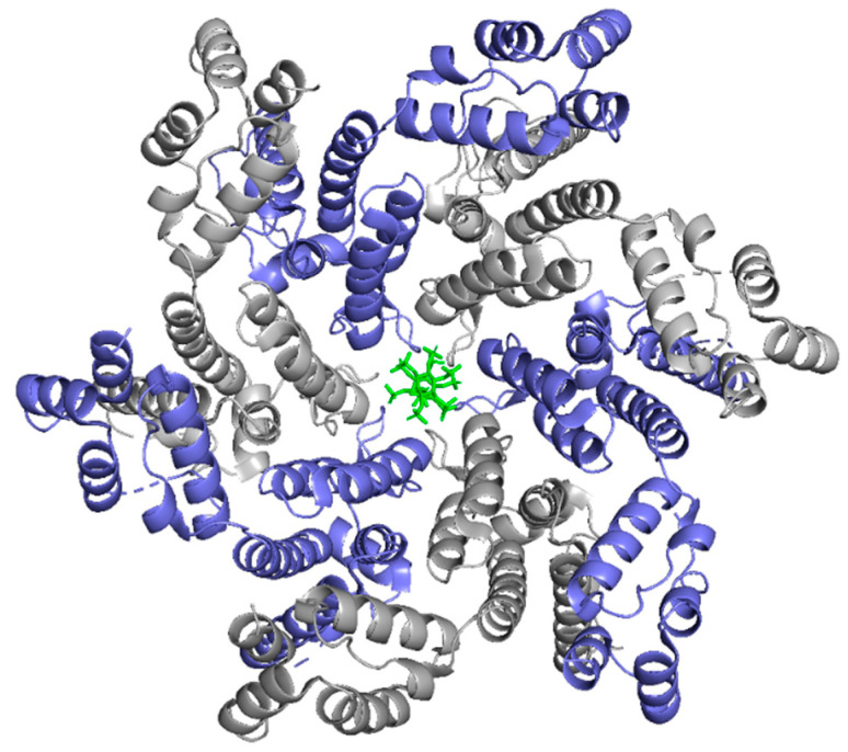
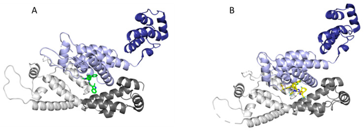
The capsid (CA) protein of the human immunodeficiency virus type 1 (HIV-1) is an essential structural component of a virion and facilitates many crucial life cycle steps through interactions with host cell factors. Capsid shields the reverse transcription complex from restriction factors while it enables trafficking to the nucleus by hijacking various adaptor proteins, such as FEZ1 and BICD2. In addition, the capsid facilitates the import and localization of the viral complex in the nucleus through interaction with NUP153, NUP358, TNPO3, and CPSF-6. In the later stages of the HIV-1 life cycle, CA plays an essential role in the maturation step as a constituent of the Gag polyprotein. In the final phase of maturation, Gag is cleaved, and CA is released, allowing for the assembly of CA into a fullerene cone, known as the capsid core. The fullerene cone consists of ~250 CA hexamers and 12 CA pentamers and encloses the viral genome and other essential viral proteins for the next round of infection. As research continues to elucidate the role of CA in the HIV-1 life cycle and the importance of the capsid protein becomes more apparent, CA displays potential as a therapeutic target for the development of HIV-1 inhibitors.

Keywords: HIV-1/AIDS; assembly; capsid; host proteins; post-entry events; restriction factors; virus-host interactions.


Conflict of interest statement

The authors declare no conflict of interest. The funders had no role in the study’s design, in the analyses or interpretation of data; in the writing of the manuscript, or in the decision to publish the manuscript.

Figures

Figure 1
Figure 1
A diagram of the 9.8 kb HIV-1 genome. The Gag portion of the genome is transcribed into the Gag polyprotein, consisting of the matrix (MA), capsid (CA), nucleocapsid (NC), P6, and two spacer peptides (SP1 and SP2). During maturation, the polyprotein is cleaved into its constituent parts. Image created with BioRender.com.
Figure 2
Figure 2
A schematic of the HIV-1 virion. Envelope proteins, GP41 and GP120, surround the host-derived membrane surface, which is lined internally with a layer of matrix protein. Inside the virion are viral proteins and the CA core containing the HIV-1 genome and proteins essential for infection. Image created with BioRender.com.
Figure 3
Figure 3
The life cycle of the HIV-1 virus. The early-stage begins with the recognition of host cell receptors (1), resulting in the fusion of the virus and release of the viral core into the cytoplasm of the host cell (2). This is followed by the trafficking of the core through the cytoplasm (3) as reverse transcription and uncoating begins to take place (4). Once at the nuclear pore, the viral contents are imported into the nucleus and localized (5) to transcriptionally active chromatin while uncoating and reverse transcription are completed (6). Following uncoating and reverse transcription, integration occurs (7). After the viral genome is integrated into the host cell, viral genes are transcribed (8) and translated (9) into the Gag polyprotein. The Gag polyprotein then localizes to the host cell membrane (10), where budding occurs (11), followed by the release of an immature virion (12). The final step in the HIV-1 lifecycle is maturation (13), where the viral protease cleaves the Gag polyprotein into its constituent, functional proteins. Image created with BioRender.com.
Figure 4
Figure 4
HIV-1 CA monomers oligomerize into hexamers and pentamers that assemble to form the capsid fullerene-cone core. (A) Shows the CA monomer with the amino-terminus colored blue and the carboxyl-terminus colored tan (PDB 3H47). (B) Shows the CA hexamer with each subunit colored differently (PDB 3H47). (C) Shows the hexamer with the amino-terminus of each subunit colored blue and the carboxyl terminus colored tan (PDB 3H47). (D) Shows the CA pentamer with each subunit colored differently (PDB 3P05). (E) Shows the pentamer with the amino-terminus of each subunit colored blue and the carboxyl terminus colored tan (PDB 3P05). (F) The complete capsid core structure containing approximately 250 hexamer oligomers and 12 pentamer oligomers (PDB 3J3Q). Image created with the PyMOL Molecular Graphics System, Version 2.4 Schrödinger, LLC.
Figure 5
Figure 5
The HIV-1 CA hexamer with the R18 ring expanded. The amino-terminus is colored blue, the carboxyl-terminus is colored tan, and each subunit’s R18 residue is colored in cyan. This is referred to as the R18 ring. This pore allows for the diffusion of materials needed for reverse transcription while sequestering reverse transcription machinery from host cell restriction factors. (PDB 3H47) Image created with the PyMOL Molecular Graphics System, Version 2.4 Schrödinger, LLC.
Figure 6
Figure 6
Here, two adjacent CA protomers of the CA hexamer form a functional interprotomer pocket. N-terminal domains (NTDs) are colored in shades of blue, and C-terminal domains (CTDs) are colored in shades of orange. Interacting domains are marked in dark blue and bright orange. Interacting residues within the interaction interface are magnified and colored in green. (PDB 6MQA) Image created with the PyMOL Molecular Graphics System, Version 2.4 Schrödinger, LLC.
Figure 7
Figure 7
The HIV-1 CA hexamer with regions important for higher-order oligomeric assembly are highlighted. The amino-terminus is colored blue, and the carboxyl-terminus is colored tan. Residues A14 and E45 between monomeric subunits are magnified and colored green. The regions of hydrophobic residues within the three-helix bundle center are marked with red boxes. (PDB 6MQA) Image created with the PyMOL Molecular Graphics System, Version 2.4 Schrödinger, LLC.
Figure 8
Figure 8
CypA binds to three CA protomers from two adjacent CA hexamers. One protomer of the CA protein is shown with the NTD in dark blue and the CTD in light blue. In total, two protomers from an adjacent CA hexamer are shown with the NTD in dark purple and light grey and the CTD in light pink and light grey. CypA, shown in green, binds to all three protomers through the canonical CypA binding loop (blue protomer) and the non-canonical binding sites (purple and grey). (PDB 6Y9W). Image created with the PyMOL Molecular Graphics System, Version 2.4 Schrödinger, LLC.
Figure 9
Figure 9
The NUP153 peptide binds between two protomers of the CA hexamer in the NTD-CTD interface pocket. The NTD of protomer A is in light grey, and the CTD is in dark grey. The NTD of protomer B is in light purple, and the CTD is in dark purple. The NUP153 peptide is shown in green. (PDB 5TSX). Image created with the PyMOL Molecular Graphics System, Version 2.4 Schrödinger, LLC.
Figure 10
Figure 10
The CPSF6 peptide binds between two protomers of the CA hexamer in the NTD-CTD interface pocket. The NTD of protomer A is in light grey, and the CTD is in dark grey. The NTD of protomer B is in light purple, and the CTD is in dark purple. The CPSF6 peptide is shown in green. (PDB 4WYM). Image created with the PyMOL Molecular Graphics System, Version 2.4 Schrödinger, LLC.
Figure 11
Figure 11
IP6 binds in the R18 pore of the CA Hexamer. Protomers are in alternating colors, slate and grey. IP6 is shown in green interacting with the CA R18 pore. (PDB 6ES8). Image created with the PyMOL Molecular Graphics System, Version 2.4 Schrödinger, LLC.
Figure 12
Figure 12
Binding of inhibitors in the interprotomer binding pocket of the CA hexamer. The NTD of protomer A is in light grey and the CTD is in dark grey. The NTD of protomer B is in light purple and the CTD is in dark purple. Inhibitors (A) PF74 (PDB 4XFZ) and (B) GS-6207 (PDB 6V2F) are shown in green and yellow, respectively. The binding of these inhibitors in the NTD-CTD interface pocket prevents necessary host cell factors CPSF6 and NUP153 from binding to this pocket. Image created with the PyMOL Molecular Graphics System, Version 2.4 Schrödinger, LLC.

References

    1. Clements J.E., Zink M.C. Molecular biology and pathogenesis of animal lentivirus infections. Clin. Microbiol. Rev. 1996;9:100–117. doi: 10.1128/CMR.9.1.100-117.1996. - DOI - PMC - PubMed
    1. Bbosa N., Kaleebu P., Ssemwanga D. HIV subtype diversity worldwide. Curr. Opin. HIV AIDS. 2019;14:153–160. doi: 10.1097/COH.0000000000000534. - DOI - PubMed
    1. German Advisory Committee Blood, Subgroup ‘Assessment of Pathogens Transmissible by Blood’ Human Immunodeficiency Virus (HIV) Transfus Med. Hemother. 2016;43:203–222. doi: 10.1159/000445852. - DOI - PMC - PubMed
    1. Cenker J.J., Stultz R.D., McDonald D. Brain Microglial Cells Are Highly Susceptible to HIV-1 Infection and Spread. AIDS Res. Hum. Retrovir. 2017;33:1155–1165. doi: 10.1089/aid.2017.0004. - DOI - PMC - PubMed
    1. Douek D.C., Brenchley J.M., Betts M.R., Ambrozak D.R., Hill B.J., Okamoto Y., Casazza J.P., Kuruppu J., Kunstman K., Wolinsky S., et al. HIV preferentially infects HIV-specific CD4+ T cells. Nature. 2002;417:95–98. doi: 10.1038/417095a. - DOI - PubMed

LinkOut - more resources