. 2021 Jan 29;9(2):128.
doi: 10.3390/biomedicines9020128.

Incorporation of Calcium Sulfate Dihydrate into a Mesoporous Calcium Silicate/Poly-ε-Caprolactone Scaffold to Regulate the Release of Bone Morphogenetic Protein-2 and Accelerate Bone Regeneration

Affiliations

Incorporation of Calcium Sulfate Dihydrate into a Mesoporous Calcium Silicate/Poly-ε-Caprolactone Scaffold to Regulate the Release of Bone Morphogenetic Protein-2 and Accelerate Bone Regeneration

Kuo-Hao Huang et al. Biomedicines. .

Abstract

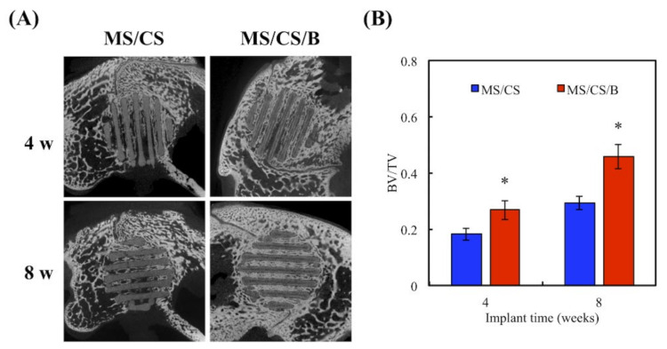
Tissue engineering and scaffolds play an important role in tissue regeneration by supporting cell adhesion, proliferation, and differentiation. The design of a scaffold is critical in determining its feasibility, and it is critical to note that each tissue is unique in terms of its morphology and composition. However, calcium-silicate-based scaffolds are undegradable, which severely limits their application in bone regeneration. In this study, we developed a biodegradable mesoporous calcium silicate (MS)/calcium sulfate (CS)/poly-ε-caprolactone (PCL) composite and fabricated a composite scaffold with 3D printing technologies. In addition, we were able to load bone morphogenetic protein-2 (BMP-2) into MS powder via a one-step immersion procedure. The results demonstrated that the MS/CS scaffold gradually degraded within 3 months. More importantly, the scaffold exhibited a gradual release of BMP-2 throughout the test period. The adhesion and proliferation of human dental pulp stem cells on the MS/CS/BMP-2 (MS/CS/B) scaffold were significantly greater than that on the MS/CS scaffold. It was also found that cells cultured on the MS/CS/B scaffold had significantly higher levels of alkaline phosphatase activity and angiogenic-related protein expression. The MS/CS/B scaffold promoted the growth of new blood vessels and bone regeneration within 4 weeks of implantation in rabbits with induced critical-sized femoral defects. Therefore, it is hypothesized that the 3D-printed MS/CS/B scaffold can act both as a conventional BMP-2 delivery system and as an ideal osteoinductive biomaterial for bone regeneration.

Keywords: 3D printing; bone morphogenetic protein-2; calcium silicate; calcium sulfate; osteogenesis.


Conflict of interest statement

The authors declare no conflict of interest.

Figures

Figure 1
Figure 1
The microstructure and composition of mesoporous calcium silicate (MS) and calcium sulfate (CS) powders and composites. The scale bar of the SEM photograph is 1 µm.
Figure 2
Figure 2
(A) Field emission–scanning electron microscopy (FE-SEM) photographs and (B) X-ray diffractometry (XRD) of an MS/CS scaffold after soaking in SBF. The scale bar of the SEM photograph is 1 µm.
Figure 3
Figure 3
The weight loss of MS/CS scaffolds after soaking in SBF for various time periods.
Figure 4
Figure 4
Stress–strain curve of the MS/CS scaffold after immersion in SBF for 7, 30, and 180 days.
Figure 5
Figure 5
Bone morphogenetic protein-2 (BMP-2) release from an MS/CS/B and MS/B scaffold after immersion in Dulbecco’s modified Eagle’s medium (DMEM) at 37 °C for 6 months.
Figure 6
Figure 6
(A) The proliferation of the human dental pulp cells (hDPSCs) cultured on scaffolds for 1, 3, 7, and 14 days. * indicates a significant difference (p < 0.05) when compared to MS/CS. (B) F-actin filament (green) staining of hDPSCs cultured on different scaffolds for 3 and 7 days. The scale bar of the photograph is 100 µm.
Figure 7
Figure 7
(A) Immunodetection of anti-bone morphogenetic protein receptor type II (BMP2R), anti-extracellular signal-regulated kinase 1/2 (ERK 1/2), anti-phospho extracellular signal-regulated kinase 1/2 (p-ERK 1/2), alkaline phosphatase (ALP), osteocalcin (OC), and β-actin protein expression in hDPSCs cultured on scaffolds for 3 days. Quantification of (B) BMP2R, (C) anti-phospho SMAD1/5/8 (p-SMAD1/5/8), (D) p-ERK 1/2, (E) ALP, and (F) OC. * indicates a significant difference (p < 0.05) when compared to MS/CS.
Figure 8
Figure 8
(A) Western blotting of knockdown by pairs of BMPR2-specific siRNAs in hDPSCs. (B) The effects on siRNA–BMPR2 transfection of proliferation in hDPSCs cultured on scaffolds for 1 and (B) 7 days. * indicates a significant difference (p < 0.05) when compared to MS/CS.
Figure 9
Figure 9
The osteogenic and angiogenic protein expression of (A) the hDPSCs and (B) the siRNA–BMPR2 transfected hDPSCs on scaffolds for 7 days. * indicates a significant difference (p < 0.05) when compared to MS/CS.
Figure 10
Figure 10
(A) Alizarin red S staining and (B) quantification of hDPSC calcium mineral deposits cultured on specimens for 7, 10, and 14 days. * indicates a significant difference (p < 0.05) when compared to MS/CS. The scale bar of the Optical microscope photograph is 400 µm.
Figure 11
Figure 11
(A) Micro-CT image showing the morphology of bone growth for a fixed-sized critical lesion after it underwent 8-week regeneration with scaffolds; (B) data analysis of relative bone mass volume (bone volume/tissue volume (BV/TV) ratio) for a fixed-sized critical lesion after it underwent 8 weeks of regeneration with scaffolds. * indicates a significant difference (p < 0.05) when compared to MS/CS.
Figure 12
Figure 12
Histological analysis of new bone regeneration around and within the scaffolds in the rabbit femoral defect model. Left, hematoxylin and eosin (HE) stain; middle, Masson’s trichrome (MT) stain; and right, von Kossa (VK) stain of regenerated bone mass after 8 weeks of regeneration in the in vivo experiment. Purple: tissue morphology. Blue: collagen and bone matrix. Brown: bone mineralization. The scale bar of the histological photograph is 400 µm.

Similar articles

Cited by

References

    1. Zhao H., Liang G., Liang W., Li Q., Huang B., Li A., Qiu D., Jin D. In vitro and in vivo evaluation of the pH-neutral bioactive glass as high performance bone grafts. Mater. Sci. Eng. C Mater. Biol. Appl. 2020;116:111249. doi: 10.1016/j.msec.2020.111249. - DOI - PubMed
    1. Ho-Shui-Ling A., Bolander J., Rustom L.E., Johnson A.W., Luyten F.P., Picart C. Bone regeneration strategies: Engineered scaffolds, bioactive molecules and stem cells current stage and future perspectives. Biomaterials. 2018;180:143–162. doi: 10.1016/j.biomaterials.2018.07.017. - DOI - PMC - PubMed
    1. Wang W.C., Nune K.C., Tan L., Zhang N., Dong J., Yan J., Misra R.D.K., Yang K. Bone regeneration of hollow tubular magnesium-strontium scaffolds in critical-size segmental defects: Effect of surface coatings. Mater. Sci. Eng. C Mater. Biol. Appl. 2019;100:297–307. doi: 10.1016/j.msec.2019.02.067. - DOI - PubMed
    1. Tu M.G., Lee K.X., Lin Y.H., Huang T.H., Ho C.C., Shie M.Y. Caffeic acid-coated nanolayer on Mineral Trioxide Aggregate potentiate the host immune responses, angiogenesis, and odontogenesis. J. Endod. 2020;46:1455–1464. doi: 10.1016/j.joen.2020.07.003. - DOI - PubMed
    1. Gómez-Lizárraga K.K., Flores-Morales C., Del Prado-Audelo M.L., Álvarez-Pérez M.A., Piña-Barba M.C., Escobedo C. Polycaprolactone- and polycaprolactone/ceramic-based 3D-bioplotted porous scaffolds for bone regeneration: A comparative study. Mater. Sci. Eng. C Mater. Biol. Appl. 2017;79:326–335. doi: 10.1016/j.msec.2017.05.003. - DOI - PubMed

LinkOut - more resources