. 2021 Jan 29;9(2):103.
doi: 10.3390/vaccines9020103.

Orientation of Antigen Display on Self-Assembling Protein Nanoparticles Influences Immunogenicity

Affiliations

Orientation of Antigen Display on Self-Assembling Protein Nanoparticles Influences Immunogenicity

Cosette G Schneider et al. Vaccines (Basel). .

Abstract

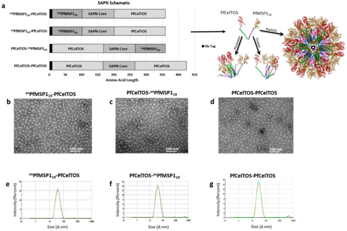
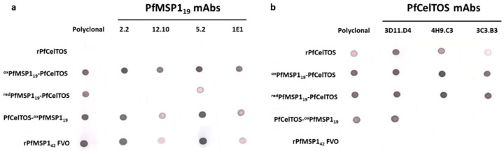
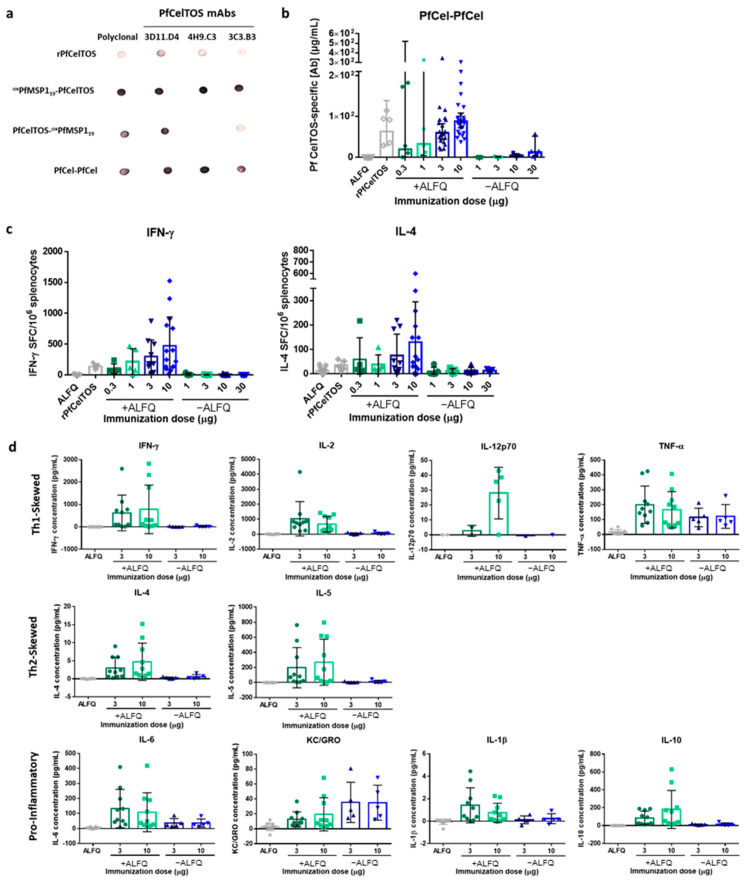
Self-assembling protein nanoparticles (SAPN) serve as a repetitive antigen delivery platform with high-density epitope display; however, antigen characteristics such as size and epitope presentation can influence the immunogenicity of the assembled particle and are aspects to consider for a rationally designed effective vaccine. Here, we characterize the folding and immunogenicity of heterogeneous antigen display by integrating (a) dual-stage antigen SAPN presenting the P. falciparum (Pf) merozoite surface protein 1 subunit, PfMSP119, and Pf cell-traversal protein for ookinetes and sporozoites, PfCelTOS, in addition to (b) a homogenous antigen SAPN displaying two copies of PfCelTOS. Mice and rabbits were utilized to evaluate antigen-specific humoral and cellular induction as well as functional antibodies via growth inhibition of the blood-stage parasite. We demonstrate that antigen orientation and folding influence the elicited immune response, and when appropriately designed, SAPN can serve as an adaptable platform for an effective multi-antigen display.

Keywords: PfCelTOS; PfMSP119; display; multi-stage; self-assembling protein nanoparticles; vaccine.


Conflict of interest statement

Peter Burkhard is the founder, co-owner, and CEO of Alpha-O Peptides AG, a company involved in nanoparticle vaccine design that holds intellectual property on the SAPN platform. The interpretations and opinions expressed herein belong to the authors and do not necessarily represent the official views of the U.S. Army, U.S. Navy, U.S. Department of Defense, or the U.S. government. Research was conducted in an AAALACi accredited facility in compliance with the Animal Welfare Act and other federal statutes and regulations relating to animals and experiments involving animals and adheres to principles stated in the Guide for the Care and Use of Laboratory Animals, NRC Publication, 2011 edition.

Figures

Figure 1
Figure 1
Self-assembling protein nanoparticles’ (SAPN) composition and particle quality. (a) Schematic diagram of SAPN with a theoretical model of PfCelTOS-oxPfMSP119 monomers assuming icosahedral symmetry. (bd) TEM of oxPfMSP119-PfCelTOS (b), PfCelTOS-oxPfMSP119 (c), and PfCelTOS-PfCelTOS (d). (eg) Hydrodynamic diameter size distribution of oxPfMSP119-PfCelTOS (e), PfCelTOS-oxPfMSP119 (f), and PfCelTOS-PfCelTOS (g) determined by dynamic light scattering (DLS).
Figure 2
Figure 2
Antigen localization on SAPN influences immunoreactivity. (a,b) Reactivity of conformation-dependent P. falciparum MSP119 (a) and P. falciparum CelTOS (b) monoclonal antibodies to the SAPN and recombinant proteins, P. falciparum MSP142 and P. falciparum CelTOS. Nitrocellulose membranes were spotted with 250 ng of folded proteins and incubated with antibodies as described in Materials and Methods.
Figure 3
Figure 3
Antigen localization and epitope density on SAPN influences immunogenicity and antibody fine specificity. (ac) BALB/c mice were immunized I.M. 3 times at a 3-week interval with redPfMSP119-PfCelTOS ((a,b), n = 39), oxPfMSP119-PfCelTOS ((a), n = 40; (b), n = 25; (c), n = 15), PfCelTOS-oxPfMSP119 ((a,b), n = 38; (c), n = 15), or 1:1 Combo ((a,b), n = 30; (c), n = 15) SAPN (0.3 µg for (a,b) and 1 µg for (c)), recombinant P. falciparum CelTOS (10 µg, n = 35), or recombinant P. falciparum MSP142 (5 µg, n = 25) in AddaVax™. The 1:1 Combo is an assembled 1:1 admixture of oxPfMSP119-PfCelTOS and PfCelTOS-oxPfMSP119. Negative controls include immunization with AddaVax™ alone ((a,b), n = 25), irrelevant SAPN (a,b), n = 25), and 0.3 µg redPfMSP119-PfCelTOS in PBS (denoted as SAPN w/o AddaVax™; (a,b), n = 5). Antigen-specific antibody concentrations against P. falciparum CelTOS (a) and P. falciparum MSP142 (b) and antigen-specific IgG titers against P. falciparum CelTOS and its N- and C-terminal subunits (c) were quantified in sera 2 weeks post-final immunization by ELISA. Final titers are shown for individual mice, with the geometric mean and 95% confidence intervals (CI). *: p < 0.05; ***: p < 0.001; ****: p < 0.0001 by the Kruskal–Wallis test with Dunn’s multiple comparisons (a,b) and the Wilcoxon matched-pairs signed rank test (c) (GraphPad Prism 6.0). Results are a meta-analysis of five independent experiments. Dotted lines separate the results for each SAPN.
Figure 4
Figure 4
SAPN induces antibodies that inhibit parasites in vitro. Two weeks following the third I.M. immunization, rabbits were exsanguinated, and serum antibody titers were measured against recombinant proteins and blood-stage parasites. (a) Fifty micrograms dose response of PfCelTOS-oxPfMSP119 (n = 3), oxPfMSP119-PfCelTOS (n = 3), and rPfMSP142 (n = 3) to the Vietnam Oak-Knoll (FVO) and 3D7 PfMSP142 antigens. (b) PfCelTOS-oxPfMSP119 dose-response relationship compared to the previous 50 μg PfCelTOS-oxPfMSP119 and rMSP142 groups. (c) Total IgG was normalized to 1.25, 2.5, and 5 mg/mL. Fifty micrograms of each SAPN construct and rPfMSP142 were tested for FVO and 3D7 parasite-specific growth inhibition by measuring the amount of parasite lactate dehydrogenase present. (d) Correlation graphs between purified IgG ELISA titer and percent inhibition at 1.25, 2.5, and 5 mg/mL with linear regression analysis. FVO strain titer and percent inhibition are represented by filled symbols and a solid trend line, while 3D7 strain titer and percent inhibition are represented by open symbols with a dash trend line. Note: there are overlapping symbols on the 2.5 mg/mL graph, where an oxPfMSP119-PfCelTOS sample has nearly identical titer (12,587, 11,760) and identical percent inhibition (23.6) for both FVO and 3D7 strains, respectively. Data are shown as the mean with +/− standard error of the mean. Statistical significance was determined using nonparametric multiple t test, the Holm–Sidak method comparing to the rPfMSP142 standard group with *: p < 0.05; **: p < 0.01. Differences in PfMSP142 strain responses were compared using Mann–Whitney tests.
Figure 5
Figure 5
Immunogenicity of N-terminal and C-terminal displayed PfCelTOS on PfCel-PfCel SAPN. (a) Reactivity of P. falciparum CelTOS antibodies to PfCel-PfCel SAPN. Nitrocellulose membrane was spotted with 250 ng of folded proteins and incubated with antibodies as described in Materials and Methods. (b) BALB/c mice were immunized I.M. 3 times at a 3-week interval with PfCel-PfCel SAPN with (0.3 µg, n = 5; 1 µg, n = 5; 3 µg, n = 20; 10 µg, n = 25) and without (1 µg, n = 5; 3 µg, n = 5; 10 µg, n = 5; 30 µg, n = 5) Army Liposome Formulation with QS-21 (ALFQ) adjuvant, recombinant PfCelTOS (10 μg, n = 5) with ALFQ, or ALFQ alone (n = 20). Antigen-specific antibody concentrations against rPfCelTOS were quantified by ELISA. Final antibody concentrations are shown for individual mice, with the geometric mean and 95% CI. (c) IFN-γ- and IL-4-secreting cells were quantified by ELISpot after stimulation of splenocytes with rPfCelTOS. The number of cytokine-specific spot-forming cells (SFC) per 106 splenocytes is shown for individual mice (n = 5 for all groups except the 3 µg and 10 µg doses with ALFQ and ALFQ alone groups, which have n = 10, n = 15, n = 15, respectively), with the mean and SD. (d) Cytokine production was quantified using the Meso Scale Discovery immunoassay. Cytokine-specific concentrations are shown for individual mice (n = 10 for groups with ALFQ, n = 5 for groups without ALFQ, except IL-12p70 for which some mice were below the level of detection), with the mean and SD. Results are representative of three independent experiments.

Similar articles

Cited by

References

    1. WHO . World Malaria Report 2020. World Health Organization; Geneva, Switzerland: 2020.
    1. Weiss D.J., Lucas T.C.D., Nguyen M., Nandi A.K., Bisanzio D., Battle K.E., Cameron E., Twohig K.A., Pfeffer D.A., Rozier J.A., et al. Mapping the global prevalence, incidence, and mortality of Plasmodium falciparum, 2000–2017: A spatial and temporal modelling study. Lancet. 2019;394:322–331. doi: 10.1016/S0140-6736(19)31097-9. - DOI - PMC - PubMed
    1. Bhatt S., Weiss D.J., Cameron E., Bisanzio D., Mappin B., Dalrymple U., Battle K., Moyes C.L., Henry A., Eckhoff P.A., et al. The effect of malaria control on Plasmodium falciparum in Africa between 2000 and 2015. Nature. 2015;526:207–211. doi: 10.1038/nature15535. - DOI - PMC - PubMed
    1. Zinszer K., Charland K., Vahey S., Jahagirdar D., Rek J.C., Arinaitwe E., Nankabirwa J., Morrison K., Sadoine M.L., Tutt-Guerette M.A., et al. The Impact of Multiple Rounds of Indoor Residual Spraying on Malaria Incidence and Hemoglobin Levels in a High-Transmission Setting. J. Infect. Dis. 2020;221:304–312. doi: 10.1093/infdis/jiz453. - DOI - PMC - PubMed
    1. Kenangalem E., Poespoprodjo J.R., Douglas N.M., Burdam F.H., Gdeumana K., Chalfein F., Prayoga, Thio F., Devine A., Marfurt J., et al. Malaria morbidity and mortality following introduction of a universal policy of artemisinin-based treatment for malaria in Papua, Indonesia: A longitudinal surveillance study. PLoS Med. 2019;16:e1002815. doi: 10.1371/journal.pmed.1002815. - DOI - PMC - PubMed

LinkOut - more resources