. 2021 Jan 29;12(2):142.
doi: 10.3390/mi12020142.

Potential Whole-Cell Biosensors for Detection of Metal Using MerR Family Proteins from Enterobacter sp. YSU and Stenotrophomonas maltophilia OR02

Affiliations

Potential Whole-Cell Biosensors for Detection of Metal Using MerR Family Proteins from Enterobacter sp. YSU and Stenotrophomonas maltophilia OR02

Georgina Baya et al. Micromachines (Basel). .

Abstract

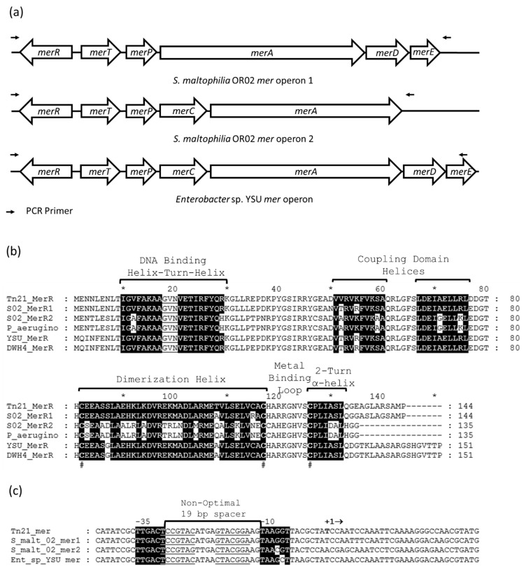
Cell-based biosensors harness a cell's ability to respond to the environment by repurposing its sensing mechanisms. MerR family proteins are activator/repressor switches that regulate the expression of bacterial metal resistance genes and have been used in metal biosensors. Upon metal binding, a conformational change switches gene expression from off to on. The genomes of the multimetal resistant bacterial strains, Stenotrophomonas maltophilia Oak Ridge strain 02 (S. maltophilia 02) and Enterobacter sp. YSU, were recently sequenced. Sequence analysis and gene cloning identified three mercury resistance operons and three MerR switches in these strains. Transposon mutagenesis and sequence analysis identified Enterobacter sp. YSU zinc and copper resistance operons, which appear to be regulated by the protein switches, ZntR and CueR, respectively. Sequence analysis and reverse transcriptase polymerase chain reaction (RT-PCR) showed that a CueR switch appears to activate a S. maltophilia 02 copper transport gene in the presence of CuSO4 and HAuCl4·3H2O. In previous studies, genetic engineering replaced metal resistance genes with the reporter genes for β-galactosidase, luciferase or the green fluorescence protein (GFP). These produce a color change of a reagent, produce light, or fluoresce in the presence of ultraviolet (UV) light, respectively. Coupling these discovered operons with reporter genes has the potential to create whole-cell biosensors for HgCl2, ZnCl2, CuSO4 and HAuCl4·3H2O.

Keywords: CuSO4; CueR; Enterobacter; HAuCl4·3H2O; HgCl2; MerR family protein; Stenotrophomonas maltophilia; ZnCl2; ZntR; bacterial metal resistance; whole-cell biosensor.


Conflict of interest statement

The authors declare no conflict of interest.

Figures

Figure 1
Figure 1
The Tn21 mercury resistance (mer) operon and MerR protein activator/repressor switch. (a) The genes merTPCAD are transcribed to the right and merR is transcribed to the left. The MerR protein binds as a dimer to the DNA operator region (underlined) between the −10 and −35 RNA polymerase binding regions (highlighted in black). The non-optimal 19 base pair (bp) spacer between the −10 and −35 regions prevents RNA polymerase from initiating transcription efficiently. In the absence of Hg(II), MerR binding to the DNA acts as an off switch to repress transcription. In the presence of Hg(II), MerR acts as an on switch by shortening the distance between and aligning the −10 and −35 regions so that RNA polymerase can initiate transcription of the merTPCAD genes efficiently. Transcription begins at the nucleotide base (bold) below + in +1. The first three base pairs, CAT, on the far left marks the beginning of MerR translation. The last three base pairs, ATG, on the far right marks the beginning of MerT translation. (b) The Tn21 MerR amino acid residue primary sequence. The helix-turn-helix domain binds to the operator region of the promoter in the DNA. The coupling domain links the DNA binding domain to the metal binding domain. The metal binding domain consists of the dimerization helix, metal binding loop and 2-turn α-helix. The dimerization helix links two identical MerR polypeptides to form an antiparallel coiled coil. The # below the sequences designates conserved cysteine amino acid residues which bind to Hg(II). The * and numbers above the MerR primary sequence denote the position of every 10 amino acid residues.
Figure 2
Figure 2
The S. maltophilia 02 and Enterobacter sp. YSU mercury resistance (mer) operons and MerR protein activator/repressor switches. (a) Mercury resistance operons in S. maltophilia 02 and Enterobacter sp. YSU. The genes between the primers were copied by polymerase chain reaction (PCR), cloned and tested for the Hg(II) resistance phenotype. (b) Prediction of the S. maltophilia 02 and Enterobacter sp. YSU MerR secondary structure using the Tn21, Pseudomonas aeruginosa, and Enterobacter cloacae plasmid DWH4 MerR amino acid residue primary sequences. Each contain a DNA binding domain, a coupling domain and a metal binding domain. The # below the sequences designates conserved cysteine amino acid residues which bind to Hg(II). The * and numbers above the MerR primary sequences denote the position of every 10 amino acid residues. (c) Prediction of the S. maltophilia 02 and Enterobacter sp. YSU mer promoter regions using the Tn21 mer promoter. All appear to contain non-optimal 19-bp spacers and MerR binding regions between the −10 and −35 region. Identical nucleotides in the −10 and −35 regions are highlighted in black. Transcription begins at the nucleotide base (bold) below + in +1. The first three base pairs, CAT, on the far left marks the beginning of MerR translation. The last three base pairs, ATG, on the far right marks the beginning of MerT translation.
Figure 3
Figure 3
The Enterobacter sp. YSU zinc resistance (znt) operon and the ZntR activator/repressor switch. (a) The Enterobacter sp. YSU zntA gene and its predicted promoter region using the E. coli zntA promoter region. The gene for zntR is not adjacent to zntA and is located on a different region of the chromosome. An arrow indicates the Tn5 transposon insertion sites in zntA. It inserted in almost the same site for both mutants. The promoter region contains a non-optimal 20 bp spacer region and a ZntR binding site (underlined arrows) between the predicted −10 and −35 regions. Identical nucleotides in the −10 and −35 regions are highlighted in black. The last three base pairs, ATG, on the far right marks the beginning of ZntA translation. (b) Prediction of the Enterobacter sp. YSU ZntR secondary structure using the E. coli ZntR amino acid residue primary sequence. Like MerR, the ZntR helix-turn-helix domain binds to the operator region of the promoter in the DNA. The coupling domain links the DNA binding domain to the metal binding domain. The metal binding domain consists of a dimerization helix, a metal binding loop and a 2-turn α-helix. The amino acids residues that are highlighted in black are identical to amino acid residues in the helices of E. coli ZntR. The # below the sequences designates cysteine and histidine amino acid residues which bind to Zn(II). The * and numbers above the ZntR primary sequences denote the position of every 10 amino acid residues.
Figure 4
Figure 4
The Enterobacter sp. YSU copper resistance operon and the CueR activator/repressor switch. (a) The Enterobacter sp. YSU copA gene and its predicted promoter region using the E. coli copA promoter region. The gene for cueR is adjacent to copA in Enterobacter sp. YSU but not in E. coli. An arrow indicates the Tn5 transposon insertion site in copA. The promoter region contains a non-optimal 19 bp spacer region and a CueR binding site (underlined) between the predicted −10 and −35 regions. Identical nucleotides in the −10 and −35 regions are highlighted in black. The first three base pairs, CAT, on the far left of the YSU_copA sequence marks the beginning of CueR translation in Enterobacter sp. YSU. The last three base pairs, ATG, on the far right marks the beginning of CopA translation in both strains. (b) Prediction of the Enterobacter sp. YSU CueR secondary structure using the E. coli CueR amino acid residue primary sequence. Like MerR, the CueR helix-turn-helix domain binds to the operator region of the promoter in the DNA. The coupling domain links the DNA binding domain to the metal binding domain. The metal binding domain consists of a dimerization helix, metal binding loop and 2-turn α-helix. The amino acids residues that are highlighted in black are identical to amino acid residues in the helices of E. coli CueR. The # below the sequences designates cysteine amino acid residues which bind to Cu(I). The * and numbers above the CueR primary sequences denote the position of every 10 amino acid residues.
Figure 5
Figure 5
The S. maltophilia 02 copper translocating P-type ATPase gene and CueR activator/repressor switch. (a) The S. maltophilia 02 copper translocating P-type ATPase gene and its predicted promoter region using the S. enterica golT, and the E. coli copA promoter regions. The gene for cueR is adjacent to and transcribed after the ATPase gene in S. maltophilia 02. The promoter region contains a non-optimal 19 bp spacer region and a CueR binding site (underlined) between the predicted −10 and −35 regions. Identical nucleotides in the −10 and −35 regions are highlighted in black. The last three base pairs, ATG, on the far right marks the beginning of ATPase translation. (b) Prediction of the S. maltophilia 02 CueR secondary structure using the S. enterica GolS and the E. coli CueR amino acid residue primary sequences. Like MerR, the CueR helix-turn-helix domain binds to the operator region of the promoter in the DNA. The coupling domain links the DNA binding domain to the metal binding domain. The metal binding domain consists of a dimerization helix, metal binding loop and 2-turn α-helix. The amino acids residues that are highlighted in black are identical to amino acid residues in the helices of E. coli CueR. The # below the sequences designates cysteine amino acid residues which bind to Au(I) and Cu(I). The * and numbers above the Gols and CueR primary sequences denote the position of every 10 amino acid residues.
Figure 6
Figure 6
Expression of the S. maltophilia 02 copper translocating P-type ATPase gene and cueR. During exponential growth, S. maltophilia 02 was exposed to 1 mM CuSO4, 200 µM HAuCl4·3H2O or no metal in three separate cultures. One hour after exposure, total RNA was purified from each culture and converted to cDNA using reverse transcriptase. The cDNA was then PCR amplified using primers specific for the copper P-type ATPase translocation gene, the cueR gene and the glyceraldehyde-3-phosphate dehydrogenase (GAPDH) gene. The PCR reactions were separated using a 2% agarose gel. (a) The copper P-type ATPase translocation gene, (b) cueR and (c) GAPDH. (Lane 1) no metal, (Lane 2) copper and (Lane 3) gold. The more intense bands for copper and gold suggest that expression of the ATPase and cueR genes increases in response to copper and gold.

Similar articles

Cited by

References

    1. Checa S.K., Zurbriggen M.D., Soncini F.C. Bacterial signaling systems as platforms for rational design of new generations of biosensors. Curr. Opin. Biotechnol. 2012;23:766–772. doi: 10.1016/j.copbio.2012.05.003. - DOI - PubMed
    1. Hakkila K., Maksimow M., Karp M., Virta M. Reporter genes lucFF, luxCDABE, gfp, and dsred have different characteristics in whole-cell bacterial sensors. Anal. Biochem. 2002;301:235–242. doi: 10.1006/abio.2001.5517. - DOI - PubMed
    1. Jouanneau S., Durand M.-J., Courcoux P., Blusseau T., Thouand G. Improvement of the identification of four heavy metals in environmental samples by using predictive decision tree models coupled with a set of five bioluminescent bacteria. Environ. Sci. Technol. 2011;45:2925–2931. doi: 10.1021/es1031757. - DOI - PubMed
    1. Grinsted J., de la Cruz F., Schmitt R. The Tn21 subgroup of bacterial transposable elements. Plasmid. 1990;24:163–189. doi: 10.1016/0147-619X(90)90001-S. - DOI - PubMed
    1. Shewchuk L.M., Verdine G.L., Walsh C.T. Transcriptional switching by the metalloregulatory MerR protein: Initial characterization of DNA and mercury (II) binding activities. Biochemistry. 1989;28:2331–2339. doi: 10.1021/bi00431a052. - DOI - PubMed

LinkOut - more resources