

Altered Cogs of the Clock: Insights into the Embryonic Etiology of Spondylocostal Dysostosis
- PMID: 33572886
- PMCID: PMC7930992
- DOI: 10.3390/jdb9010005
Altered Cogs of the Clock: Insights into the Embryonic Etiology of Spondylocostal Dysostosis
Abstract
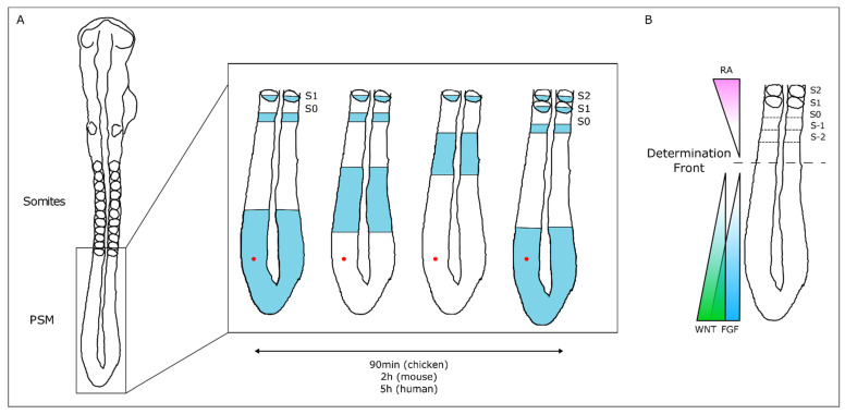
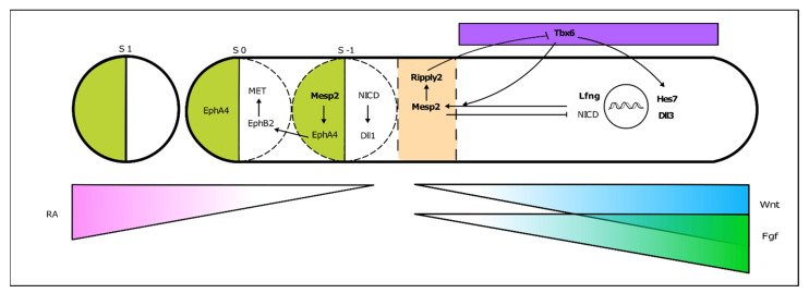
Spondylocostal dysostosis (SCDO) is a rare heritable congenital condition, characterized by multiple severe malformations of the vertebrae and ribs. Great advances were made in the last decades at the clinical level, by identifying the genetic mutations underlying the different forms of the disease. These were matched by extraordinary findings in the Developmental Biology field, which elucidated the cellular and molecular mechanisms involved in embryo body segmentation into the precursors of the axial skeleton. Of particular relevance was the discovery of the somitogenesis molecular clock that controls the progression of somite boundary formation over time. An overview of these concepts is presented, including the evidence obtained from animal models on the embryonic origins of the mutant-dependent disease. Evidence of an environmental contribution to the severity of the disease is discussed. Finally, a brief reference is made to emerging in vitro models of human somitogenesis which are being employed to model the molecular and cellular events occurring in SCDO. These represent great promise for understanding this and other human diseases and for the development of more efficient therapeutic approaches.
Keywords: DLL3; HES7; LFNG; MESP2; RIPPLY2; TBX6; somite formation; somitogenesis clock; spondylocostal dysostosis.
Conflict of interest statement
The authors declare no conflict of interest.
Figures


References
-
- UNICEF. WHO. World Bank UN DESA Levels & Trends in Child Mortality 2019. UN IGME Rep. 2019:52.
-
- Krauss R.S., Hong M. Gene–environment interactions and the etiology of birth defects. Curr. Top. Dev. Biol. 2016;116:569–580. - PubMed
-
- Berdon W.E., Lampl B.S., Cornier A.S., Ramirez N., Turnpenny P.D., Vitale M.G., Seimon L.P., Cowles R.A. Clinical and radiological distinction between spondylothoracic dysostosis (Lavy-Moseley syndrome) and spondylocostal dysostosis (Jarcho-Levin syndrome) Pediatr. Radiol. 2011;41:384–388. doi: 10.1007/s00247-010-1928-8. - DOI - PubMed
-
- Jarcho S. Hereditary malformation of the vertebral bodies. Bull. Johns Hopkins Hosp. 1938;62:216–226.
Publication types
Grants and funding
LinkOut - more resources
Full Text Sources
Other Literature Sources
