Review
. 2021 Jan 29;11(2):78.
doi: 10.3390/jpm11020078.

Personalized Endoscopy in Complex Malignant Hilar Biliary Strictures

Affiliations
Review

Personalized Endoscopy in Complex Malignant Hilar Biliary Strictures

Ivo Boškoski et al. J Pers Med. .

Abstract

Malignant hilar biliary obstruction (HBO) represents a complex clinical condition in terms of diagnosis, surgical and medical treatment, endoscopic approach, and palliation. The main etiology of malignant HBO is hilar cholangiocarcinoma that is considered an aggressive biliary tract's cancer and has still today a poor prognosis. Endoscopy plays a crucial role in malignant HBO from the diagnosis to the palliation. This technique allows the collection of cytological or histological samples, direct visualization of the suspect malignant tissue, and an echoendoscopic evaluation of the primary tumor and its locoregional staging. Because obstructive jaundice is the most common clinical presentation of malignant HBO, endoscopic biliary drainage, when indicated, is the preferred treatment over the percutaneous approach. Several endoscopic techniques are today available for both the diagnosis and the treatment of biliary obstruction. The choice among them can differ for each clinical scenario. In fact, a personalized endoscopic approach is mandatory in order to perform the proper procedure in the singular patient.

Keywords: confocal laser endomicroscopy; endoscopic retrograde cholangiopancreatography; endoscopic ultrasound; malignant hilar strictures; peroral cholangioscopy; personalized endoscopy; plastic biliary stents; self-expandable metal stents.


Conflict of interest statement

Professor Guido Costamagna: Consultant for and food and beverage compensation from Cook Medical, Boston Scientific, and Olympus. Dr. Ivo Boškoski: Consultant for Apollo Endosurgery, Cook Medical, and Boston Scientific; board member for Endo Tools; research grant recipient from Apollo Endosurgery; food and beverage compensation from Apollo Endosurgery, Cook Medical, Boston Scientific, and Endo Tools. All the other authors have nothing to declare.

Figures

Figure 1
Figure 1
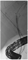
Peroral cholangioscopy of (A) a malignant hilar stricture and (B) target biopsy under direct visualization.
Figure 2
Figure 2
Confocal laser endomicroscopy showing a thickened reticular structure representing a criterion for malignancy.
Figure 3
Figure 3
Abdominal MRI showing a hilar cholangiocarcinoma (arrow) with left liver lobe atrophy (arrowhead).
Figure 4
Figure 4
Three percutaneous drainages in a patient with hilar cholangiocarcinoma. (A) Bile spillage from the drainages (B) with skin erythema.
Figure 5
Figure 5
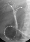
(A) Bismuth type II malignant hilar stricture (B) drained with two plastic stents. (C) Bismuth type IIIa malignant hilar stricture (D) drained with three self-expandable metal stents.
Figure 6
Figure 6
Endoscopic intervention for occluded self-expandable metal stent; sludge removal with a Fogarty balloon.
Figure 7
Figure 7
Magnetic resonance cholangiography (MRC) showing (A) Bismuth type IIIa malignant hilar stricture (B) confirmed by Endoscopic Retrograde Cholangio-Pancreatography (ERCP); (C) three guidewires are placed and (D) three plastic stents are inserted, obtaining complete drainage of all the liver segments. (E) MRC of a Bismuth type II malignant hilar stricture; (F) during ERCP, only the left hepatic ducts are opacified and a single plastic stent is inserted, obtaining incomplete drainage of the liver.
Figure 8
Figure 8
Radiofrequency ablation of hilar cholangiocarcinoma after self-expandable metal stents placement.
Figure 9
Figure 9
Three self-expandable metal stents with two nasobiliary drains with catheters for intraluminal brachytherapy in a case of hilar cholangiocarcinoma.
Figure 10
Figure 10
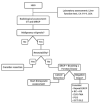
Diagnostic algorithm (HBO: hilar biliary obstruction; Ca 19-9: carbohydrate antigen 19-9; CEA: carcinoembryonic antigen; CT: computed tomography; MRCP: magnetic resonance cholangiopancreatography; ERCP: endoscopic retrograde cholangiopancreatography; BC: biliary brushing; FB: endoscopic forceps biopsy; EUS-FNA: echoendoscopic fine-needle aspiration; PCS: peroral cholangioscopy; OCT: optical coherence tomography; CLE: confocal laser endomicroscopy).
Figure 11
Figure 11
Therapeutic algorithm (MHBO: malignant hilar biliary obstruction; FLR: future liver remnant; PVE: portal vein embolization; PTBD: percutaneous transhepatic biliary drainage; PS: plastic stent; NBD: nasobiliary drainage; EUS-BD: echoendoscopic biliary drainage; U-SEMS: uncovered self-expandable metal stent; SEMS: self-expandable metal stent; ERCP: endoscopic retrograde cholangiopancreatography; RFA: radiofrequency ablation; PDT: photodynamic therapy).

References

    1. Chamberlain R.S., Blumgart L.H. Hilar Cholangiocarcinoma: A Review and Commentary. Ann. Surg. Oncol. 2000;7:55–66. doi: 10.1007/s10434-000-0055-4. - DOI - PubMed
    1. Koshiol J., Ferreccio C., Devesa S.S., Roa J.C., Fraumeni J.F. Schottenfeld and Fraumeni Cancer Epidemiology and Prevention. 4th ed. Oxford University Press (OUP); Oxford, UK: 2017. Biliary Tract Cancer.
    1. Bismuth H., Corlette M.B. Intrahepatic cholangioenteric anastomosis in carcinoma of the hilus of the liver. Surg. Gynecol. Obstet. 1975;140:170–178. - PubMed
    1. Squadroni M., Tondulli L., Gatta G., Mosconi S., Beretta G., Labianca R. Cholangiocarcinoma. Crit. Rev. Oncol. 2017;116:11–31. doi: 10.1016/j.critrevonc.2016.11.012. - DOI - PubMed
    1. Bridgewater J., Galle P.R., Khan S.A., Llovet J.M., Park J.-W., Patel T., Pawlik T.M., Gores G.J. Guidelines for the diagnosis and management of intrahepatic cholangiocarcinoma. J. Hepatol. 2014;60:1268–1289. doi: 10.1016/j.jhep.2014.01.021. - DOI - PubMed

LinkOut - more resources