Review
. 2021 Jan 29;13(2):205.
doi: 10.3390/v13020205.

Integrase Strand Transfer Inhibitors Are Effective Anti-HIV Drugs

Affiliations
Review

Integrase Strand Transfer Inhibitors Are Effective Anti-HIV Drugs

Steven J Smith et al. Viruses. .

Abstract

Integrase strand transfer inhibitors (INSTIs) are currently recommended for the first line treatment of human immunodeficiency virus type one (HIV-1) infection. The first-generation INSTIs are effective but can select for resistant viruses. Recent advances have led to several potent second-generation INSTIs that are effective against both wild-type (WT) HIV-1 integrase and many of the first-generation INSTI-resistant mutants. The emergence of resistance to these new second-generation INSTIs has been minimal, which has resulted in alternative treatment strategies for HIV-1 patients. Moreover, because of their high antiviral potencies and, in some cases, their bioavailability profiles, INSTIs will probably have prominent roles in pre-exposure prophylaxis (PrEP). Herein, we review the current state of the clinically relevant INSTIs and discuss the future outlook for this class of antiretrovirals.

Keywords: HIV; INSTIs; antiviral therapy; drug resistance; integration.


Conflict of interest statement

The content of this publication does not necessarily reflect the views or policies of the Department of Health and Human Services or the National Institutes of Health, nor does the mention of trade names, commercial products, or organizations imply endorsement by the U.S. government.

Figures

Figure 1
Figure 1
Chemical structures of the clinically relevant integrase strand transfer inhibitors (INSTIs). Chemical structures of the clinically relevant INSTIs are shown. The chelating motifs on the centralized pharmacophores that interact with Mg2+ cofactors in the integrase (IN) active site are highlighted with a blue circle. The halobenzyl moieties, which are connected to the centralized pharmacophore by a linker group, are circled in red.
Figure 2
Figure 2
RAL in the active site of the PFV intasome. RAL (cyan) is shown chelating the Mg2+ cofactors in the active site of the PFV intasome. PFV IN is in white, and the penultimate cytosine near the 3′ end of the viral DNA is in orange; the adenine at the 3′ end of the viral DNA is omitted for clarity. PFV IN active residues D128, D185, and E221 are labeled (the corresponding HIV-1 IN residues are shown in parentheses). Additional IN residues that undergo resistance mutations are labeled with their corresponding HIV-1 IN residues given in parentheses.
Figure 3
Figure 3
EVG in the active site of the PFV intasome. EVG (cyan) binding in the active site of the PFV intasome, chelating the Mg2+ cofactors (gray). PFV IN is in white, and the penultimate cytosine near the 3′ end of the viral DNA is in orange; the adenine on the 3′ end of the viral DNA is omitted for clarity. PFV IN active residues D128, D185, and E221 are labeled (corresponding HIV-1 IN residues are given in parentheses). Additional IN residues that undergo resistance mutations are labeled with the corresponding HIV-1 IN residues given in parentheses.
Figure 4
Figure 4
RAL overlaid onto DTG in the active site of the PFV intasome. The structures of both RAL (cyan) and DTG (purple) bound in the active site of the PFV intasomes [29]. IN active site residues D128, D185, and E221 are shown (the corresponding HIV-1 IN residues are given in parentheses). The penultimate cytosine near the 3′ end of the viral DNA end is labeled and colored orange as is IN residue Y212 (shown light blue; this residue is Y143 in HIV-1 IN) where mutations conferring resistance commonly arise. The halogenated benzyl moieties are highlighted by a red circle. The terminal adenine on the 3′ end of the viral DNA is omitted for clarity.
Figure 5
Figure 5
BIC in the active site of the HIV-1 intasome. Panel (A): BIC (cyan) is bound in the active site in the HIV-1 intasome. G118 of the β4α2 loop (labeled) is highlighted using a space filled model to reveal the close contact with the “A” ring of BIC. Panel (B): BIC (cyan) is shown in the HIV-1 intasome. The mutated amino acid, R118, is shown in a space filling model which shows how this amino acid substitution could disrupt the binding of BIC in the active site of the HIV-1 intasome. In both panels, the catalytic residues are shown and labeled. The penultimate cytosine is labeled and shown in orange, and the Mg2+ cofactors are gray. The terminal adenine on the 3′ end of the viral DNA end is omitted for clarity.
Figure 6
Figure 6
Modeling CAB onto the structure of BIC in the active site of the HIV-1 intasome. CAB (purple) is docked onto the structure of BIC (cyan) in the active site of the HIV-1 intasome. HIV-1 IN is shown; its catalytic residues are shown and labeled. The penultimate cytosine is shown (labeled), as are the Mg2+ cofactors (shown in gray and labeled). The terminal adenine on the viral DNA end is omitted for clarity.
Figure 7
Figure 7
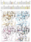
Comparison of HIV-1, SIV, and PFV intasomes. (a) Structure-guided alignment of the INs from HIV-1 (PDB ID: 6PUT), SIVrcm (6rcm), and PFV (3s3m), spanning HIV-1 IN residues M50-E157. Yellow stripes depict HIV IN residues, and the corresponding homologs in SIV and PFV, that are involved in drug resistance. Red stripes highlight the catalytic DDE triad responsible for IN enzymatic activity. (b) Comparison of the catalytic cores of HIV-1 IN (6put, first panel in tan), SIVrcm (6rcm, second panel in blue), and PFV (3s3m, third panel in pink) showing 20 residues that are involved in HIV drug resistance. All three structures are superimposed in the fourth panel. In the second and third panel, residues that are not conserved when HIV-1 IN is compared with either SIVrcm IN (second panel) or PFV IN (third panel) are labeled with a star.

Similar articles

Cited by

References

    1. Volberding P.A., Deeks S.G. Antiretroviral therapy and management of HIV infection. Lancet. 2010;376:49–62. doi: 10.1016/S0140-6736(10)60676-9. - DOI - PubMed
    1. U.S. Department of Health and Human Services . Guidelines for the Use of Antiretroviral Agents in Adults and Adolescents with HIV. G-1. The U.S. Department of Health and Human Services Guidelines, AIDSinfo; Washington, DC, USA: 2019.
    1. Kearney M.F., Spindler J., Shao W., Yu S., Anderson E.M., O’Shea A., Rehm C., Poethke C., Kovacs N., Mellors J.W., et al. Lack of detectable HIV-1 molecular evolution during suppressive antiretroviral therapy. PLoS Pathog. 2014;10:e1004010. doi: 10.1371/journal.ppat.1004010. - DOI - PMC - PubMed
    1. Fida M., Rizza S.A., Temesgen Z. Dolutegravir plus lamivudine dual therapy—A new option for initial antiretroviral therapy. Drugs Today. 2019;55:297–304. doi: 10.1358/dot.2019.55.5.2962947. - DOI - PubMed
    1. Cahn P., Madero J.S., Arribas J.R., Antinori A., Ortiz R., Clarke A.E., Hung C.C., Rockstroh J.K., Girard P.M., Sievers J., et al. Dolutegravir plus lamivudine versus dolutegravir plus tenofovir disoproxil fumarate and emtricitabine in antiretroviral-naive adults with HIV-1 infection (GEMINI-1 and GEMINI-2): Week 48 results from two multicentre, double-blind, randomised, non-inferiority, phase 3 trials. Lancet. 2019;393:143–155. doi: 10.1016/S0140-6736(18)32462-0. - DOI - PubMed

Publication types

LinkOut - more resources