. 2021 Jan 29;22(3):1363.
doi: 10.3390/ijms22031363.

Monokaryotic Pleurotus sapidus Strains with Intraspecific Variability of an Alkene Cleaving DyP-Type Peroxidase Activity as a Result of Gene Mutation and Differential Gene Expression

Affiliations

Monokaryotic Pleurotus sapidus Strains with Intraspecific Variability of an Alkene Cleaving DyP-Type Peroxidase Activity as a Result of Gene Mutation and Differential Gene Expression

Nina-Katharina Krahe et al. Int J Mol Sci. .

Abstract

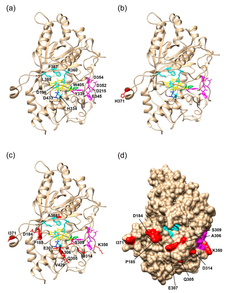
The basidiomycete Pleurotus sapidus produced a dye-decolorizing peroxidase (PsaPOX) with alkene cleavage activity, implying potential as a biocatalyst for the fragrance and flavor industry. To increase the activity, a daughter-generation of 101 basidiospore-derived monokaryons (MK) was used. After a pre-selection according to the growth rate, the activity analysis revealed a stable intraspecific variability of the strains regarding peroxidase and alkene cleavage activity of PsaPOX. Ten monokaryons reached activities up to 2.6-fold higher than the dikaryon, with MK16 showing the highest activity. Analysis of the PsaPOX gene identified three different enzyme variants. These were co-responsible for the observed differences in activities between strains as verified by heterologous expression in Komagataella phaffii. The mutation S371H in enzyme variant PsaPOX_high caused an activity increase alongside a higher protein stability, while the eleven mutations in variant PsaPOX_low resulted in an activity decrease, which was partially based on a shift of the pH optimum from 3.5 to 3.0. Transcriptional analysis revealed the increased expression of PsaPOX in MK16 as reason for the higher PsaPOX activity in comparison to other strains producing the same PsaPOX variant. Thus, different expression profiles, as well as enzyme variants, were identified as crucial factors for the intraspecific variability of the PsaPOX activity in the monokaryons.

Keywords: Pleurotus sapidus; alkene cleavage; basidiomycota; biocatalysis; dikaryon; dye-decolorizing peroxidase (DyP); gene expression; gene mutation; intraspecific variability; monokaryon.


Conflict of interest statement

The authors declare no conflict of interest. The funders had no role in the design of the study; in the collection, analyses, or interpretation of data; in the writing of the manuscript, or in the decision to publish the results.

Figures

Figure 1
Figure 1
Alkene cleavage of trans-anethole into p-anisaldehyde by the dye-decolorizing peroxidase PsaPOX.
Figure 2
Figure 2
Distribution of vegetative growth rate of the P. sapidus monokaryons grown on standard nutrient liquid (SNL) agar plates. The strains were categorized as slow-, moderate-, and fast-growing strains according to the growth rate.
Figure 3
Figure 3
Alkene cleavage activity in comparison to peroxidase activity and growth rate of selected monokaryons and the parental dikaryon (yellow bar or circle) of P. sapidus. (a) Enzymatically generated concentration of p-anisaldehyde after conversion of trans-anethole (33.5 mM) using 30 mg/mL finely ground lyophilized P. sapidus mycelium in the presence of 25 mM MnSO4, 100 µM H2O2, and 50 mM sodium acetate buffer pH 3.5 for 16 h at RT and 200 rpm. (b) Comparison of alkene cleavage activity for the lyophilized P. sapidus mycelium after submerged cultivation and radial growth rate for the cultivation on SNL agar plates. (c) Peroxidase activity of the lyophilized mycelium. (d) Comparison of alkene cleavage and peroxidase activity of the lyophilized mycelium of the P. sapidus strains. p-Anisaldehyde concentrations are the average of duplicate experiments, and peroxidase activities the average of triplicate experiments of two independent biological replicates (with standard deviations shown as error bars in (a,b)). Growth rates are the average of two independent biological replicates cultivated under the same conditions.
Figure 4
Figure 4
Stability of alkene cleavage activity towards trans-anethole of selected monokaryons and the parental dikaryon over five serial generations. Values are the average of duplicate experiments of two independent biological replicates with standard deviations shown as error bars.
Figure 5
Figure 5
Alignment of the amino acid sequence of the PsaPOX variants. DK: PsaPOX_DK; low: PsaPOX_low; high: PsaPOX_high. Inverted triangles show amino acids important for heme binding (H334 (red) functions as ligands for heme and the four other amino acid residues form a hydrogen peroxide binding pocket). Aspartic acid (D433), which forms a hydrogen bond with histidine to stabilize compound I (oxidized heme after transfer of two electrons to H2O2), is indicated by a cross. The black box indicates the GXXDG motif containing the catalytic aspartic acid residue (D196, yellow inverted triangle), which cleaves H2O2 heterolytically with the help of the neighboring arginine (R360, blue inverted triangle) to form compound I, and the circle presents an exposed tryptophan (W405) potentially involved in a long-range electron transfer. Important amino acids for Mn2+-oxidation (D215, Y339, E345, D352, and D354) are marked by squares. Secondary structure elements are shown above the alignment (arrow: β-sheets, barrel: α-helices, dashed line: random coil) and were predicted by the SWISS-MODEL server [37] using the X-ray crystal structure of Pleos-DyP4 from P. ostreatus (PDB-ID 6fsk). Asterisks indicate conserved residues, colons indicate equivalent residues, and dots indicate partial residue conservations. Observed mutations compared to the parental sequence (DK) were highlighted in black with white font. The alignment was performed with Clustal Omega (European Bioinformatics Institute, Hinxton, UK) [38].
Figure 6
Figure 6
Comparison of the PsaPOX variants regarding their peroxidase and alkene cleavage activity. (a) Specific peroxidase activity. Values are the average of triplicate measurements using three independent replicates with standard deviations shown as error bars (b) p-Anisaldehyde concentration after bioconversion of trans-anethole (6.7 mM) using 0.25 mg/mL PsaPOX variant in the presence of 25 mM MnSO4, 100 µM H2O2, and 50 mM sodium acetate buffer pH 3.5 for 16 h at RT and 200 rpm. Values are the average of duplicate independent experiments with standard deviations shown as error bars. DK: PsaPOX_DK; high: PsaPOX_high; low: PsaPOX_low.
Figure 7
Figure 7
Structural homology models of the three PsaPOX variants. The models were generated with the SWISS-MODEL server using the X-ray crystal structure of Pleos-DyP4 (PDB-ID 6fsk). (a) Overall fold of PsaPOX_DK showing the typical ferredoxin-like fold with the heme cofactor (shown in yellow, the heme iron is highlighted in orange) sandwiched between distal and proximal sides. Conserved amino acids of the active site/hydrogen peroxide pocket (proximal H334, distal D196, R360, L385, and F387) are shown in cyan. D433, which forms a hydrogen bond with H334 to stabilize compound I (oxidized heme after transfer of two electrons to H2O2), is presented in blue, and the exposed W405 potentially involved in a long-range electron transfer is shown in green. Important amino acids for Mn2+-oxidation (D215, Y339, E345, D352, and D354) are highlighted in magenta. Overall fold of (b) PsaPOX_high, and (c) PsaPOX_low. Mutations present in the enzyme variants are shown in red. (d) Solvent access surface of PsaPOX_low, showing the heme active site in cyan, the Mn2+-oxidation site in magenta, and the position of the surface-exposed amino acid exchanges in red.
Figure 8
Figure 8
Influence of pH and temperature on activity and stability of the PsaPOX variants. (a) pH optimum and (b) temperature optimum. Relative peroxidase activity was defined as the percentage of activity detected with respect to the highest activity of the corresponding enzyme variant in each experiment. pH stability (c) was determined after incubation of PsaPOX in Britton–Robinson buffer, ranging from pH 2.0 to 9.5 for 1 h at RT, and temperature stability (d) was determined after incubation at 20–90 °C and pH 3.5 for 1 h. Residual activities were determined at pH 3.5 and 40 °C. Values are the average of triplicate experiments with standard deviations shown as error bars. DK: PsaPOX_DK; high: PsaPOX_high; low: PsaPOX_low.
Figure 9
Figure 9
Effect of hydrogen peroxide concentration on the activity of the PsaPOX variants (a), and relative peroxidase activity of the PsaPOX variants during biotransformation of trans-anethole over 16 h (b). Relative peroxidase activity was defined as the percentage of activity detected normalized to the highest activity (a) or to the starting activity (b) of the corresponding enzyme variant in each experiment. Values are the average of triplicate experiments with standard deviations shown as error bars. DK: PsaPOX_DK; high: PsaPOX_high; low: PsaPOX_low.
Figure 10
Figure 10
Relative expression of the PsaPOX gene (a) in comparison to the alkene cleavage activity, and (b) of selected monokaryons and the dikaryotic P. sapidus strain. Expression rates were normalized to the expression of the parental dikaryon. Values are the average of three (expression) or two (alkene cleavage activity) independent biological replicates with standard deviations shown as error bars. P. sapidus strains were grouped according to their PsaPOX sequence (blue: PsaPOX_DK; red: PsaPOX_high; green: PsaPOX_low).

Similar articles

Cited by

References

    1. Fahlbusch K.-G., Hammerschmidt F.-J., Panten J., Pickenhagen W., Schatkowski D., Bauer K., Garbe D., Surburg H. Ullmann’s Encyclopedia of Industrial Chemistry. Wiley-VCH Verlag GmbH & Co. KGaA; Weinheim, Germany: 2003. Flavors and Fragrances; pp. 73–140.
    1. Rajagopalan A., Lara M., Kroutil W. Oxidative Alkene Cleavage by Chemical and Enzymatic Methods. Adv. Synth. Catal. 2013;355:3321–3335. doi: 10.1002/adsc.201300882. - DOI
    1. Spannring P., Bruijnincx P.C.A., Weckhuysen B.M., Klein Gebbink R.J.M. Transition metal-catalyzed oxidative double bond cleavage of simple and bio-derived alkenes and unsaturated fatty acids. Catal. Sci. Technol. 2014;4:2182–2209. doi: 10.1039/c3cy01095c. - DOI
    1. Berger R.G. Biotechnology of flavours—The next generation. Biotechnol. Lett. 2009;31:1651–1659. doi: 10.1007/s10529-009-0083-5. - DOI - PubMed
    1. Bel-Rhlid R., Berger R.G., Blank I. Bio-mediated generation of food flavors—Towards sustainable flavor production inspired by nature. Trends Food Sci. Technol. 2018;78:134–143. doi: 10.1016/j.tifs.2018.06.004. - DOI

Supplementary concepts

LinkOut - more resources