. 2021 Jan 29;22(3):1375.
doi: 10.3390/ijms22031375.

Extracellular Vesicles Do Not Mediate the Anti-Inflammatory Actions of Mouse-Derived Adipose Tissue Mesenchymal Stem Cells Secretome

Affiliations

Extracellular Vesicles Do Not Mediate the Anti-Inflammatory Actions of Mouse-Derived Adipose Tissue Mesenchymal Stem Cells Secretome

María Carmen Carceller et al. Int J Mol Sci. .

Abstract

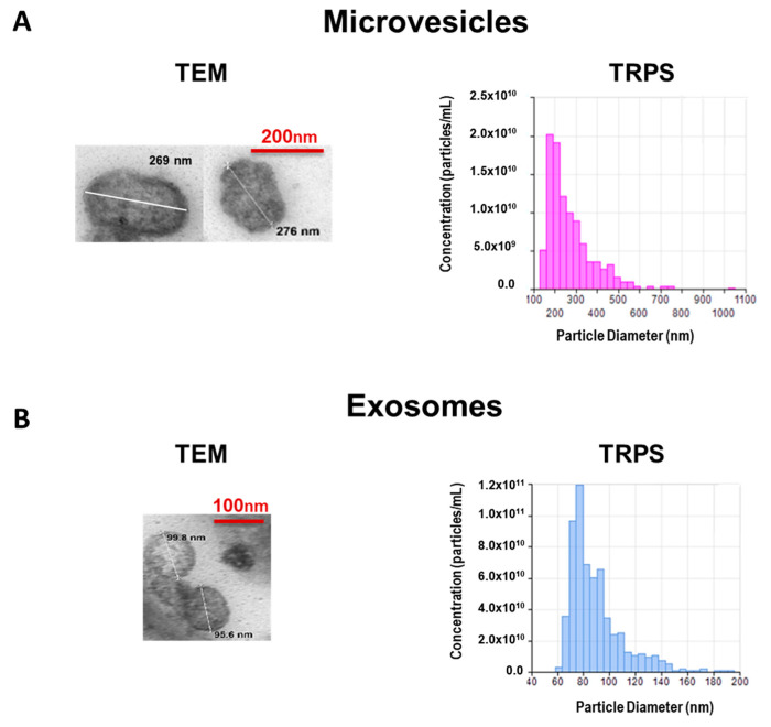
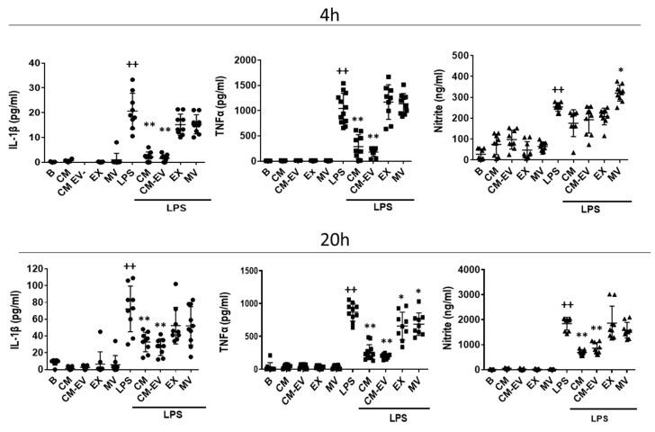
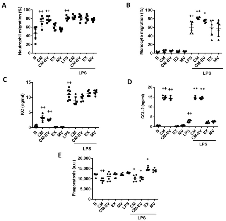
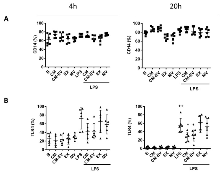
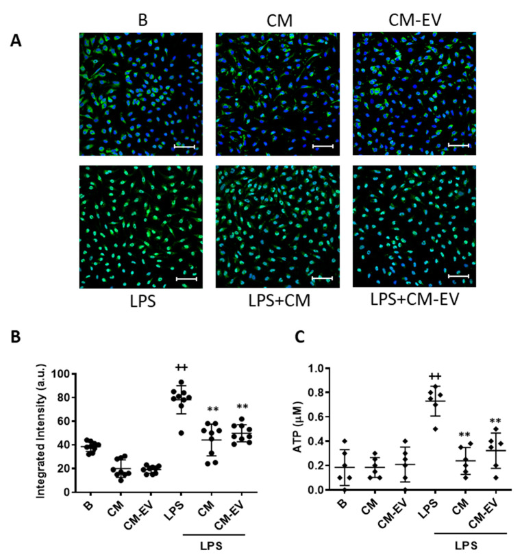
Adipose tissue represents an abundant source of mesenchymal stem cells (MSC) for therapeutic purposes. Previous studies have demonstrated the anti-inflammatory potential of adipose tissue-derived MSC (ASC). Extracellular vesicles (EV) present in the conditioned medium (CM) have been shown to mediate the cytoprotective effects of human ASC secretome. Nevertheless, the role of EV in the anti-inflammatory effects of mouse-derived ASC is not known. The current study has investigated the influence of mouse-derived ASC CM and its fractions on the response of mouse-derived peritoneal macrophages against lipopolysaccharide (LPS). CM and its soluble fraction reduced the release of pro-inflammatory cytokines, adenosine triphosphate and nitric oxide in stimulated cells. They also enhanced the migration of neutrophils or monocytes, in the absence or presence of LPS, respectively, which is likely related to the presence of chemokines, and reduced the phagocytic response. The anti-inflammatory effect of CM may be dependent on the regulation of toll-like receptor 4 expression and nuclear factor-κB activation. Our results demonstrate the anti-inflammatory effects of mouse-derived ASC secretome in mouse-derived peritoneal macrophages stimulated with LPS and show that they are not mediated by EV.

Keywords: extracellular vesicles; inflammation; macrophage; mesenchymal stem cells secretome; mouse-derived adipose tissue.


Conflict of interest statement

The authors declare no conflict of interest.

Figures

Figure 1
Figure 1
Characterization of microvesicles (A) and exosomes (B) isolated from CM of AMSC. Representative transmission electron microscopy (TEM) images with estimated size. Microparticle size distribution and concentration were determined by tunable resistive pulse sensing (TRPS).
Figure 2
Figure 2
Release of pro-inflammatory mediators by mouse-derived peritoneal macrophages. Cytokines were measured by ELISA and nitrite was determined by fluorometry in cell supernatants at 4 h and 20 h of incubation with CM, CM-EV, EX, or MV in the presence or absence of LPS. B: cells incubated with control medium and not stimulated with LPS. Data are presented as mean ± SD (n = 9) from 3 independent experiments. One-way analysis of variance followed by Tukey’s post hoc test; ++ p < 0.001 versus B; * p < 0.05, ** p < 0.01 versus LPS control.
Figure 3
Figure 3
Macrophage functions. Neutrophil migration (A), monocyte migration (B), CXCL-1 levels (C), CCL-2 levels (D) and phagocytosis (E). For migration experiments, macrophages were incubated with CM, CM-EV, EX, or MV in the presence or absence of LPS for 20 h. Then, neutrophils or monocytes were added to the upper compartment of the transwells and after 4 h, the migrated cells were quantified by flow cytometry. CXCL-1 and CCL-2 were measured by ELISA in the supernatants of neutrophil or monocyte migration assays, respectively. Macrophage phagocytosis of fluorescent polystyrene beads was determined by flow cytometry. B: cells incubated with control medium and not stimulated with LPS. Data are presented as mean ± SD (n = 6) from 3 independent experiments. One-way analysis of variance followed by Tukey’s post hoc test; ++ p < 0.001 versus B; * p < 0.05, ** p < 0.01 versus LPS control.
Figure 4
Figure 4
Expression of CD14 (A) and TLR4 (B). Macrophages were incubated with CM, CM-EV, EX, or MV in the presence or absence of LPS for 4 h or 20 h. Protein expression of CD14 and TLR4 was determined by flow cytometry. B: cells incubated with control medium and not stimulated with LPS. Data are presented as mean ± SD (n = 6) from 3 independent experiments. One-way analysis of variance followed by Tukey’s post hoc test; ++ p < 0.001 versus B; * p < 0.05 versus LPS control.
Figure 5
Figure 5
NF-κB nuclear translocation and ATP release. For NF-κB translocation, macrophages were incubated with CM or CM-EV for 30 min and immunofluorescence was determined by confocal microscopy using an NF-κB p65 XP® Rabbit (Alexa Fluor® 488 Conjugate). (A) Representative images. Microscopic magnification of the objective lens 40 x. Bar = 50 µm. (B) Quantification of nuclear translocation. For ATP release (C), macrophages were incubated with CM or CM-EV in the presence or absence of LPS for 20 h. ATP levels were determined by luminescence in the medium. B: cells incubated with control medium and not stimulated with LPS. Data are presented as mean ± SD (n = 6–9) from 3 independent experiments. One-way analysis of variance followed by Tukey’s post hoc test; ++ p < 0.001 versus B; ** p < 0.01 versus LPS control.

Similar articles

Cited by

References

    1. Chamberlain G., Fox J., Ashton B., Middleton J. Concise review: Mesenchymal stem cells: Their phenotype, differentiation capacity, immunological features, and potential for homing. Stem Cells. 2007;25:2739–2749. doi: 10.1634/stemcells.2007-0197. - DOI - PubMed
    1. Shen C., Lie P., Miao T., Yu M., Lu Q., Feng T., Li J., Zu T., Liu X., Li H. Conditioned medium from umbilical cord mesenchymal stem cells induces migration and angiogenesis. Mol. Med. Rep. 2015;12:20–30. doi: 10.3892/mmr.2015.3409. - DOI - PMC - PubMed
    1. Abumaree M., Al J.M., Pace R.A., Kalionis B. Immunosuppressive properties of mesenchymal stem cells. Stem Cell Rev. 2012;8:375–392. doi: 10.1007/s12015-011-9312-0. - DOI - PubMed
    1. Caplan A.I., Dennis J.E. Mesenchymal stem cells as trophic mediators. J. Cell Biochem. 2006;98:1076–1084. doi: 10.1002/jcb.20886. - DOI - PubMed
    1. Zhang M., Mal N., Kiedrowski M., Chacko M., Askari A.T., Popovic Z.B., Koc O.N., Penn M.S. SDF-1 expression by mesenchymal stem cells results in trophic support of cardiac myocytes after myocardial infarction. FASEB J. 2007;21:3197–3207. doi: 10.1096/fj.06-6558com. - DOI - PubMed

Substances

LinkOut - more resources