Review
. 2021 Jan 30;11(2):105.
doi: 10.3390/life11020105.

Molecules and Mechanisms to Overcome Oxidative Stress Inducing Cardiovascular Disease in Cancer Patients

Affiliations
Review

Molecules and Mechanisms to Overcome Oxidative Stress Inducing Cardiovascular Disease in Cancer Patients

Francesco Sabbatino et al. Life (Basel). .

Abstract

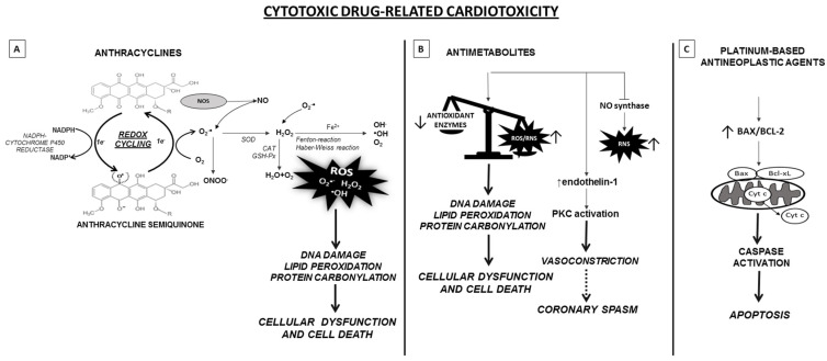
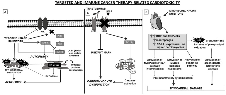
Reactive oxygen species (ROS) are molecules involved in signal transduction pathways with both beneficial and detrimental effects on human cells. ROS are generated by many cellular processes including mitochondrial respiration, metabolism and enzymatic activities. In physiological conditions, ROS levels are well-balanced by antioxidative detoxification systems. In contrast, in pathological conditions such as cardiovascular, neurological and cancer diseases, ROS production exceeds the antioxidative detoxification capacity of cells, leading to cellular damages and death. In this review, we will first describe the biology and mechanisms of ROS mediated oxidative stress in cardiovascular disease. Second, we will review the role of oxidative stress mediated by oncological treatments in inducing cardiovascular disease. Lastly, we will discuss the strategies that potentially counteract the oxidative stress in order to fight the onset and progression of cardiovascular disease, including that induced by oncological treatments.

Keywords: ROS; antioxidative treatment; cardiotoxicity; cardiovascular disease; endothelial dysfunction; oncological treatments; oxidative stress.


Conflict of interest statement

The authors declare no conflict of interest.

Figures

Figure 1
Figure 1
Schematic representation of the molecular mechanisms underlying cytotoxic drugs-related cardiotoxicity. (A) Anthracyclines undergo redox cycling catalyzed by NADPH-Cytochrome P-450 reductase. The one-electron (1e) reduction of the quinone compound leads to the formation of semiquinone intermediate, that in presence of molecular oxygen auto-oxides generating the parent anthracycline and O2∙. O2∙ is either transformed to RNS or dismutated to H2O2 by SOD. Subsequently, H2O2 is eliminated by CAT and GSH-Px or may react, in presence of O2∙, with the ion Fe2+, via the Fenton and Haber-Weiss reaction, giving rise to reactive oxygen species (ROS). ROS lead to DNA, lipid and protein damage and apoptosis induction. In addition, anthracyclines can bind to NOS, causing an increase of RNS, particularly the ONOO, highly harmful for cells. (B) Antimetabolites increase the levels of ROS/RNS mainly in cardiomyocytes and endothelial cells. In addition, antimetabolites can reduce the enzymatic activities of antioxidant enzymes. As a result, there is an imbalance between the production of ROS/RNS and the availability of antioxidants. This generates oxidative stress causing the oxidation of macromolecules, thus disturbing cellular functions. Antimetabolites, especially 5-FU, also lead to NOS dysregulation with reduced NO availability and increased RNS along with endothelin-1 upregulation and the activation of protein kinase C. These processes lead to endothelium-dependent and -independent vasoconstriction, and potentially to coronary spasm. (C) Finally, platinum-based antineoplastic agents induce oxidative stress by impairment of mitochondrial metabolism and nuclear activity, promoting apoptosis and cell death by BAX induction. In cardiomyocites, platinum-derivatives, primarily cisplatin, cause mitochondria dysfunction associated to increased caspase activity, ultimately leading to apoptosis. Abbreviations: CAT, catalase; GSH-Px, glutathione peroxidase; H2O2, hydrogen peroxide; NOS, nitrous oxide synthase; O2∙, superoxide anion radical; ONOO, peroxy-nitrite; RNS, reactive nitrogen species; ROS, reactive oxygen species; SOD, superoxide dismutase.
Figure 2
Figure 2
Schematic representation of molecular mechanisms of targeted and immune cancer therapy-related cardiotoxicity. (A) TKI administration induces cardiotoxicity via induction of ER stress and mitochondrial dysfunction, leading to unfolded protein accumulation, Ca2+ release, ROS generation, AMPK activation and mTOR inhibition. The interplay among these cellular pathways results in autophagy and apoptosis. (B) In heart, the cardioactive growth factor NRG-1 triggers HER-4/HER-2 heterodimerization to induce protective pathways in response to stress. Blockade of NRG-1/HER-4/HER-2 axis by trastuzumab results in the inhibition of MAPK and PI3K/AKT pathways which cause alterations of structure and functions of cardiomyocytes. In addition, blocking of HER2 is correlated with a change in the antiapoptotic/proapoptotic proteins ratio, due to an upregulation of BCL-XS (proapoptotic) and downregulation of BCL-XL (antiapoptotic), which leads to contractile dysfunction of cardiac cell. (C) ICI can induce cardiotoxicity by increasing in heart tissue infiltration of activated T lymphocytes (CD4+/CD8+ T cells) and macrophages and increased expression levels of PD-L1. This leads to pro-inflammatory and cardiotoxic effects and the activation of specific pathways, including NLRP3, Myd88, p65/NF-kb and arachidonate-leukotriene pathways. The latter probably is regulated by the high production of ROS and phospholipid oxidation due to accumulation of lymphocytes and macrophages. Abbreviations: AMPK, AMP-activated protein kinase; ER, endoplasmic reticulum; MAPK, mitogen-activated protein kinase; Myd88, myeloid differentiation primary response 88; mTOR, mammalian target of rapamycin; NLRP3, NLR Family Pyrin Domain Containing 3; NRG-1, neuregulin-1; PDL-1, Programmed death-ligand 1; ROS, Reactive oxygen species; RTK, receptor tyrosine kinase; TKIs, Tyrosine kinase inhibitors.

Similar articles

Cited by

References

    1. Valko M., Leibfritz D., Moncol J., Cronin M.T.D., Mazur M., Telser J. Free Radicals and Antioxidants in Normal Physiological Functions and Human Disease. Int. J. Biochem. Cell Biol. 2007;39:44–84. doi: 10.1016/j.biocel.2006.07.001. - DOI - PubMed
    1. Nordberg J., Arnér E.S. Reactive Oxygen Species, Antioxidants, and the Mammalian Thioredoxin System. Free Radic. Biol. Med. 2001;31:1287–1312. doi: 10.1016/S0891-5849(01)00724-9. - DOI - PubMed
    1. Ristow M., Schmeisser K. Mitohormesis: Promoting Health and Lifespan by Increased Levels of Reactive Oxygen Species (ROS) Dose Response. 2014;12:288–341. doi: 10.2203/dose-response.13-035.Ristow. - DOI - PMC - PubMed
    1. Cao S.S., Kaufman R.J. Endoplasmic Reticulum Stress and Oxidative Stress in Cell Fate Decision and Human Disease. Antioxid. Redox Signal. 2014;21:396–413. doi: 10.1089/ars.2014.5851. - DOI - PMC - PubMed
    1. Conti V., Corbi G., Simeon V., Russomanno G., Manzo V., Ferrara N., Filippelli A. Aging-Related Changes in Oxidative Stress Response of Human Endothelial Cells. Aging Clin. Exp. Res. 2015;27:547–553. doi: 10.1007/s40520-015-0357-9. - DOI - PubMed

LinkOut - more resources