. 2021 Jan 30;13(3):531.
doi: 10.3390/cancers13030531.

The Effect of Neddylation Blockade on Slug-Dependent Cancer Cell Migration Is Regulated by p53 Mutation Status

Affiliations

The Effect of Neddylation Blockade on Slug-Dependent Cancer Cell Migration Is Regulated by p53 Mutation Status

Yelee Kim et al. Cancers (Basel). .

Abstract

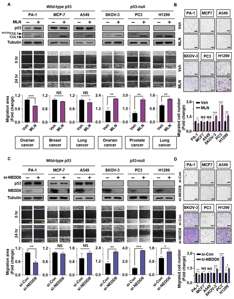
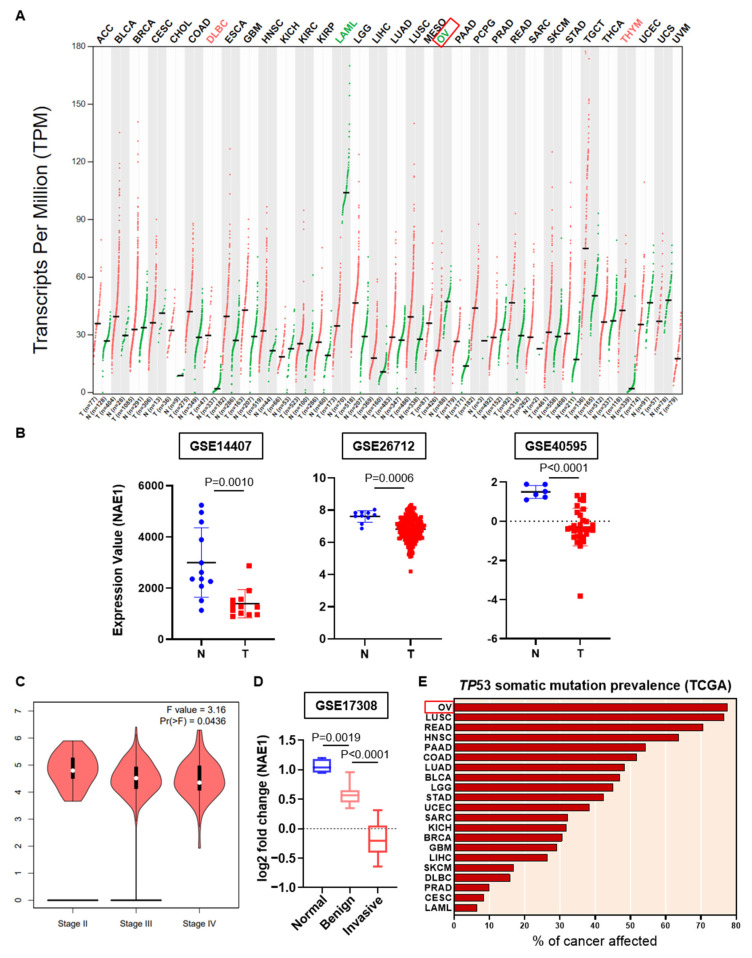
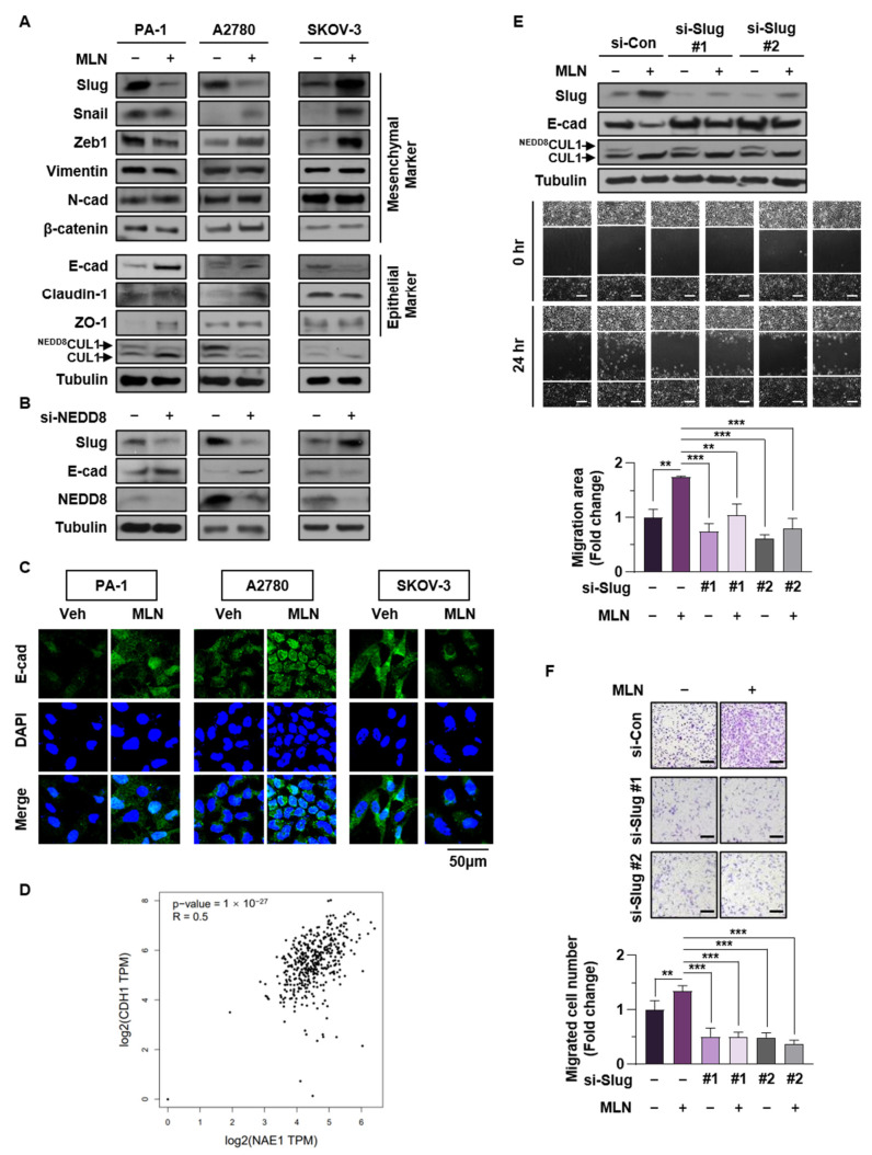
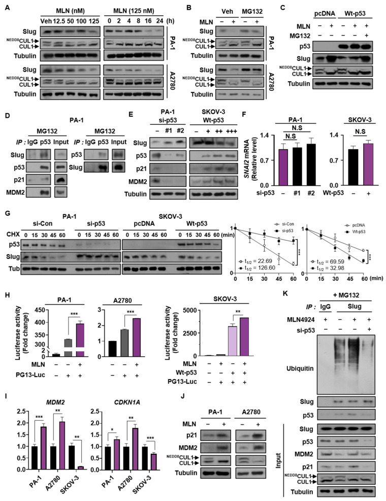
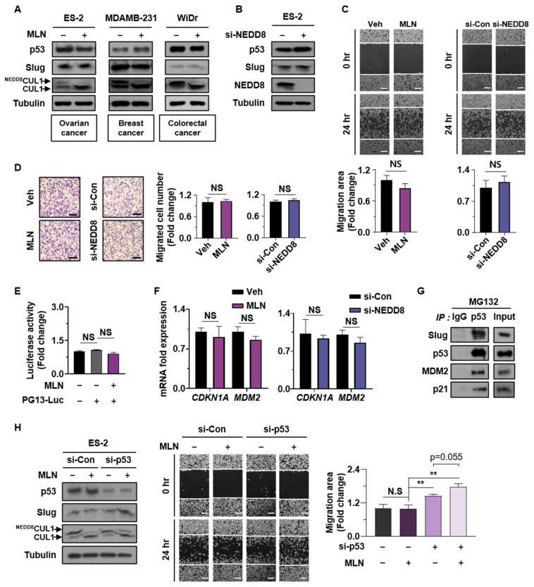
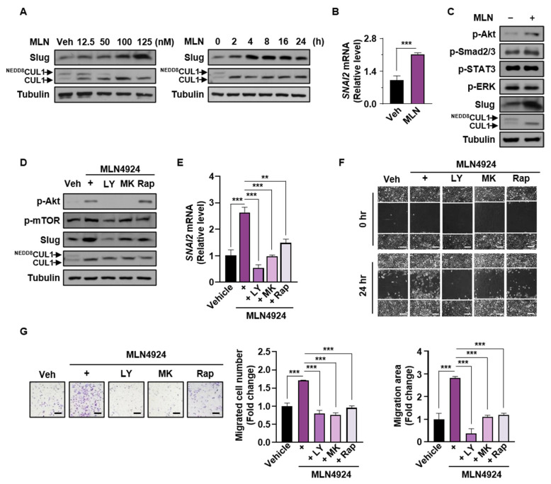
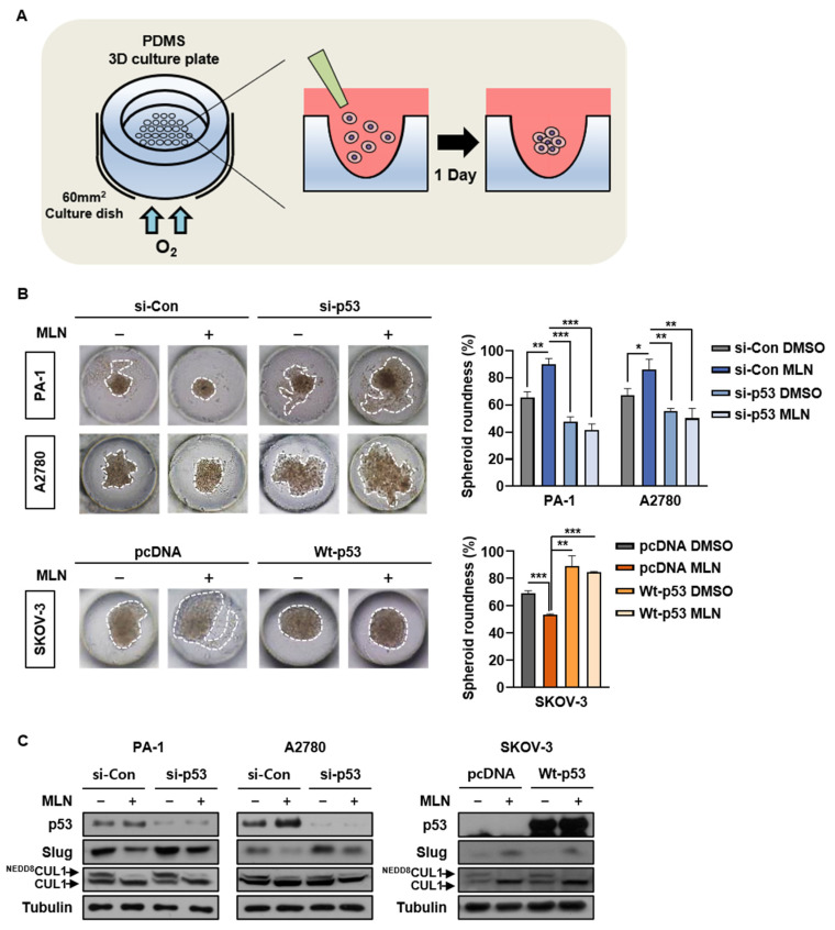
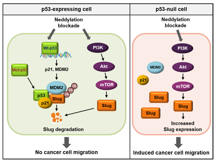
The tumor suppressor protein p53 is frequently inactivated in human malignancies, in which it is associated with cancer aggressiveness and metastasis. Because p53 is heavily involved in epithelial-mesenchymal transition (EMT), a primary step in cell migration, p53 regulation is important for preventing cancer metastasis. p53 function can be modulated by diverse post-translational modifications including neddylation, a reversible process that conjugates NEDD8 to target proteins and inhibits the transcriptional activity of p53. However, the role of p53 in cancer migration by neddylation has not been fully elucidated. In this study, we reported that neddylation blockade induces cell migration depending on p53 status, specifically via the EMT-promoting transcription factor Slug. In cancer cell lines expressing wild type p53, neddylation blockade increased the transcriptional activity of p53 and expression of its downstream genes p21 and MDM2, eventually promoting proteasomal degradation of Slug. In the absence of p53, neddylation blockade increased cell migration by activating the PI3K/Akt/mTOR/Slug signaling axis. Because mutant p53 was transcriptionally inactivated but maintained the ability to bind to Slug, neddylation blockade did not affect the migration of cells expressing mutant p53. Our findings highlight how the p53 expression status influences neddylation-mediated cell migration in multiple cancer cell lines via Slug.

Keywords: PI3K/Akt/mTOR signaling; Slug; epithelial-mesenchymal transition (EMT); migration; neddylation; p53.


Conflict of interest statement

The authors declare no conflict of interests. The funders had no role in the design of the study; in the collection, analyses, or interpretation of data; in the writing of the manuscript; or in the decision to publish the results.

Figures

Figure 1
Figure 1
Neddylation blockade induces cancer cell migration according to p53 status. (A,C) Wild type p53 (PA-1, MCF-7, and A549) and p53-null (SKOV-3, PC3, and H1299) cancer cell lines were subjected to wound healing assays with or without MLN4924 (125 nM) for 24 h or were transfected with si-Control or si-NEDD8 and analyzed by Western blotting. Scale bar: 200 μm. Whole areas were measured using ImageJ software. The upper band of CUL1 indicates neddylated CUL1, and the lower band indicates CUL1. (B,D) Cancer cells treated with or without MLN4924 (125 nM) for 24 h or transfected with si-Control or si-NEDD8 were subjected to Transwell assays. The number of cells in four randomly chosen fields was counted. Data are presented as the means ± standard deviation (n = 3). * p < 0.05; ** p < 0.01; *** p < 0.001; NS, not significant.
Figure 2
Figure 2
Associations of NAE1 expression, the pathological stage, and TP53 mutation prevalence in ovarian tumors. (A) Expression level (transcript per million: log2(TPM + 1)) of NAE1 between TCGA tumors (red dots) and TCGA normal, and GTEx normal data (green dots) in 27 cancer types using Gene Expression Profiling Interactive Analysis (GEPIA). T: Tumor; N: Normal; n: number. (B) Distribution of NAE1 expression in normal ovarian surface epithelium (blue circles) and serous ovarian cancer epithelium (red squares) (GSE14407, GSE26712, GSE40595). (C) Violin plot indicating the correlation between NAE1 expression and pathological stage in patients with ovarian cancer (GEPIA). The p-value was set at 0.05. (D) Log2 fold change of NAE1 mRNA expression in ovarian surface epithelium (blue box), benign ovarian serous tumors (pink box), and invasive ovarian serous tumors (red box) (GSE17308). (E) TP53 somatic mutation prevalence in TCGA data were downloaded from the International Cancer Genome Consortium (ICGC) web portal.
Figure 3
Figure 3
Slug plays a major role in regulating neddylation blockade-mediated EMT in cancer cells (A,B) PA-1, A2780, and SKOV-3 cells were treated with or without MLN4924 (125 nM) or transfected with si-Control or si-NEDD8. Protein levels of representative EMT markers were verified by western blot assays. (C) After cells had been treated with or without MLN4924 for 24 h in serum free medium, the expression of E-cadherin (green) was visualized by immunofluorescence. Scale bar: 50 μm. The results shown are representative of three experiments with similar results. (D) Correlation between expression of NAE1 and CDH1 in ovarian cancer, analyzed using the GEPIA database. (E) SKOV-3 cells transfected with si-Control or si-Slug#1, 2 and/or treated with MLN4924 (125 nM) were subjected to wound healing assays. Cell lysates were then subjected to western blot assays to verify protein levels of Slug and E-cadherin. Scale bar: 200 μm. Whole areas were measured using ImageJ software, and data are presented as the means ± standard deviation (n = 3). (F) SKOV-3 cells transfected with si-Control or si-Slug#1, 2 and/or treated with MLN4924 (125 nM) were subjected to Transwell assay. The number of cells in four randomly chosen fields was counted. ** p < 0.01; *** p < 0.001; NS, not significant.
Figure 4
Figure 4
Neddylation blockade promotes proteasomal degradation of Slug in cancer cells expressing wild type p53. (A) PA-1 and A2780 cells were treated with MLN4924 at different doses and times. Slug expression was verified by Western blot analysis. (B) Cells treated with or without MLN4924 for 24 h were incubated with or without MG132 for 6 h, and the cell lysates were subjected to Western blot assays. (C) SKOV-3 cells transfected with pcDNA or Wt-p53 were incubated with or without MLN4924 and MG132, and the cell lysates were subjected to Western blot assays (D) PA-1 cells were treated with MG132 for 6 h, and the cell lysates were assessed by immunoprecipitation using anti-p53 or anti-Slug antibodies. Precipitated proteins were analyzed by Western blot assays. (E) PA-1 cells transfected with si-Control or si-p53 RNAs (#1, #2) and SKOV-3 cells transfected with different doses of wild type p53 DNA were subjected to immunoblotting. (F) PA-1 cells transfected with si-Control, si-p53#1, and si-p53#2, and SKOV-3 cells transfected with pcDNA and wild type p53 were subjected to RT-qPCR. (G) si-Control or si-p53 transfected PA-1 cells and pcDNA or wild type p53 transfected SKOV-3 cells were treated with cycloheximide for the indicated time, and then cell lysates were subjected to Western blotting. Band intensities (mean ± SD, n = 3) on blots were analyzed using ImageJ and plotted. (H) Cells were co-transfected with luciferase plasmid and the other plasmid shown and then subjected to luciferase reporter assays. The results were normalized to β-galactosidase activity. (I,J) Cells were treated with or without MLN4924 and subjected to RT-qPCR and Western blot assays to analyze mRNA and protein levels of MDM2 and p21. Data are expressed as means ± standard deviation (n = 3). * p < 0.05; ** p < 0; *** p < 0.001; NS, not significant. (K) A2780 cells transfected with si-Control or si-p53 #1 were treated with or without MLN4924 for 24 h and then with MG132 for 6 h. Cell lysates were then precipitated using an anti-Slug antibody, and the precipitates were analyzed by western blot assays.
Figure 5
Figure 5
Interaction of mutant p53 with Slug neutralizes the effect of neddylation on cell migration. (A,B) ES-2, MDAMB-231, and WiDr cells treated with vehicle or MLN4924, and ES-2 cells transfected with si-Control or si-NEDD8 were subjected to Western blot assays. (C,D) ES-2 cells were subjected to wound healing and Transwell assays with or without MLN4924 or transfected with si-Control or si-NEDD8. Scale bar: 200 μm. Whole areas were measured using ImageJ software. The numbers of cells in four randomly chosen fields were counted. Data are presented as the means ± standard deviation (n = 3). (E) ES-2 cells were co-transfected with luciferase plasmid and the other plasmid shown and then subjected to luciferase reporter assays. The results were normalized to β-galactosidase activity. Data are expressed as means ± standard deviation (n = 3). (F) ES-2 cells were treated with or without MLN4924 for 16 h and subjected to RT-qPCR to analyze mRNA levels of MDM2 and p21. Data are expressed as means ± standard deviation (n = 3). (G) ES-2 cells treated with MG132 for 6 h, and the cell lysates were assessed by immunoprecipitation using an anti-p53 antibody. Precipitated proteins were analyzed by Western blot assays. (H) ES-2 cells transfected with si-Control or si-p53 and treated with or without MLN4924 were subjected to wound healing assay and western blot. Scale bar: 200 μm. Whole areas were measured using ImageJ software. The numbers of cells in four randomly chosen fields were counted. Data are presented as the means ± standard deviation (n = 3). ** p < 0.01; NS, not significant.
Figure 6
Figure 6
Neddylation blockade enhances expression of Slug via the PI3K/Akt/mTOR/Slug axis in p53-null cancer cells. (A) SKOV-3 cells were treated with MLN4924 at different doses and times and then subjected to Western blot analysis. (B) SKOV-3 cells treated with or without MLN4924 were subjected to RT-qPCR. Data are expressed as means ± standard deviation (n = 3). ** p < 0.01; *** p < 0.001; NS, not significant. (C) SKOV-3 cells were treated with vehicle or MLN4924, and the proteins shown were analyzed by Western blot assays. (D,E) SKOV-3 cells were treated with MLN4924 and the indicated inhibitors for and then subjected to Western blot and RT-qPCR assays. Data are expressed as means ± standard deviation (n = 3). ** p < 0.01; *** p < 0.001; NS, not significant. (F) SKOV-3 cells were treated with MLN4924 and the indicated inhibitors and then subjected to wound healing assays. Scale bar: 200 μm. Whole areas were measured using ImageJ software. (G) SKOV-3 cells incubated with MLN4924 and the indicated inhibitors were subjected to Transwell assays. Scale bar: 200 μm. Data are presented as the means ± standard deviation (n = 3). ** p < 0.01; *** p < 0.001; NS, not significant.
Figure 7
Figure 7
Presence of p53 determines neddylation blockade-mediated cancer cell migration. (A) Schematic of polydimethylsiloxane three-dimensional spheroid cultures. (B,C) PA-1 and A2780 cells transfected with si-Control or si-p53 and SKOV-3 cells transfected with pcDNA or wild type p53 were incubated in PDMS 3D culture chips with culture medium containing vehicle or MLN4924. Cell lysates were then subjected to western blotting. Representative optical microscopy images were obtained on day 5. The average spheroid diameter was measured using ImageJ, and data are presented as the means ± standard deviation (n = 3). * p < 0.05; ** p < 0.01; *** p < 0.001; NS, not significant.
Figure 8
Figure 8
Schematic illustrating the different roles of neddylation blockade according to p53 status.

Similar articles

Cited by

References

    1. Lambert A.W., Pattabiraman D.R., Weinberg R.A. Emerging Biological Principles of Metastasis. Cell. 2017;168:670–691. doi: 10.1016/j.cell.2016.11.037. - DOI - PMC - PubMed
    1. Weiss L. Metastasis of cancer: A conceptual history from antiquity to the 1990s. Cancer Metastasis Rev. 2000;19:214. - PubMed
    1. Pecorelli S. Revised FIGO staging for carcinoma of the vulva, cervix, and endometrium. Int. J. Gynaecol. Obstet. 2009;105:103–104. doi: 10.1016/j.ijgo.2009.02.012. - DOI - PubMed
    1. Schroeder A., Heller D.A., Winslow M.M., Dahlman J.E., Pratt G.W., Langer R., Jacks T., Anderson D.G. Treating metastatic cancer with nanotechnology. Nat. Rev. Cancer. 2011;12:39–50. doi: 10.1038/nrc3180. - DOI - PubMed
    1. Powell E., Piwnica-Worms D., Piwnica-Worms H. Contribution of p53 to metastasis. Cancer Discov. 2014;4:405–414. doi: 10.1158/2159-8290.CD-13-0136. - DOI - PMC - PubMed

LinkOut - more resources