. 2021 Jan 30;13(2):215.
doi: 10.3390/v13020215.

Virus Etiology, Diversity and Clinical Characteristics in South African Children Hospitalised with Gastroenteritis

Affiliations

Virus Etiology, Diversity and Clinical Characteristics in South African Children Hospitalised with Gastroenteritis

Esmari Rossouw et al. Viruses. .

Abstract

Background: Viral gastroenteritis remains a major cause of hospitalisation in young children. This study aimed to determine the distribution and diversity of enteric viruses in children ≤5 years, hospitalised with gastroenteritis at Kalafong Provincial Tertiary Hospital, Pretoria, South Africa, between July 2016 and December 2017.

Methods: Stool specimens (n = 205) were screened for norovirus GI and GII, rotavirus, sapovirus, astrovirus and adenovirus by multiplex RT-PCR. HIV exposure and FUT2 secretor status were evaluated. Secretor status was determined by FUT2 genotyping.

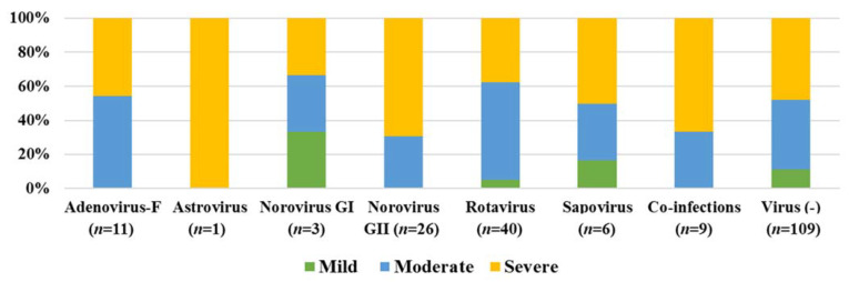
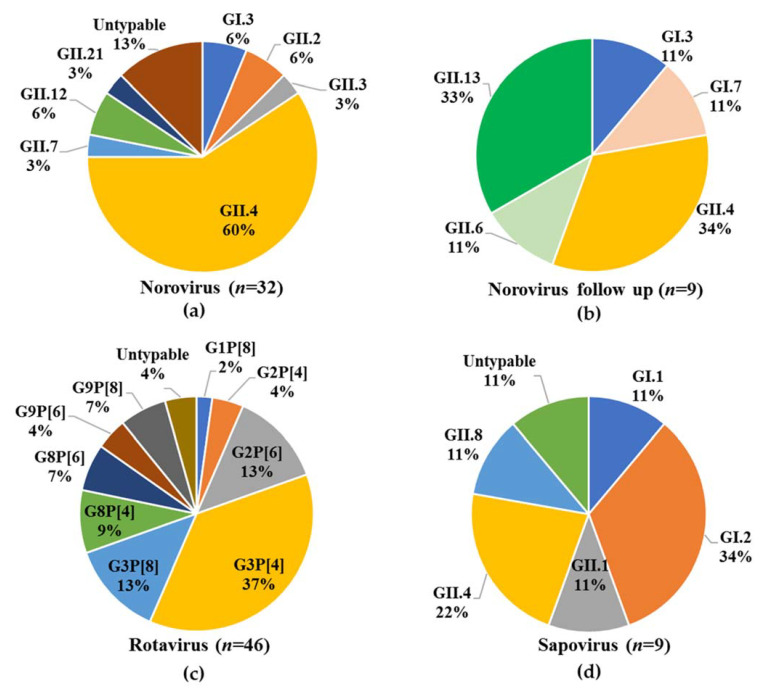
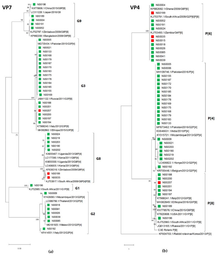
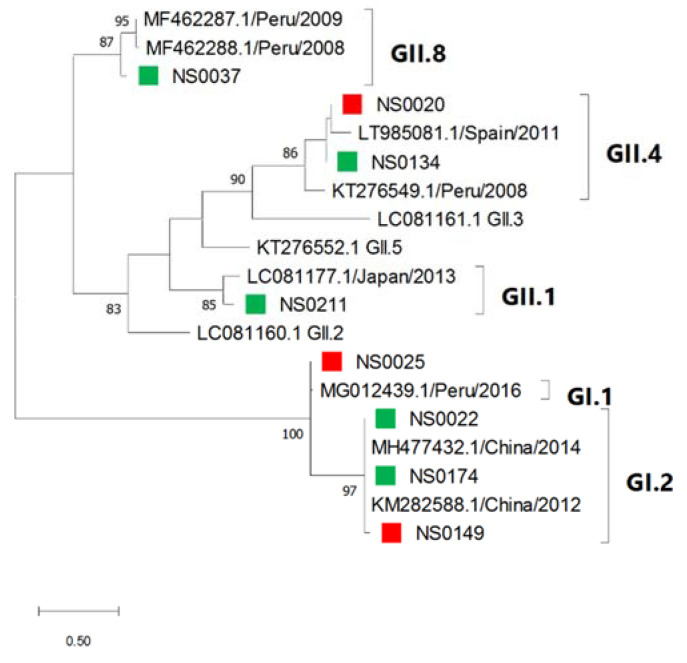
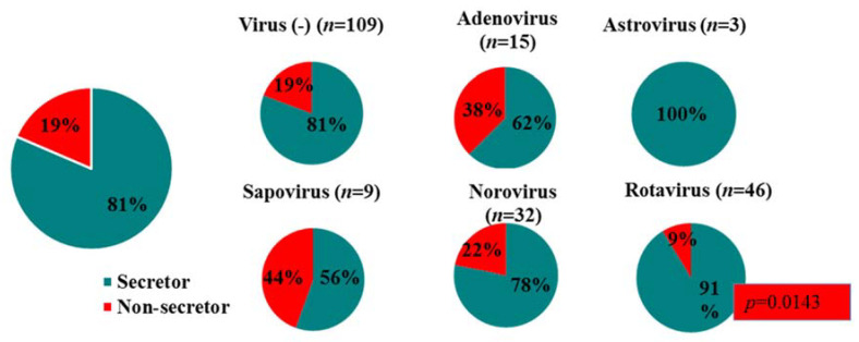
Results: At least one gastroenteritis virus was detected in 47% (96/205) of children. Rotavirus predominated (46/205), followed by norovirus (32/205), adenovirus (15/205), sapovirus (9/205) and astrovirus (3/205). Norovirus genotypes GI.3, GII.2, GII.3, GII.4, GII.7, GII.12, GII.21, and rotavirus strains G1P[8], G2P[4], G2P[6], G3P[4], G3P[8], G8P[4], G8P[6], G9P[6], G9P[8] and sapovirus genotypes GI.1, GI.2, GII.1, GII.4, GII.8 were detected; norovirus GII.4[P31] and rotavirus G3P[4] predominated. Asymptomatic norovirus infection (GI.3, GI.7, GII.4, GII.6, GII.13) was detected in 22% of 46 six-week follow up stools. HIV exposure (30%) was not associated with more frequent or severe viral gastroenteritis hospitalisations compared to unexposed children. Rotavirus preferentially infected secretor children (p = 0.143) and norovirus infected 78% secretors and 22% non-secretors.

Conclusion: Rotavirus was still the leading cause of gastroenteritis hospitalisations, but norovirus caused more severe symptoms.

Keywords: FUT2 secretor status; HIV exposure; norovirus; paediatric gastroenteritis; rotavirus.


Conflict of interest statement

The authors declare no conflict of interest. The funders had no role in the design of the study; in the collection, analyses, or interpretation of data; in the writing of the manuscript, or in the decision to publish the results.

Figures

Figure 1
Figure 1
Virus distribution in (a) 96 virus infected patients and (b) within different age groups of the 205 children ≤5 years hospitalised with gastroenteritis at Kalafong Provincial Tertiary Hospital (KPTH) from July 2016 to December 2017.
Figure 2
Figure 2
Disease severity observed during gastroenteritis virus infections (96) and virus unrelated gastroenteritis episodes (109) in children ≤5 years, hospitalised with gastroenteritis at KPTH from July 2016 to December 2017.
Figure 3
Figure 3
Virus diversity in children (≤5 years) hospitalised with gastroenteritis at KPTH from July 2016 to December 2017. (a) Norovirus genotype distribution in 32/205 children, (b) Norovirus genotype distribution in asymptomatic children (n = 9) 6 weeks after their initial hospitalisation with gastroenteritis, (c) Rotavirus genotype distribution in 46/205 children, (d) Sapovirus genotype distribution in 9/205 children.
Figure 4
Figure 4
Maximum likelihood phylogenetic tree of (a) the partial capsid gene (GI—222 bp, GII—210 bp) and (b) the partial RdRp gene (198 bp) of the norovirus strains detected in children hospitalised with gastroenteritis at KPTH between July 2016 and December 2017. Closely related strains from GenBank are indicated by accession numbers. Bootstrap support of >70% is shown. The scale bar represents nucleotide substitutions per site. Secretor status of infected child is indicated by green (secretor) and red (non-secretor) blocks.
Figure 5
Figure 5
Comparison of norovirus strains detected in symptomatic and asymptomatic children. Maximum likelihood phylogenetic tree of the partial capsid gene (GI—288 bp; GII—276 bp) of the norovirus strains detected in children six weeks after initial hospitalisation with gastroenteritis at KPTH between July 2016 and December 2017 (blue blocks). The norovirus strains that were detected in two children at initial hospitalisation and at 6-week follow up are shown in matched colours (NS0017—yellow; NS0173—red). Closely related strains from GenBank are indicated by accession numbers. Bootstrap support of >70% is shown. The scale bar represents 0.5 nucleotide substitutions per site over the indicated region.
Figure 6
Figure 6
Rotavirus G and P types identified by maximum likelihood phylogenetic analysis of (a) the VP7 gene (585 bp) and (b) the VP4 gene (555 bp) of the rotavirus strains detected in children hospitalised with gastroenteritis at KPTH between July 2016 and December 2017. Closely related strains from GenBank are indicated by accession numbers. Bootstrap support of >70% is shown. The scale bar represents nucleotide substitutions per site. Secretor status of infected child is indicated by green (secretor) and red (non-secretor) blocks.
Figure 7
Figure 7
Sapovirus genotypes identified by maximum likelihood phylogenetic analysis of the partial capsid gene (207 bp) of sapovirus strains detected in children hospitalised with gastroenteritis at KPTH between July 2016 and December 2017. Closely related strains from GenBank were included. Bootstrap support of >70% is shown. The scale bar represents 0.5 nucleotide substitutions per site. Secretor status of infected child is indicated by green (secretor) and red (non-secretor) blocks.
Figure 8
Figure 8
Secretor/non-secretor ratios for children infected with the different gastroenteritis viruses.

References

    1. Institute for Health Metrics and Evaluation (IHME) Findings from the Global Burden of Disease Study 2017. IHME; Seattle, WA, USA: 2018.
    1. Troeger C., Khalil I.A., Rao P.C., Cao S., Blacker B.F., Ahmed T., Armah G., Bines J.E., Brewer T.G., Colombara D.V. Rotavirus vaccination and the global burden of rotavirus diarrhea among children younger than 5 years. JAMA Pediatr. 2018;172:958–965. doi: 10.1001/jamapediatrics.2018.1960. - DOI - PMC - PubMed
    1. Parashar U.D., Burton A., Lanata C., Boschi-Pinto C., Shibuya K., Steele D., Birmingham M., Glass R.I. Global mortality associated with rotavirus disease among children in 2004. J. Infect. Dis. 2009;200(Suppl. 1):S9–S15. doi: 10.1086/605025. - DOI - PubMed
    1. Burnett E., Jonesteller C.L., Tate J.E., Yen C., Parashar U.D. Global impact of rotavirus vaccination on childhood hospitalizations and mortality from diarrhea. J. Infect. Dis. 2017;215:1666–1672. doi: 10.1093/infdis/jix186. - DOI - PMC - PubMed
    1. Hallowell B.D.P., Parashar U.D., Curns A., DeGroote N.P., Tate J.E. Trends in the laboratory detection of rotavirus before and after implementation of routine rotavirus vaccination—United States, 2000–2018. MMWR. 2019;68:539–543. - PMC - PubMed

Publication types