. 2021 Feb 11;18(1):42.
doi: 10.1186/s12974-020-02052-4.

Cell-free oxidized hemoglobin drives reactive oxygen species production and pro-inflammation in an immature primary rat mixed glial cell culture

Affiliations

Cell-free oxidized hemoglobin drives reactive oxygen species production and pro-inflammation in an immature primary rat mixed glial cell culture

Alex Adusei Agyemang et al. J Neuroinflammation. .

Abstract

Background: Germinal matrix intraventricular hemorrhage (GM-IVH) is associated with deposition of redox active cell-free hemoglobin (Hb), derived from hemorrhagic cerebrospinal fluid (CSF), in the cerebrum and cerebellum. In a recent study, using a preterm rabbit pup model of IVH, intraventricularly administered haptoglobin (Hp), a cell-free Hb scavenger, partially reversed the damaging effects observed following IVH. Together, this suggests that cell-free Hb is central in the pathophysiology of the injury to the immature brain following GM-IVH. An increased understanding of the causal pathways and metabolites involved in eliciting the damaging response following hemorrhage is essential for the continued development and implementation of neuroprotective treatments of GM-IVH in preterm infant.

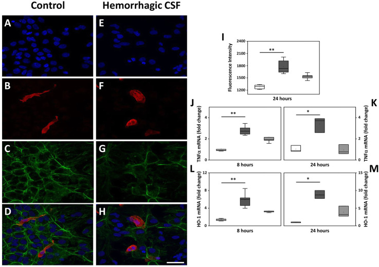
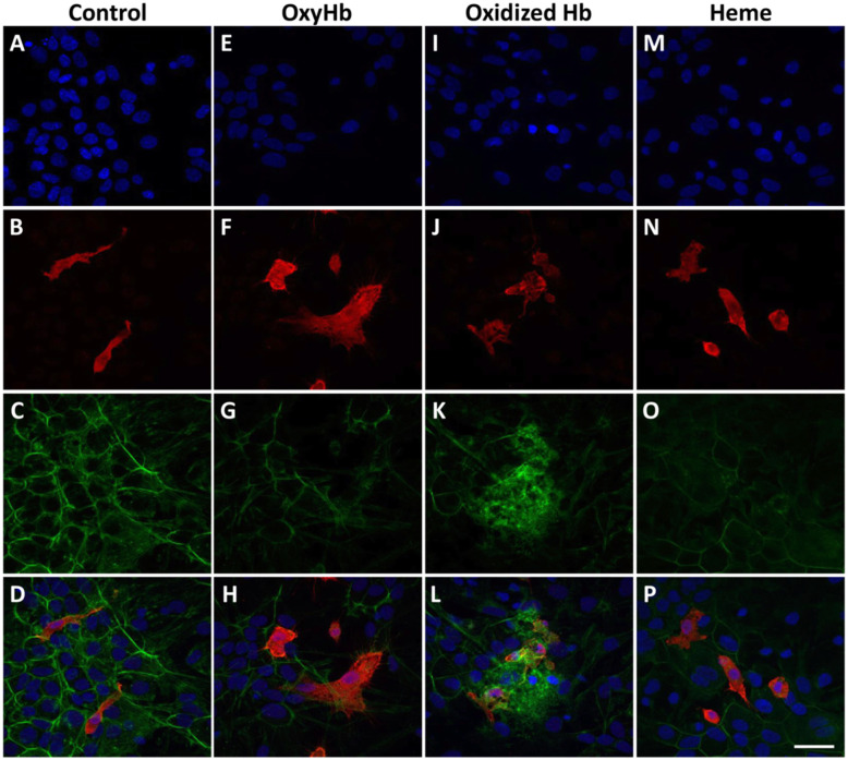
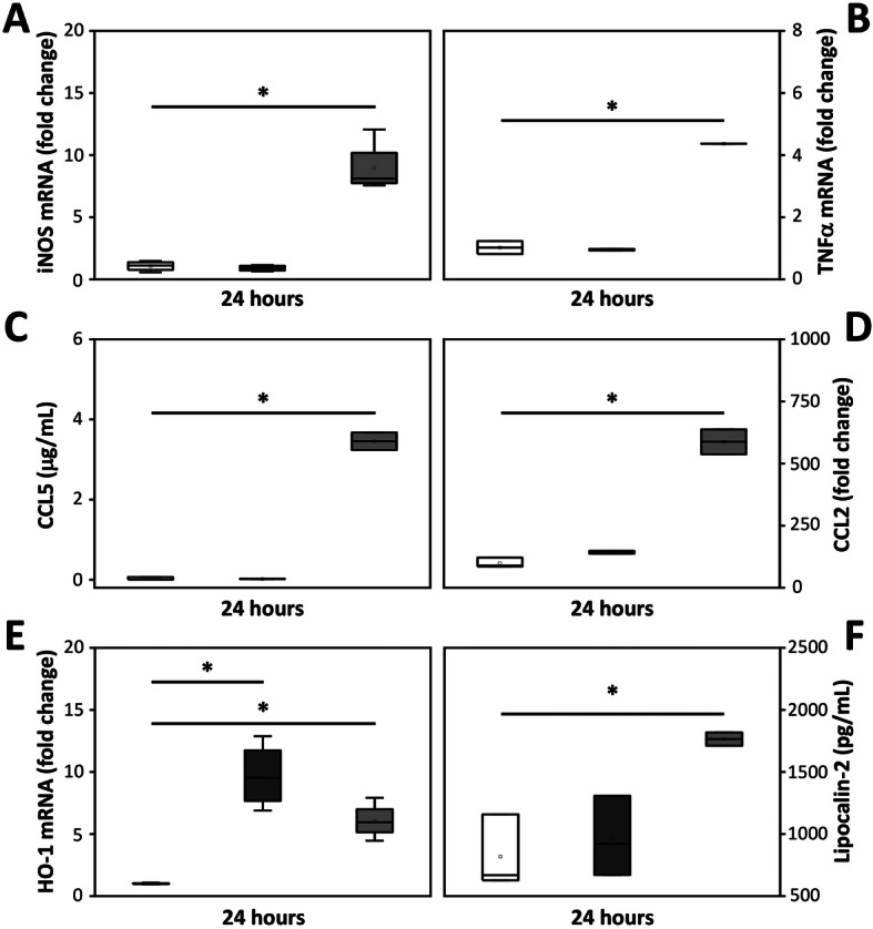
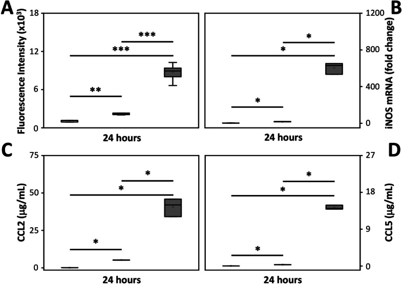
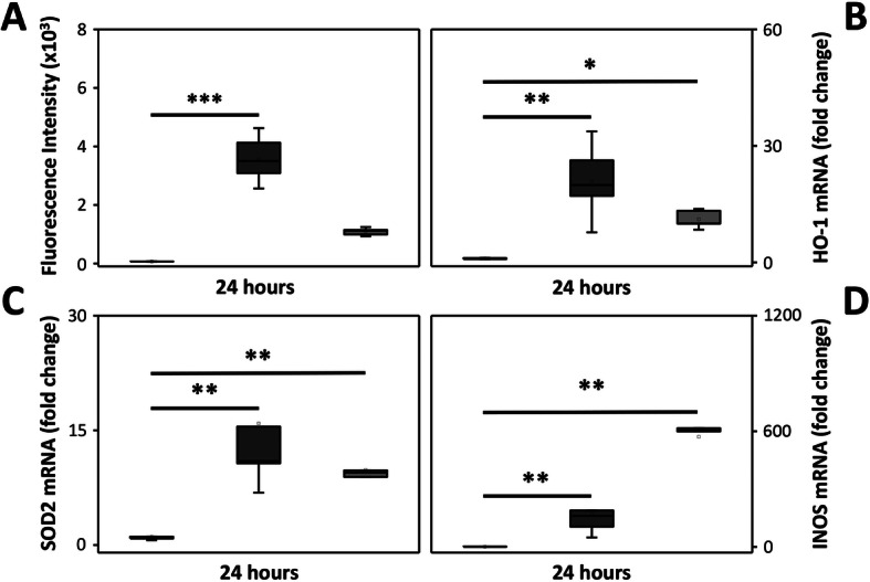
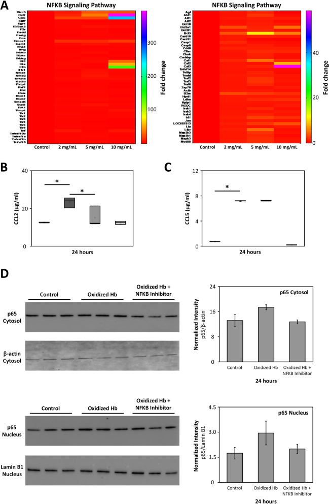
Methods: We exposed immature primary rat mixed glial cells to hemorrhagic CSF obtained from preterm human infants with IVH (containing a mixture of Hb-metabolites) or to a range of pure Hb-metabolites, incl. oxidized Hb (mainly metHb with iron in Fe3+), oxyHb (mainly Fe2+), or low equivalents of heme, with or without co-administration with human Hp (a mixture of isotype 2-2/2-1). Following exposure, cellular response, reactive oxygen species (ROS) generation, secretion and expression of pro-inflammatory cytokines and oxidative markers were evaluated.

Results: Exposure of the glial cells to hemorrhagic CSF as well as oxidized Hb, but not oxyHb, resulted in a significantly increased rate of ROS production that positively correlated with the rate of production of pro-inflammatory and oxidative markers. Congruently, exposure to oxidized Hb caused a disintegration of the polygonal cytoskeletal structure of the glial cells in addition to upregulation of F-actin proteins in microglial cells. Co-administration of Hp partially reversed the damaging response of hemorrhagic CSF and oxidized Hb.

Conclusion: Exposure of mixed glial cells to oxidized Hb initiates a pro-inflammatory and oxidative response with cytoskeletal disintegration. Early administration of Hp, aiming to minimize the spontaneous autoxidation of cell-free oxyHb and liberation of heme, may provide a therapeutic benefit in preterm infant with GM-IVH.

Keywords: Haptoglobin; Hemoglobin metabolites; Hemorrhagic cerebrospinal fluid; Intraventricular hemorrhage; Mixed glial cells; Redox.


Conflict of interest statement

NB and TG are employed by CSL Behring. This does not present any conflict of interest.

The authors AA, SVK, MI, NO, BH, DL, and MG declare that they have no competing interests.

Figures

Fig. 1
Fig. 1
Cellular response following exposure to hemorrhagic CSF. ah Representative images are from mixed glial cell cultures following exposure to 3% hemorrhagic CSF (containing 7 μM cell-free Hb) from preterm human infants with GM-IVH. Images illustrate the detected IF labeling of Iba1 (red; b, d, f, h), F-actin cytoskeletal protein (phalloidin, green; c, d, g, h) and DAPI nuclear staining (blue; a, d, e, h), in the wells of mixed glial cells exposed to culture media only (control; ad) and in wells exposed to hemorrhagic CSF (eh). d, h Merged images of DAPI, Iba1, and phalloidin. Scale bar in h indicates 200 μm and is representative for ah. i ROS production was investigated (by performing the DCFDA assay as described in materials and method) following a 24 h exposure of mixed glial cells to 3% hemorrhagic CSF (containing 7 µM cell-free Hb). j-m TNFα and HO-1 mRNA expression were analyzed at 8 and 24 h after exposure to 3% hemorrhagic CSF (containing 7 µM cell-free Hb). Hemorrhagic CSF (dark gray bars, n = 6), co-administration with Hp2-2/2-1 (10 µM, light gray bars, n = 6) and control cells (white bars, n = 6). Results are presented as box plots displaying medians and 25th and 75th percentiles. Differences between hemorrhagic CSF vs. control and hemorrhagic CSF with co-administered Hp vs. control were analyzed using the Kruskal–Wallis test followed by pairwise comparison with significance values adjusted for multiple comparisons. *P < 0.05, **P < 0.01
Fig. 2
Fig. 2
Cellular response following exposure to pure Hb-metabolites. Representative images are from mixed glial cell cultures following exposure to the Hb-metabolites oxyHb (2.5 mg/ml), oxidized Hb (2.5 mg/ml), and heme (15 μM). Images illustrate the detected IF labeling of Iba1 (red; b, d, f, h, j, l, n, p), F-actin cytoskeletal protein (phalloidin, green; c, d, g, h, k, l, o, p) and DAPI nuclear staining (blue; a, d, e, h, i, l, m, p), in the wells of mixed glial cells exposed to culture media only (control; ad) and in wells exposed to pure Hb-metabolites (oxyHb 2.5 mg/ml, eh; oxidized Hb 2.5 mg/ml, il; and heme 15 μM, mp). d, h, l, p Merged images of DAPI, Iba1, and phalloidin. Scale bar in p indicates 200 μm and is representative for ap
Fig. 3
Fig. 3
Oxidized Hb induces a pro-inflammatory response. mRNA expression and protein secretion of pro-inflammatory and oxidative stress markers, iNOS (a), TNFα (b), CCL5 (c), CCL2 (d), HO-1 (e), and lipocalin-2 (f) were investigated following exposure of mixed glial cell cultures to pure Hb-metabolites (oxyHb 2.5 mg/ml, dark gray bar, n = 4; oxidized Hb 2.5 mg/ml, light gray bar, n = 4) or culture medium only (control, white bar, n = 4). Results are presented as box plots displaying medians and 25th and 75th percentiles. Differences between oxyHb vs. control, oxidized Hb vs control, and oxidized Hb vs oxyHb were analyzed using the Kruskal–Wallis test followed by pairwise comparison with significance values adjusted for multiple comparisons. *P < 0.05
Fig. 4
Fig. 4
Dose-dependent effect of oxidized Hb. Mixed glial cells were exposed to increasing concentration of oxidized Hb (5 mg/ml, dark grey bars, n = 6; 10 mg/ml, light grey bars, n = 6) or culture medium only (control, white bar, n = 6) for 24 h. Subsequently, ROS production (a), mRNA expression of iNOS (b) and protein secretion of CCL2 (c) and CCL5 (d) were analyzed. Results are presented as box plots displaying medians and 25th and 75th percentiles. Differences between 10 mg/ml oxidized Hb vs. control, 5 mg/ml oxidized Hb vs. control, and 10 mg/ml oxidized Hb vs 5 mg/ml oxidized Hb were analyzed using the Kruskal–Wallis test followed by pairwise comparison with significance values adjusted for multiple comparisons. *P < 0.05, **P < 0.01, ***P < 0.001
Fig. 5
Fig. 5
Effect of Hp on the damage induced by oxidized Hb. Mixed glial cells were exposed to oxidized Hb (10 mg/ml, dark grey bars, n = 6) with or without co-administration of Hp2-2/2-1 (40 mg/ml, corresponding to a 1:1 molar ratio, light grey bars, n = 6) or culture medium only (control, white bars, n = 6) for 24 h. Subsequently, ROS production (a), mRNA expression of HO-1 (b), SOD2 (c), and iNOS (d) were analyzed. Results are presented as box plots displaying medians and 25th and 75th percentiles. Differences between oxidized Hb vs. control and oxidized Hb with co-administered Hp vs. control were analyzed using the Kruskal–Wallis test followed by pairwise comparison with significance values adjusted for multiple comparisons. **P < 0.01, ***P < 0.001
Fig. 6
Fig. 6
Oxidized Hb induces NFKB signaling pathway. Mixed glial cells were exposed to increasing concentration of oxidized Hb (2–10 mg/ml) or culture medium only (control) for 24 h. a A heat map revealing a concentration-dependent upregulation by oxidized Hb in mRNA expression of activators, transcriptional factors, and pro-inflammatory cytokines associated with the NFKB pathway. Addition of the NFKB activation inhibitor VI benzoxathiole (3.0 μM) in the presence of oxidized Hb (2.5 mg/ml, grey bar, n = 5) partly inhibited the oxidized Hb (dark grey bar, n = 5) induced protein level of CCL2 (b), but not CCL5 (c). Results of b and c are presented as box plots displaying medians and 25th and 75th percentiles. Control (white bar, n = 5) and NFKB activation inhibitor VI benzoxathiole only (light grey, n = 5). Protein levels of the NFKB transcription factor p65 were assessed in the cytosolic and nuclear fraction using western blot. A tendency towards increased level of p65, in both the cytosolic and the nuclear fraction, was observed following exposure to oxidized Hb (2.5 mg/ml). Co-administration of the NFKB inhibitor VI benzoxathiole displayed a trend of reduced p65 protein levels (d). β-actin (cytosolic fraction) and Lamin B1 (nuclear fraction) were used as protein loading controls and analyzed on the same material as p65. All samples were applied to one membrane. Membranes are from a representative analysis. Densitometric quantification was performed and mean normalized intensity, normalized for respective β-actin (cytosolic) or Lamin B1 (nuclear) intensity, is presented. Differences between oxidized Hb vs. control, oxidized Hb + NFKB inhibitor vs. control, and oxidized Hb + NFKB inhibitor vs. oxidized Hb were analyzed using the Kruskal–Wallis test followed by pairwise comparison with significance values adjusted for multiple comparisons. *P < 0.05

References

    1. Ballabh P. Intraventricular hemorrhage in premature infants: mechanism of disease. Pediatr Res. 2010;67:1–8. doi: 10.1203/PDR.0b013e3181c1b176. - DOI - PMC - PubMed
    1. Agyemang AA, Sveinsdóttir K, Vallius S, Sveinsdóttir S, Bruschettini M, Romantsik O, et al. Cerebellar exposure to cell-free hemoglobin following preterm intraventricular hemorrhage: causal in cerebellar damage? Transl Stroke Res. 2017;8(5):461. doi: 10.1007/s12975-017-0539-1. - DOI - PMC - PubMed
    1. Ley D, Romantsik O, Vallius S, Sveinsdóttir K, Sveinsdóttir S, Agyemang AA, et al. High presence of extracellular hemoglobin in the periventricular white matter following preterm intraventricular hemorrhage. Front Physiol. 2016;7:330. doi: 10.3389/fphys.2016.00330. - DOI - PMC - PubMed
    1. Lee JY, Keep RF, He Y, Sagher O, Hua Y, Xi G. Hemoglobin and iron handling in brain after subarachnoid hemorrhage and the effect of deferoxamine on early brain injury. J Cereb Blood Flow Metab. 2010;30:1793–1803. doi: 10.1038/jcbfm.2010.137. - DOI - PMC - PubMed
    1. Lok J, Leung W, Murphy S, Butler W, Noviski N, Lo EH. Intracranial hemorrhage: mechanisms of secondary brain injury. Acta Neurochir Suppl. 2011;111:63–69. doi: 10.1007/978-3-7091-0693-8_11. - DOI - PMC - PubMed

MeSH terms