Review
. 2021 Feb 12;371(6530):eabd0951.
doi: 10.1126/science.abd0951.

Communicating clocks shape circadian homeostasis

Affiliations
Review

Communicating clocks shape circadian homeostasis

Kevin B Koronowski et al. Science. .

Abstract

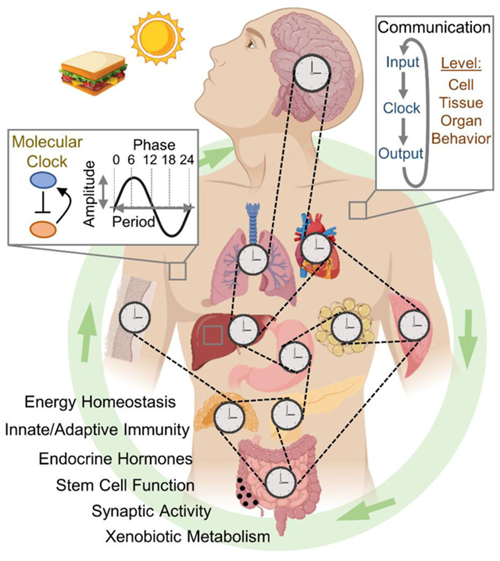
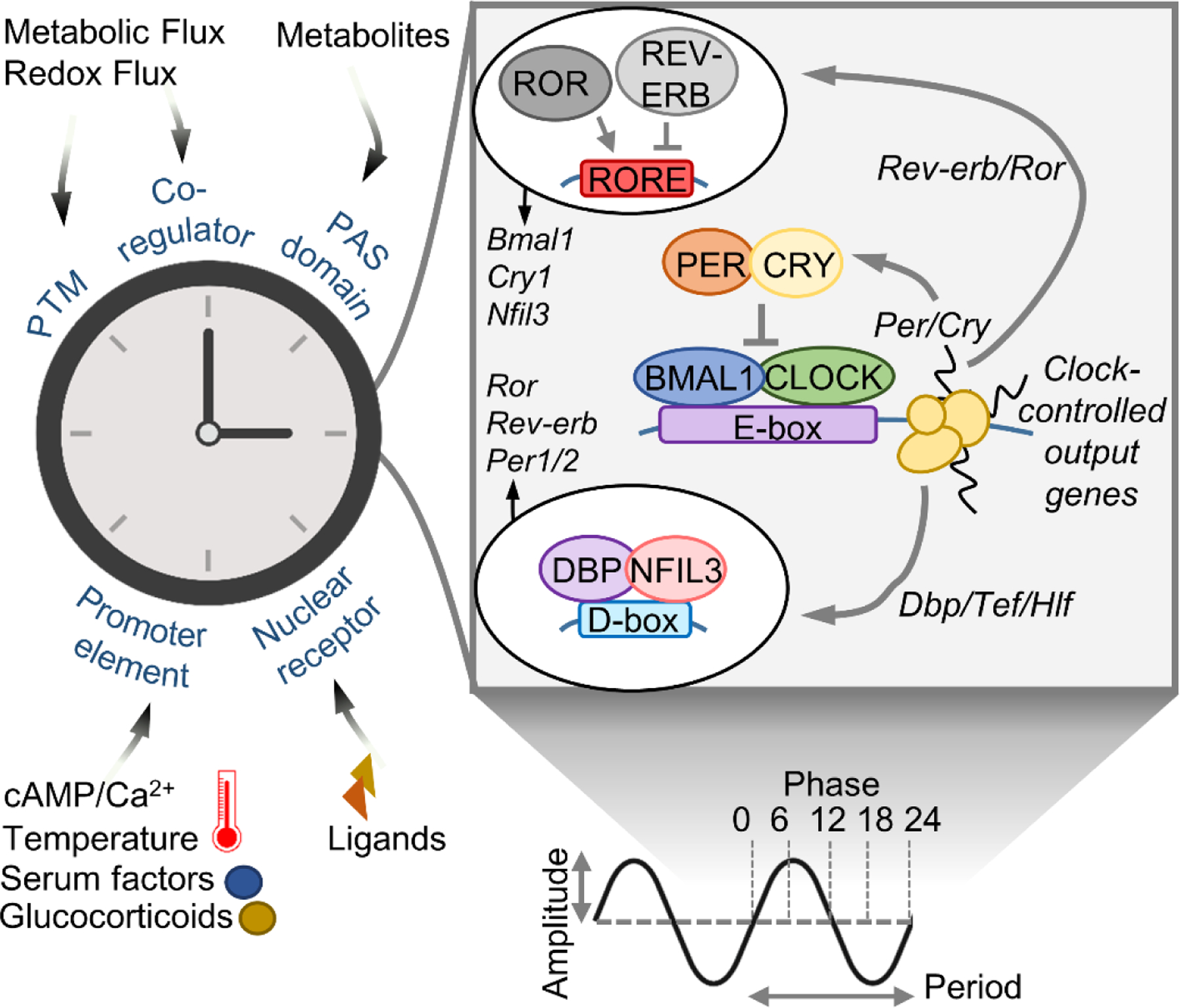
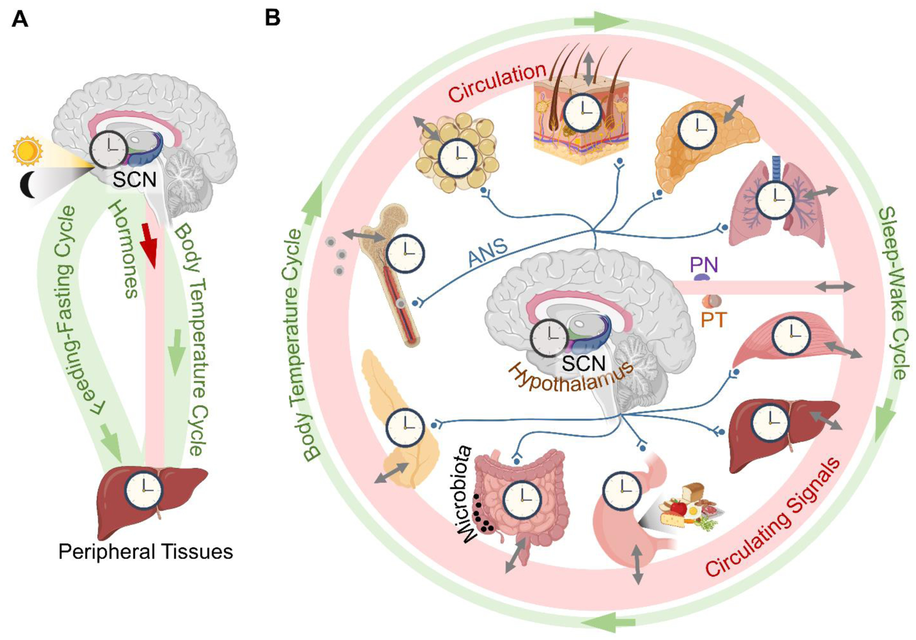
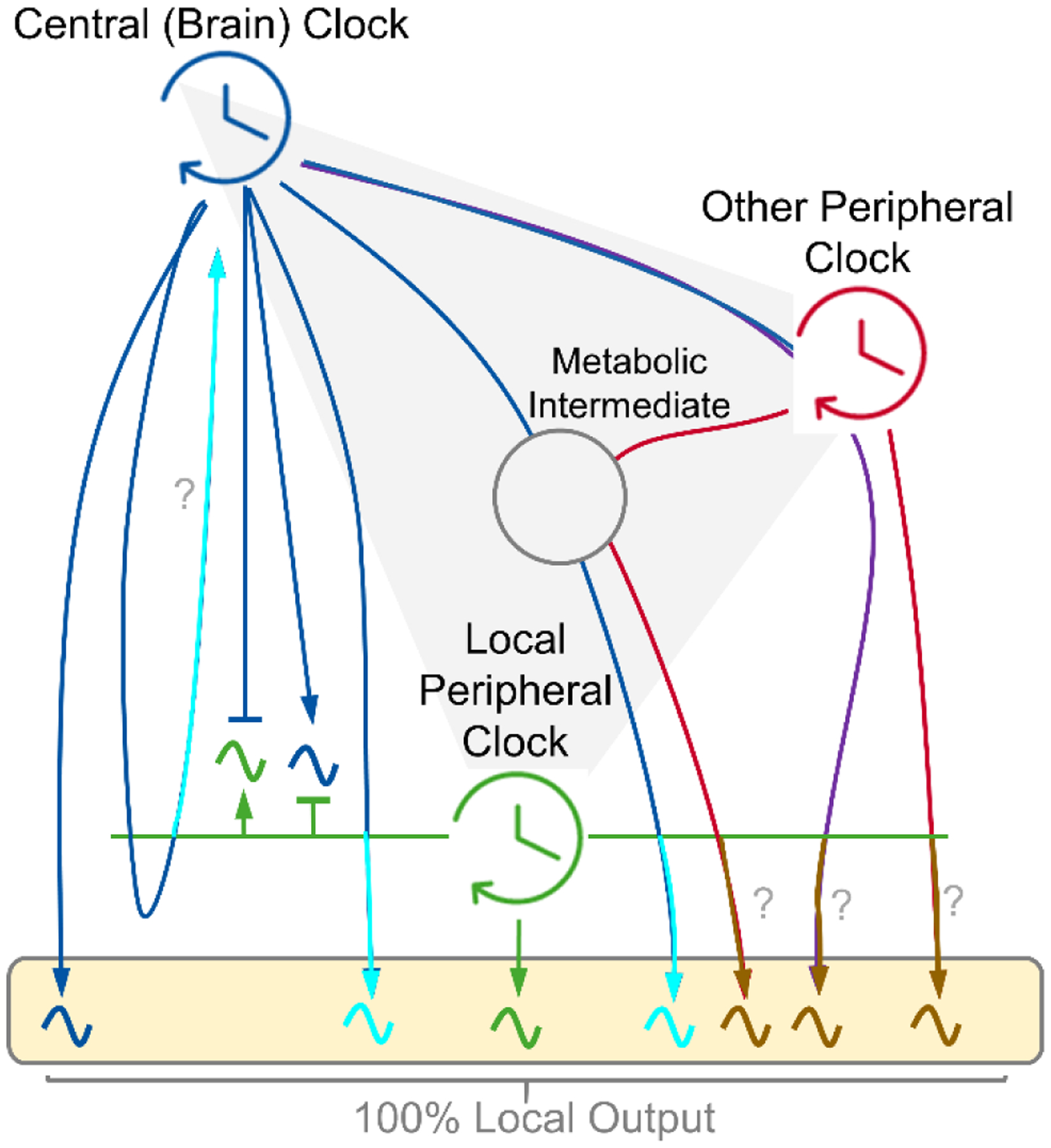
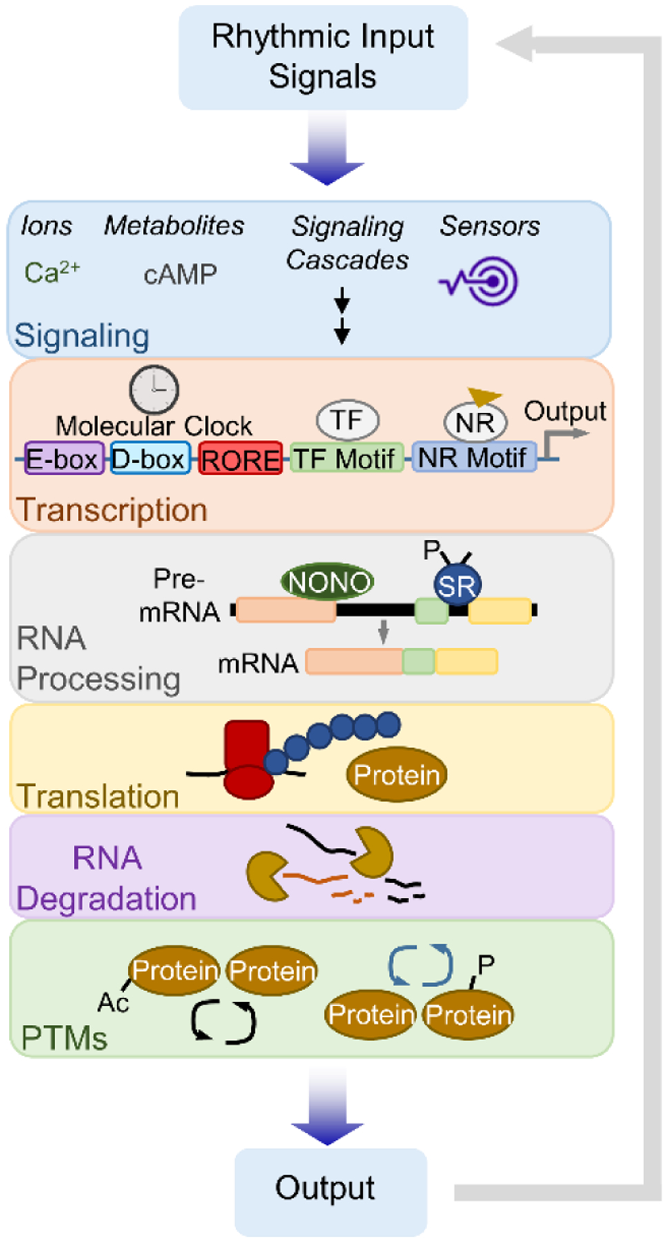
Circadian clocks temporally coordinate physiology and align it with geophysical time, which enables diverse life-forms to anticipate daily environmental cycles. In complex organisms, clock function originates from the molecular oscillator within each cell and builds upward anatomically into an organism-wide system. Recent advances have transformed our understanding of how clocks are connected to achieve coherence across tissues. Circadian misalignment, often imposed in modern society, disrupts coordination among clocks and has been linked to diseases ranging from metabolic syndrome to cancer. Thus, uncovering the physiological circuits whereby biological clocks achieve coherence will inform on both challenges and opportunities in human health.


Conflict of interest statement

Competing interests: The authors have no competing interests.

Figures

Figure Caption 0.
Figure Caption 0.. Cellular to Organismal Timekeeping: Communication Between Clocks.
The mammalian circadian clock is a coupled network of cell and tissue clocks. Light and food are predominant cues, pushing and pulling on the phase, enhancing or attenuating the amplitude and activating or inhibiting functional rhythms. In a tissue-specific manner, clocks receive input signals and convert them into timed functional outputs, many of which in turn act as inputs, effectively connecting the network.
Figure 1.
Figure 1.. Extracellular signals and the molecular clock.
Simplified scheme of the molecular clock/oscillator. Right – the core transcriptional activators circadian locomotor output cycles protein kaput (CLOCK) and brain and muscle ARNT-like 1 (BMAL1), together with the repressors period (PER) and cryptochrome (CRY) and interlocked auxiliary loops (regulating D-boxes and ROR elements), control a near 24-hr cycle of each other’s expression as well as clock controlled output genes (CCGs). Left – extracellular signals that engage the clock through various means to set period, phase, amplitude and output. PTM – posttranslational modification; cAMP – cyclic adenosine monophosphate; PAS – Per-Arnt-Sim; ROR –RAR-related orphan receptor; REV-ERB – nuclear receptor subfamily 1 group D; DBP – albumin D-box binding protein; Tef – thyrotroph embryonic factor; Hlf – hepatic leukemia factor; NFIL3 – nuclear factor, interleukin 3 regulated.
Figure 2.
Figure 2.. Coupling of clocks in neurons and astrocytes.
Astrocytes harbor autonomous clocks that operate in anti-phase to neurons. During daytime, astrocytic glutamate release is suppressed, which inhibits presynaptic terminals to alleviate GABA, stimulating electrical activity and Ca2+-induced CREB activation leading to Per2 gene expression. At nighttime, astrocytes release glutamate, which acts on presynaptic terminals to enhance GABA, in effect inhibiting electrical activity and Per2. Regulation of Per2 in this manner links rhythmic astrocytic signals to the clock in neurons. Other components implicated in this process are GAT3, an astrocytic GABA transporter, and EphA4, an adhesion molecule that physically connects astrocytes and neurons and the connexin 43 (Cx43) hemichannel. Upper right inlet – the sleep-wake cycle drives oscillation of synaptic proteins and their phosphorylation, another layer of circadian regulation. GABA – γ-aminobutyric acid; cAMP – cyclic adenosine monophosphate; CREB – cAMP response element-binding protein; Per2 – period 2; GAT3 – GABA transporter type 3; EphA4 – EPH receptor A4; Glu – glutamate; ERK – extracellular signal-regulated kinase; NT – neurotransmitter; NMDAR2C – N-methyl-D-aspartate receptor 2 c; GABAR – GABA receptor.
Figure 3.
Figure 3.. Connections of the mammalian clock system.
(A) A conceptual model based on knowledge from the turn of the century. (B) A current model of the system. In addition to the suprachiasmatic nucleus (SCN), other distinct hypothalamic clocks control daily energy homeostasis. The autonomic nervous system (ANS) differentially innervates peripheral tissues and modulates their clocks by adjusting sympathetic tone. All clocks are connected via the circulatory system by an ever-growing list of synchronizing factors, many of which are under local circadian control in peripheral tissues. See also Table 1 for circadian factors, their signaling pathways and clock targets within the system. PN – pineal gland; PT – pituitary gland.
Figure 4.
Figure 4.. Inputs determining peripheral clock output.
Conceptual model showing the drivers of circadian output of a peripheral metabolic tissue. Arrows indicate a driving circadian signal. The box depicts 100% of the local oscillatory output. Local clocks exhibit autonomy in driving a small fraction of output (green) (54) (53). Central (brain) clock-controlled cycles, feeding-fasting and sleep-wake, can drive rhythms independently or presumably in cooperation with the local clock (blue and turquoise). Peripheral clocks may influence rhythmicity of each other in a central clock dependent or independent manner (red, purple and brown). Many circadian signals likely synergize across tissues and intermediates (e.g. gut microbiota) to shape the full rhythmic output, with mechanisms which remain elusive (gray). It is unclear if the local clock is required for certain connections (?).
Figure 5.
Figure 5.. Multi-level control of clock output and communication.
The clock can autonomously support a portion of circadian output. Nuclear receptors (NRs) regulate gene expression in tandem with clock proteins and are activated by extrinsic ligands, many of which oscillate in the bloodstream as a result of clock control in another tissue. The daily nuclear accumulation of certain transcription factors (TFs) is driven by feeding or body temperature rhythms. Systemic rhythms post-transcriptionally shape mRNA and protein oscillations. Feeding and fasting regulate RNA processing, translation and degradation while temperature fluctuations can induce alternative splicing programs that generate rhythmicity. Daily systemic energy metabolism regulates the functional status of proteins through post-translational modifications tied to metabolite levels. cAMP – cyclic adenosine monophosphate; NONO – non-POU domain-containing octamer-binding protein; SR – serine/arginine-rich splicing factor family; Ac – acetylation; P – phosphorylation.

References

    1. Meyers SR, Malinverno A, Proterozoic Milankovitch cycles and the history of the solar system. Proc Natl Acad Sci U S A 115, 6363–6368 (2018). - PMC - PubMed
    1. Mills JN, Minors DS, Waterhouse JM, The circadian rhythms of human subjects without timepieces or indication of the alternation of day and night. J Physiol 240, 567–594 (1974). - PMC - PubMed
    1. Whitmore D, Foulkes NS, Sassone-Corsi P, Light acts directly on organs and cells in culture to set the vertebrate circadian clock. Nature 404, 87–91 (2000). - PubMed
    1. Moore RY, Eichler VB, Loss of a circadian adrenal corticosterone rhythm following suprachiasmatic lesions in the rat. Brain Res 42, 201–206 (1972). - PubMed
    1. Schibler U, Sassone-Corsi P, A web of circadian pacemakers. Cell 111, 919–922 (2002). - PubMed

Publication types

LinkOut - more resources