. 2021 Feb 11;9(1):13.
doi: 10.1038/s41413-020-00130-0.

The molecular conformation of silk fibroin regulates osteogenic cell behavior by modulating the stability of the adsorbed protein-material interface

Affiliations

The molecular conformation of silk fibroin regulates osteogenic cell behavior by modulating the stability of the adsorbed protein-material interface

Yanlin Long et al. Bone Res. .

Abstract

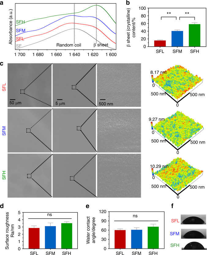
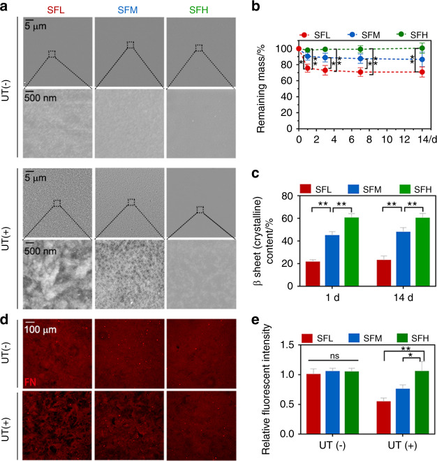
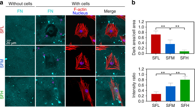
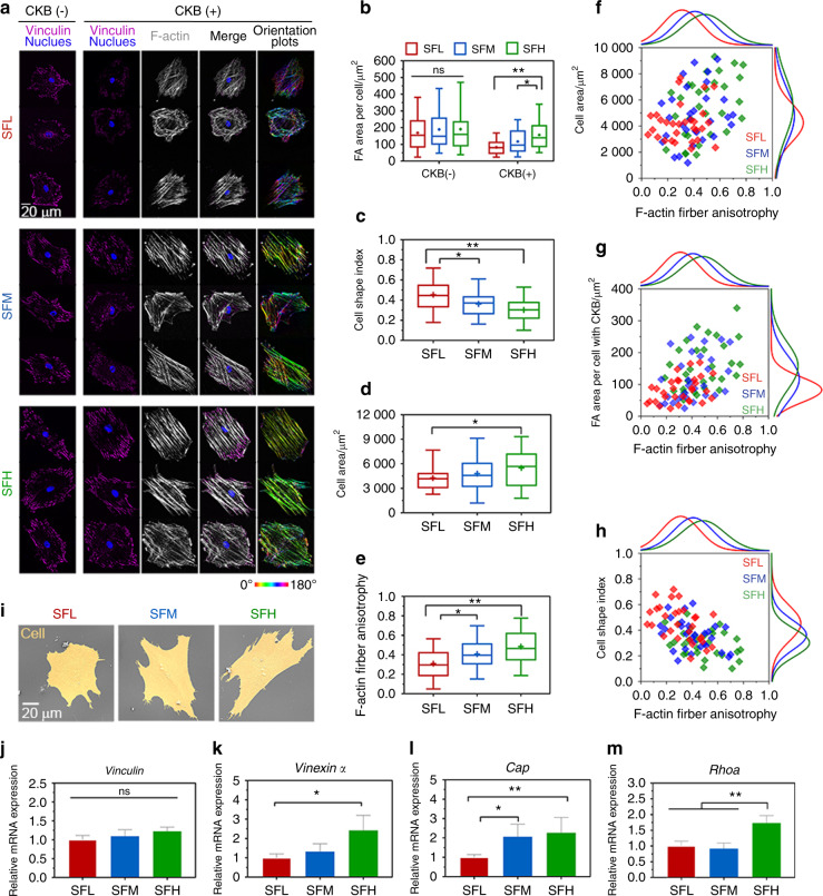
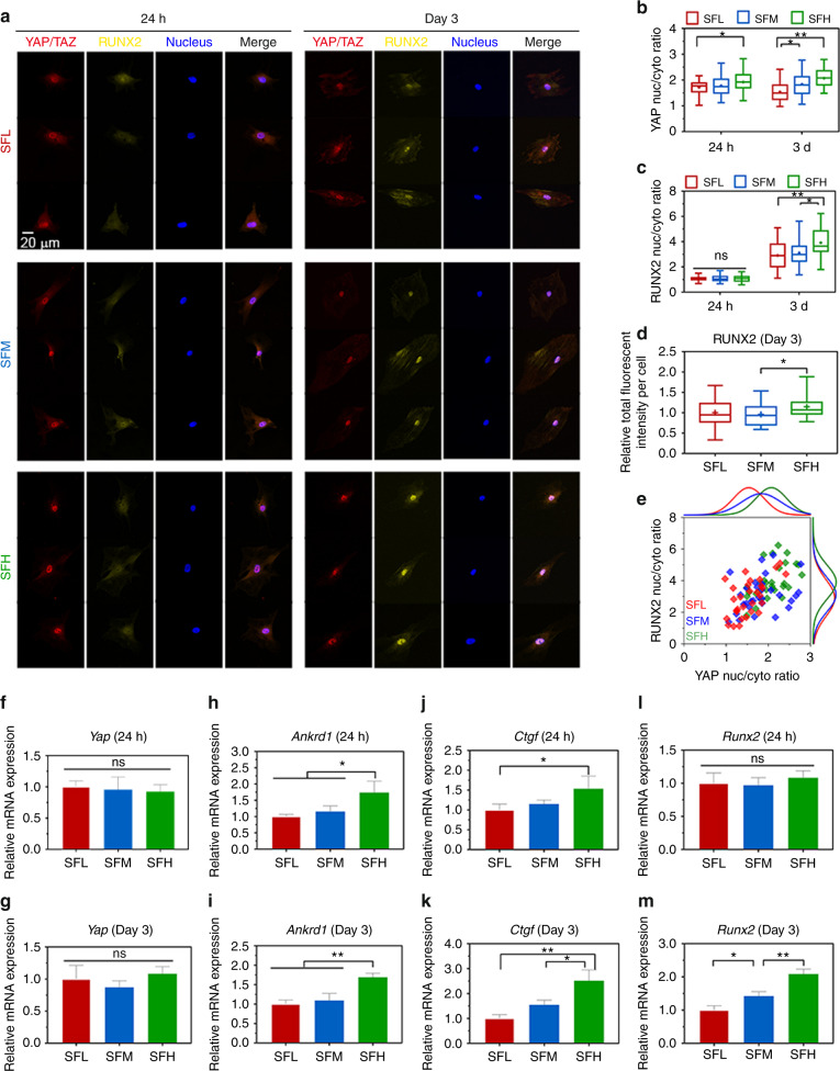
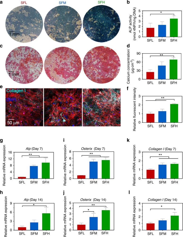
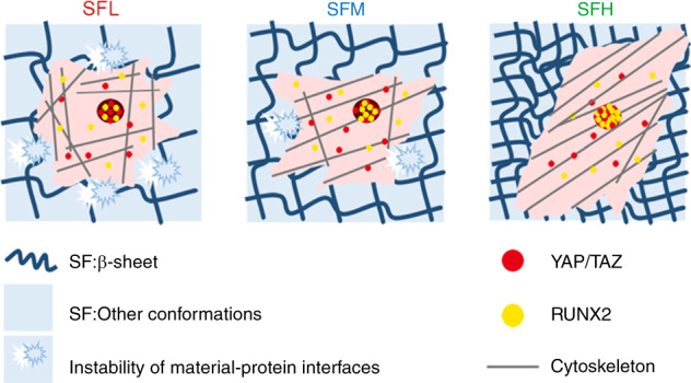
Silk fibroin (SF) can be used to construct various stiff material interfaces to support bone formation. An essential preparatory step is to partially transform SF molecules from random coils to β-sheets to render the material water insoluble. However, the influence of the SF conformation on osteogenic cell behavior at the material interface remains unknown. Herein, three stiff SF substrates were prepared by varying the β-sheet content (high, medium, and low). The substrates had a comparable chemical composition, surface topography, and wettability. When adsorbed fibronectin was used as a model cellular adhesive protein, the stability of the adsorbed protein-material interface, in terms of the surface stability of the SF substrates and the accompanying fibronectin detachment resistance, increased with the increasing β-sheet content of the SF substrates. Furthermore, (i) larger areas of cytoskeleton-associated focal adhesions, (ii) higher orders of cytoskeletal organization and (iii) more elongated cell spreading were observed for bone marrow-derived mesenchymal stromal cells (BMSCs) cultured on SF substrates with high vs. low β-sheet contents, along with enhanced nuclear translocation and activation of YAP/TAZ and RUNX2. Consequently, osteogenic differentiation of BMSCs was stimulated on high β-sheet substrates. These results indicated that the β-sheet content influences osteogenic differentiation of BMSCs on SF materials in vitro by modulating the stability of the adsorbed protein-material interface, which proceeds via protein-focal adhesion-cytoskeleton links and subsequent intracellular mechanotransduction. Our findings emphasize the role of the stability of the adsorbed protein-material interface in cellular mechanotransduction and the perception of stiff SF substrates with different β-sheet contents, which should not be overlooked when engineering stiff biomaterials.


Conflict of interest statement

The authors declare no competing interests.

Figures

Fig. 1
Fig. 1
Material characterization of SF substrates with different β-sheet contents. a, b FTIR absorbance spectra (a) of the amide I region (between 1 695 and 1 595 cm−1) obtained from different SF substrates and (b) the β-sheet contents calculated by Fourier self-deconvolution from these spectra. c Surface topography observed by SEM and AFM. d Surface roughness values analyzed by AFM. e, f Surface wettability (e) determined by water contact angle measurement and (f) representative images of water droplets. Error bars represent one standard deviation. (*P < 0.05 and **P < 0.01)
Fig. 2
Fig. 2
Interfacial stability between different SF substrates and adsorbed FN. a SEM images of different SF substrates after being immersed in PBS for 24 h, with and without ultrasonic treatment (UT). b, c Remaining mass (b) and β-sheet contents (c) of different SF substrates immersed in PBS with UT treatment at specific time points. d, e Immunofluorescence images (d) and the quantitative analysis (e) of adsorbed FN on different SF substrates after being immersed in FN solution for 24 h, with and without UT treatment. Error bars represent one standard deviation. (*P < 0.05 and **P < 0.01)
Fig. 3
Fig. 3
Interfacial stability of adsorbed FN on different substrates in a cell culture environment. a Immunofluorescence staining of adsorbed FN (cyan) on different SF substrates with and without BMSCs cultured for 24 h, together with F-actin (red) and nuclei (blue). The purple arrows indicate the detached FN underlying the cells. b Ratio of the FN dark area underlying the cell outline to the cell area. c Ratio of FN intensity underlying the cell outline to that of the background. Error bars represent one standard deviation. (*P < 0.05 and **P < 0.01)
Fig. 4
Fig. 4
Spreading behavior of BMSCs on different SF substrates. a Representative mosaic immunofluorescence images of vinculin (purple), F-actin (white), and nuclei (blue) in BMSCs without and with CKB treatment and the corresponding orientation plots for F-actin staining, where the different colors indicate different orientations of actin filaments as per the given color map. b–e Quantitative analysis of (b) FA area per cell, (c) cell shape index, (d) cell area, and (e) F-actin anisotropy in BMSCs. f–h Single-cell scatter plots of F-actin anisotropy as a function of (f) cell area, (g) FA area per cell, and (h) cell shape index. i SEM images of BMSCs’ spreading morphology. j–m The mRNA expression levels of (j) Vinculin, (k) Vinexin α, (l) Cap, and (m) Rhoa in BMSCs. For each box plot, the box boundaries represent the 25th–75th percentiles, and the whiskers represent the min and max values. The central line and cross represent the median value and mean, respectively. Error bars represent one standard deviation. (*P < 0.05 and **P < 0.01)
Fig. 5
Fig. 5
YAP/TAZ and RUNX2 nuclear translocation in BMSCs on different SF substrates. a Representative mosaic immunofluorescence images of YAP/TAZ (red), RUNX2 (yellow), and nuclei (blue) in BMSCs at 24 h and day 3. b, c The nuclear-cytoplasmic ratios of (b) YAP/TAZ and (c) RUNX2 in BMSCs analyzed from the immunofluorescence images. d Relative fluorescence intensity per cell of RUNX2 at day 3. e Single-cell scatter plots of the YAP/TAZ nuclei/cytoplasm ratio as a function of the RUNX2 nuclei/cytoplasm ratio at day 3. f–m The mRNA expression levels of (f–g) Yap, (h–i) Ankrd1, (j–k) Ctgf, and (l–m) Runx2 in BMSCs at 24 h and day 3. For each box plot, the box boundaries represent the 25th–75th percentiles, and the whiskers represent the min and max values. The central line and cross represent the median value and mean, respectively. Error bars represent one standard deviation. (*P < 0.05 and **P < 0.01)
Fig. 6
Fig. 6
Osteogenic differentiation of BMSCs on different SF substrates. a, b Images of ALP staining (a) and quantitative detection of ALP activity (b) in BMSCs at day 7. c, d Images of ARS staining (c) and quantitative analysis of calcium concentration (d) in BMSCs at day 14. e, f Immunofluorescence images of collagen Ι (e) in BMSCs and quantitative analysis of fluorescence intensity of collagen Ι (f) at day 14. g–l mRNA expression levels of representative (g–h) Alp, (i–j) Osterix, and (k–l) collagen Ι in BMSCs at days 7 and 14. Error bars represent one standard deviation. (*P < 0.05 and **P < 0.01)
Fig. 7
Fig. 7
The molecular conformation of SF regulates osteogenic cell behavior by modulating the stability of the adsorbed protein-material interface. Applying FN as a model cellular adhesive protein, the surface stability of the SF substrates and the accompanying detachment resistance of adsorbed protein on these SF substrates increased with increasing β-sheet content. Furthermore, more cytoskeleton-associated FAs, higher orders of cytoskeletal organization, and more elongated spreading shapes were observed in the BMSCs cultured on the SFH surface than in those cultured on the SFL surface, along with the enhancement of nuclear translocation and activation of YAP/TAZ and RUNX2 in intracellular mechanotransduction

References

    1. Gao C, Peng S, Feng P, Shuai C. Bone biomaterials and interactions with stem cells. Bone Res. 2017;5:1–33. doi: 10.1038/boneres.2017.59. - DOI - PMC - PubMed
    1. Perrone GS, et al. The use of silk-based devices for fracture fixation. Nat. Commun. 2014;5:3385. doi: 10.1038/ncomms4385. - DOI - PubMed
    1. Cheng X, et al. Electrodeposited assembly of additive-free silk fibroin coating from pre-assembled nanospheres for drug delivery. ACS Appl. Mater. Interfaces. 2020;12:12018–12029. doi: 10.1021/acsami.9b21808. - DOI - PMC - PubMed
    1. Han H, et al. Silk biomaterials with vascularization capacity. Adv. Funct. Mater. 2016;26:421–432. doi: 10.1002/adfm.201504160. - DOI - PMC - PubMed
    1. Sengupta S, et al. Quantifying osteogenic cell degradation of silk biomaterials. Biomacromolecules. 2010;11:3592–3599. doi: 10.1021/bm101054q. - DOI - PMC - PubMed

LinkOut - more resources