. 2021 Feb 11;12(1):950.
doi: 10.1038/s41467-021-21238-9.

Addressable nanoantennas with cleared hotspots for single-molecule detection on a portable smartphone microscope

Affiliations

Addressable nanoantennas with cleared hotspots for single-molecule detection on a portable smartphone microscope

Kateryna Trofymchuk et al. Nat Commun. .

Abstract

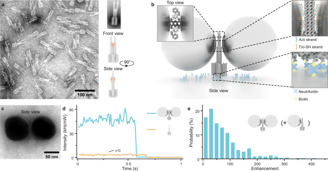
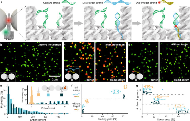
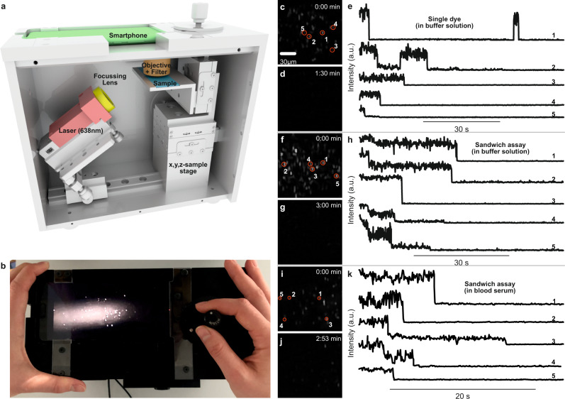
The advent of highly sensitive photodetectors and the development of photostabilization strategies made detecting the fluorescence of single molecules a routine task in many labs around the world. However, to this day, this process requires cost-intensive optical instruments due to the truly nanoscopic signal of a single emitter. Simplifying single-molecule detection would enable many exciting applications, e.g., in point-of-care diagnostic settings, where costly equipment would be prohibitive. Here, we introduce addressable NanoAntennas with Cleared HOtSpots (NACHOS) that are scaffolded by DNA origami nanostructures and can be specifically tailored for the incorporation of bioassays. Single emitters placed in NACHOS emit up to 461-fold (average of 89 ± 7-fold) brighter enabling their detection with a customary smartphone camera and an 8-US-dollar objective lens. To prove the applicability of our system, we built a portable, battery-powered smartphone microscope and successfully carried out an exemplary single-molecule detection assay for DNA specific to antibiotic-resistant Klebsiella pneumonia on the road.


Conflict of interest statement

P.T. and G.P.A. are inventors on an awarded patent of the described bottom-up method for fluorescence enhancement in molecular assays, EP1260316.1, 2012, US20130252825 A1. The remaining authors declare no competing interests.

Figures

Fig. 1
Fig. 1. Concept of the DNA origami nanoantenna with a cleared hotspot.
a TEM image (left, reproduced at least 3 times) and sketches (right) of the DNA origami structure used for the nanoantenna assembly with the position of the plasmonic hotspot indicated in red. A representative class averaged TEM image of the DNA origami used is shown on the upper right. b Schematics of NACHOS assembly: the DNA origami construct is bound to the BSA-biotin coated surface via biotin-NeutrAvidin interactions, thiolated DNA-functionalized 100 nm silver particles are attached to the DNA origami nanoantenna via polyadenine (A20) binding strands in the zipper-like geometry to minimize the distance between the origami and the nanoparticles. c TEM image of a NACHOS with 100 nm silver nanoparticles (reproduced at least 3 times). d Single-molecule fluorescence intensity transients, measured by confocal microscopy, normalized to the same excitation power of a single Alexa Fluor 647 dye incorporated in a DNA origami (orange) and in a DNA origami nanoantenna with two 100 nm silver nanoparticles (blue) excited at 639 nm e. Fluorescence enhancement distribution of Alexa Fluor 647 measured in NACHOS with 100 nm silver nanoparticles. A total number of 164 and 449 single molecules in the reference (more examples are provided in Supplementary Fig. 3) and NACHOS structures were analyzed, respectively.
Fig. 2
Fig. 2. Single-molecule diagnostic assay with NACHOS.
a Sketch of NACHOS with three capture strands in the hotspot and a green reference dye (ATTO 542) for labeling of the DNA origami. Capture strands are placed in the NACHOS hotspot region. Upon incubation, they hybridize with DNA target strands specific to Klebsiella pneumonia, exposing a specific, 17-nt long region for the hybridization with the imager strand labeled with Alexa Fluor 647. b Confocal fluorescence image of the NACHOS before incubation with DNA target and imager strands. c Confocal fluorescence images after incubation with DNA target (2 nM) and imager strands (6 nM) in buffer solution (left) and blood serum (right), scale same as in panel b; d Confocal fluorescence image of the NACHOS after incubation with only the DNA imager strand (6 nM) in buffer solution (left) and blood serum (right), scale same as in panel b. The scans in panels b-d are representative for at least 20 images. e Fluorescence enhancement histogram of the sandwich assay in NACHOS measured in buffer solution (light blue) as well as in blood serum (dark blue). The inset includes a zoom in into the enhancement histogram overlaid with an enhancement histogram obtained for non-enhanced single Alexa Fluor 647 dyes (orange). Between 127 and 273 NACHOS and reference structures were analyzed in buffer solution and blood serum, respectively. f Binding yield obtained for the full sandwich assay (2 nM target and 6 nM imager strands) and for the control experiment (imager strand only) without nanoparticles (orange) as well as with nanoparticles in buffer solution (light blue) and in blood serum (dark blue). At least 546 spots were analyzed out of at least 5 different areas for each sample. Alexa Fluor 647 was excited at 639 nm and ATTO 542 at 532 nm. g Distribution of fluorescence bleaching steps observed in fluorescence transients for NACHOS in buffer solution and in blood serum and reference structures. Over 240 structures from at least 6 different areas per sample were analyzed. The box plots in panels f and g show the 25/75 percentiles and the whisker represents the 1.5*IQR (inter quartile range) length, the center lines represent the average values.
Fig. 3
Fig. 3. Single-molecule detection on a portable smartphone microscope.
a Sketch of the portable smartphone microscope with the battery driven 638 nm laser (red), the focusing lens (f = 5 cm) (yellow), the microscope coverslip with the sample (blue), the objective lens and the emission filter (brown), and the smartphone monochrome camera as detector (green). b Top view photograph of the portable smartphone microscope. c Background corrected fluorescence image of NACHOS with 100 nm silver nanoparticles and a single Alexa Fluor 647 dye. d Fluorescence image as in c after illumination for 1:30 min. e Exemplary fluorescence transients of a single Alexa Fluor 647 in NACHOS measured on the portable microscope setup. Single bleaching steps of dyes and long-time blinking events are visible. f Background corrected fluorescence image of NACHOS equipped with a sandwich assay with 100 nm silver nanoparticles and Alexa Fluor 647 imager strands. g Fluorescence image as in f after illumination of the area for 3:00 min. h Exemplary fluorescence transients of Alexa Fluor 647 in a three-capture-strand DNA origami nanoantenna measured on the portable smartphone microscope. i Background corrected fluorescence image of NACHOS equipped with the sandwich assay with 100 nm silver nanoparticles and Alexa Fluor 647 imager strands after incubation in blood serum. j Fluorescence image as in i after illumination of the area for 2:53 min. k Exemplary fluorescence transients of Alexa Fluor 647 in a three-capture-strand NACHOS measured on the portable smartphone microscope. Fluorescence transients with one, two, and three bleaching steps (analogous to single-molecule confocal measurements) were observed. The movies represented in the panels c, d, f, g, i and j were reproduced at least 5 times. Three movies for each measurement are provided in the Supplementary Movies. The fluorescence transients shown in panels e, h and k were extracted from a single movie.

References

    1. Gill P, Ghaemi A. Nucleic acid isothermal amplification technologies: a review. Nucleosides Nucleotides Nucleic Acids. 2008;27:224–243. doi: 10.1080/15257770701845204. - DOI - PubMed
    1. Diederich B, Then P, Jügler A, Förster R, Heintzmann R. cellSTORM-Cost-effective super-resolution on a cellphone using dSTORM. PLoS ONE. 2019;14:e0209827–e0209827. doi: 10.1371/journal.pone.0209827. - DOI - PMC - PubMed
    1. Gu L, et al. Research progress on rolling circle amplification (RCA)-based biomedical sensing. Pharmaceuticals. 2018;11:35. doi: 10.3390/ph11020035. - DOI - PMC - PubMed
    1. Fakruddin M, et al. Nucleic acid amplification: alternative methods of polymerase chain reaction. J. Pharm. Bioallied. Sci. 2013;5:245–252. doi: 10.4103/0975-7406.120066. - DOI - PMC - PubMed
    1. Kühnemund M, et al. Targeted DNA sequencing and in situ mutation analysis using mobile phone microscopy. Nat. Commun. 2017;8:13913. doi: 10.1038/ncomms13913. - DOI - PMC - PubMed

Publication types

LinkOut - more resources