. 2021 Feb 11;11(1):3563.
doi: 10.1038/s41598-021-82554-0.

Morpho-functional changes of cardiac telocytes in isolated atrial amyloidosis in patients with atrial fibrillation

Affiliations

Morpho-functional changes of cardiac telocytes in isolated atrial amyloidosis in patients with atrial fibrillation

Tatyana V Sukhacheva et al. Sci Rep. .

Abstract

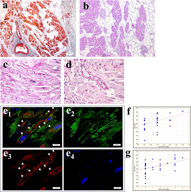
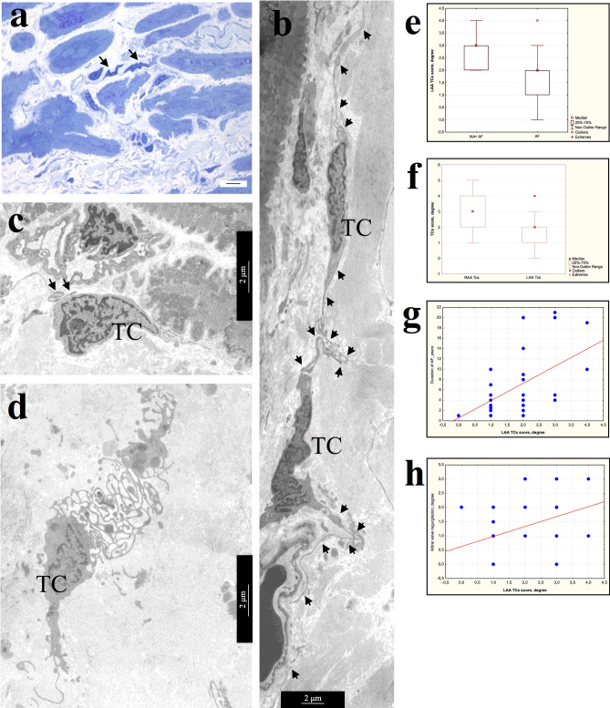
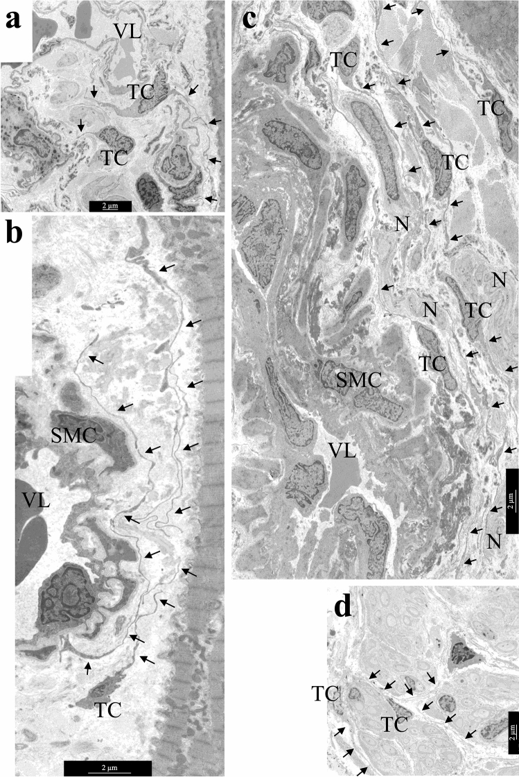
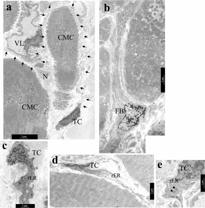
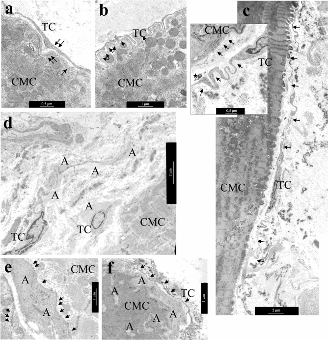
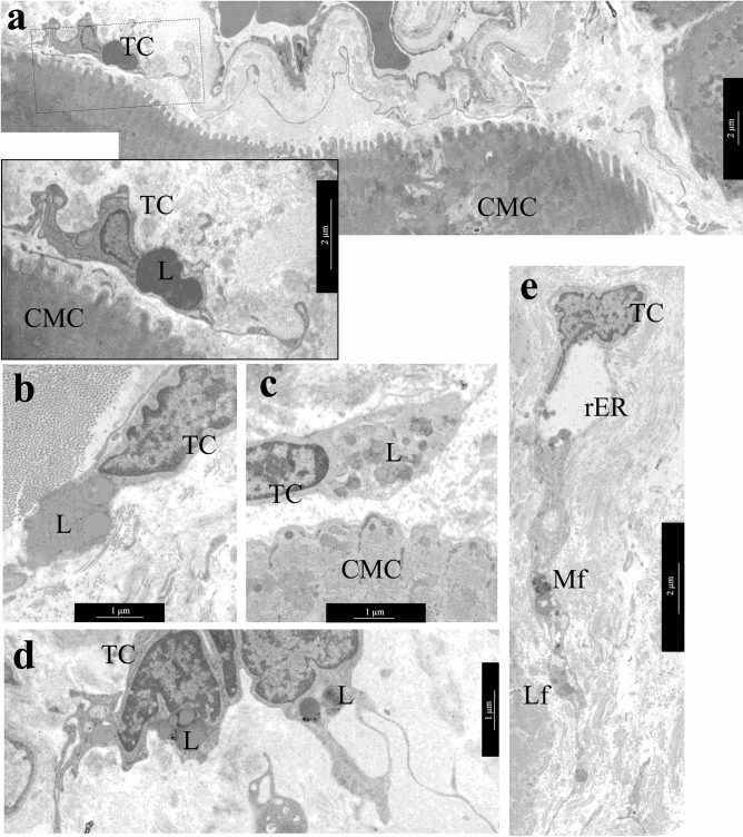
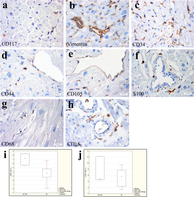
Telocytes are interstitial cells with long, thin processes by which they contact each other and form a network in the interstitium. Myocardial remodeling of adult patients with different forms of atrial fibrillation (AF) occurs with an increase in fibrosis, age-related isolated atrial amyloidosis (IAA), cardiomyocyte hypertrophy and myolysis. This study aimed to determine the ultrastructural and immunohistochemical features of cardiac telocytes in patients with AF and AF + IAA. IAA associated with accumulation of atrial natriuretic factor was detected in 4.3-25% biopsies of left (LAA) and 21.7-41.7% of right (RAA) atrial appendage myocardium. Telocytes were identified at ultrastructural level more often in AF + IAA, than in AF group and correlated with AF duration and mitral valve regurgitation. Telocytes had ultrastructural signs of synthetic, proliferative, and phagocytic activity. Telocytes corresponded to CD117+, vimentin+, CD34+, CD44+, CD68+, CD16+, S100-, CD105- immunophenotype. No significant differences in telocytes morphology and immunophenotype were found in patients with various forms of AF. CD68-positive cells were detected more often in AF + IAA than AF group. We assume that in aged AF + IAA patients remodeling of atrial myocardium provoked transformation of telocytes into "transitional forms" combining the morphological and immunohistochemical features with signs of fibroblast-, histiocyte- and endotheliocyte-like cells.


Conflict of interest statement

The authors declare no competing interests.

Figures

Figure 1
Figure 1
Interstitial area of atrial appendage myocardium of patients with AF. (a,b) Significant fibrosis and lipomatosis in the interstitium of atrial appendage myocardium. (a) Masson’s trichrome stain. (b) Hematoxylin and eosin. bar 7 µm. (c–e) Isolated atrial amyloidosis in atrial appendage myocardium. Amyloid deposition in the interstitial area on the surface of cardiomyocytes (c) and the wall of the intramural vessel (d). (c,d) Sirius red stain. bar 30 µm. e1-4—ANP+ fibrillar material in the interstitium on the surface of cardiomyocytes sarcolemma and in the walls of blood vessels (arrows). ANP+ atrial granules in the sarcoplasm of cardiomyocytes. e1—ANP/Desmin/DAPI immunohistochemistry. e2—Desmin-positive fibrils in the sarcoplasm of cardiomyocytes (Alexa 488). e3—Detection of ANP-positive atrial granules in the sarcoplasm of cardiomyocytes and ANP-positive fibrils in the interstitium (arrows) (Alexa 546). e4—DAPI-stained nuclei. Immunoconfocal laser microscopy. bar 10 µm. (f,g) Correlation of the amyloid distribution in the LAA (f) and RAA (g) myocardium with the age of patients with AF.
Figure 2
Figure 2
Morphology of TCs in atrial appendage myocardium of patients with AF. (a) Comparative morphology of TCs (arrows) and cardiomyocytes. Semi-thin sections. PAS reaction with methylene blue staining. bar 10 µm. (b) Spindle-shaped and star-shaped TCs with 2–3 processes (arrows). (c) Contact of the TC process (arrows) with the “body” of another TC. (b,c) bar 2 µm. (d) TC with numerous branching processes that form a labyrinthic system. b-d Transmission electron microscopy (TEM). (b,c) bar 2 µm. TC—telocyte. (e–h) Semi-quantitative assessment of presence TCs in atrial appendage myocardium. (e) Comparison of TCs presence in LAA myocardium of AF + IAA patients and AF patients (Mann–Whitney test, p < 0.05). (f) The ratio of TCs score in LAA and RAA myocardium (Mann–Whitney test, p < 0.05). (g,h) Positive correlation of TCs score in LAA myocardium with the duration of AF (g, r = 0.50; p = 0.0035), the degree of mitral valve regurgitation (h, r = 0.37; p = 0.033).
Figure 3
Figure 3
The location of TCs in the interstitial area—around large and small vessels, next to cardiomyocytes and nerve fibers. (a) TCs next to capillary. (b) TC near arteriole. (c) TCs in perivascular spacenext to a relatively large vessel. (d) TCs inside the nerve fiber. TCs processes are indicated by arrows. TC—telocyte. Nnerve fibers. SMC—smooth muscle cells. VL—vessel lumen. bar 2 µm.
Figure 4
Figure 4
Ultrastructural features of atrial appendage TCs. (a,b) Comparison of ultrastructural morphology of TCs and fibroblasts of atrial appendage myocardium. (a) Features of TCs morphology: spindle-shaped cells with a small amount of cytoplasm in the perinuclear region, long thin processes (arrows) of TCs surround cardiomyocytes, blood vessels, nerve fibers. (b) Features of fibroblast (FB) morphology: star-shaped cells with a large volume of cytoplasm filled with GER tanks, thick processes of fibroblast in the interstitium. (c) rER tanks, mitochondria, and electron-dense inclusions in the perinuclear zone of TC cytoplasm. (d) In some TCs rER tanks are extended, filled with electron transparent contents. (e) Two centrioles in the cytoplasm of TC (arrows), rER tanks. (a,b,e)—bar 2 µm. (c,d)—bar 1 µm. TEM. TC—telocyte, FB—fibroblast. rER—rough endoplasmic reticulum. CMC—cardiomyocyte. N—nerve fibers. SMC—smooth muscle cells. VL—vessel lumen.
Figure 5
Figure 5
Distance interactions of TCs and cardiomyocytes. (a) The caveolae in the membrane of cardiomyocyte and TC process (arrows). bar 500 nm. (b) The process of TC follows all surface curves and is distributed in the invagination of the T-system of cardiomyocytes. Note the caveolae on the sarcolemma of a cardiomyocyte (arrows). bar 1 µm. (c) TC with extended Tps (arrows) located along the cardiomyocyte surface. bar 2 µm. Insert—Several caveolae on the surface of cardiomyocyte and TC (arrows). A multivesicular body (asterisk) in the interstitium next to the process of the TC. bar 500 nm. (d–f) TCs in areas of amyloidosis. (d) TCs and their processes are surrounded by amyloid fibrils. bar 2 µm. (e) Accumulation of amyloid fibrils on the surface of the cardiomyocyte membrane. Multiple vesicles (arrows) on the inner surface of the sarcolemma of the cardiomyocyte. (f) Amyloid fibrillar deposits on the surface of the cardiomyocyte membrane and in invaginations of the T-system of the cardiomyocyte. The TC process (arrows) at a distance from the cardiomyocyte membrane. (e,f)—bar 1 µm. TEM. TC—telocyte, CMC—cardiomyocyte, A—amyloid fibrils.
Figure 6
Figure 6
Lipid inclusions and dystrophic changes in atrial appendage myocardial TCs in patients with AF. (a) Single lipid inclusions in the perinuclear zone of the TC cytoplasm. bar 1 µm. Insert—bar 2 µm. (b–d) The accumulation of multiple small and large lipid inclusions in the perinuclear zone of the TCs cytoplasm. bar 1 µm. (e) Myelin-like figures (Mf), rough endoplasmic reticulum (rER) and lipofuscin granules (Lf) in the TC cytoplasm. bar 2 µm. TEM. TC—telocyte, CMC—cardiomyocyte. L—lipid inclusions.
Figure 7
Figure 7
Immunohistochemical detection of TCs in atrial appendage myocardium of patients with AF. (a) Single CD117+ cells in the interstitium. (b) Vimentin+ TCs, endotheliocytes, and connective tissue cells. (c) CD34+ endotheliocytes, and TCs surrounding vessels, cardiomyocytes, and endotheliocytes in the interstitium. (d) CD44+ TCs in the interstitium, as well as weakly-labeled endotheliocytes. (e) CD105+ endotheliocytes. (f) S100-positively stained nerve fibers. (g) CD68+ round cells of histiocytic origin and spindle-shaped TCs in the interstitium. (h) CD16+ round cells of histiocytic origin and spindle-shaped TCs in the perivascular area. (a–h) Streptavidin–biotin complexes. bar 15 µm. (i) The number of CD68+ cells in the atrial appendage myocardium of the AF + IAA patients significantly exceeds the number of these cells in the AF group. Mann–Whitney test, p < 0.05. (j) The number of CD16+cells in the atrial appendage myocardium of the AF + IAA patients does not differ significantly from the number of these cells in the AF group. Mann–Whitney test, p > 0.05.

Similar articles

Cited by

References

    1. Popescu LM, et al. Insights into the interstitium of ventricular myocardium: interstitial Cajal-like cells (ICLC) J. Cell. Mol. Med. 2006;10:429–458. doi: 10.1111/j.1582-4934.2006.tb00410.x. - DOI - PMC - PubMed
    1. Hinescu ME, Gherghiceanu M, Mandache E, Ciontea SM, Popescu LM. Interstitial Cajal-like cells (ICLC) in atrial myocardium: ultrastructural and immunohistochemical characterization. J. Cell. Mol. Med. 2006;10:243–257. doi: 10.1111/j.1582-4934.2006.tb00306.x. - DOI - PMC - PubMed
    1. Kostin S. Myocardial telocytes: a specific new cellular entity. J. Cell. Mol. Med. 2010;14:1917–1921. doi: 10.1111/j.1582-4934.2010.01111.x. - DOI - PMC - PubMed
    1. Gherghiceanu M, Manole CG, Popescu LM. Telocytes in endocardium: electron microscope evidence. J. Cell. Mol. Med. 2010;14:2330–2334. doi: 10.1111/j.1582-4934.2010.01133.x. - DOI - PMC - PubMed
    1. Suciu L, Nicolescu MI, Popescu LM. Cardiac telocytes: serial dynamic images in cell culture. J. Cell. Mol. Med. 2010;14:2687–2692. doi: 10.1111/j.1582-4934.2010.01185.x. - DOI - PMC - PubMed

Publication types

MeSH terms