. 2021 Feb 11;11(1):3639.
doi: 10.1038/s41598-021-83058-7.

Human myelomeningocele risk and ultra-rare deleterious variants in genes associated with cilium, WNT-signaling, ECM, cytoskeleton and cell migration

Affiliations

Human myelomeningocele risk and ultra-rare deleterious variants in genes associated with cilium, WNT-signaling, ECM, cytoskeleton and cell migration

K S Au et al. Sci Rep. .

Abstract

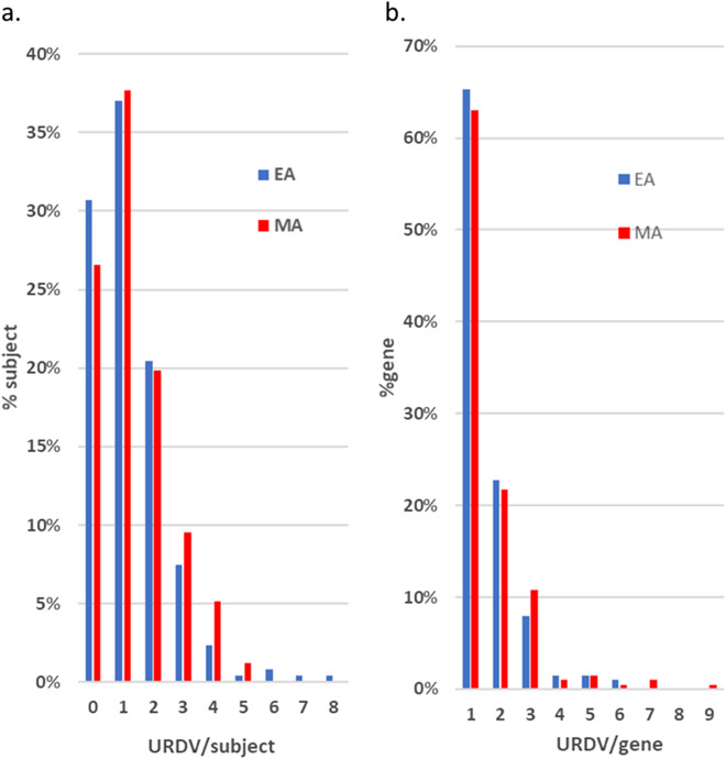
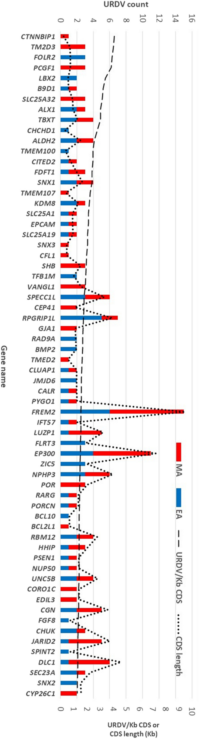
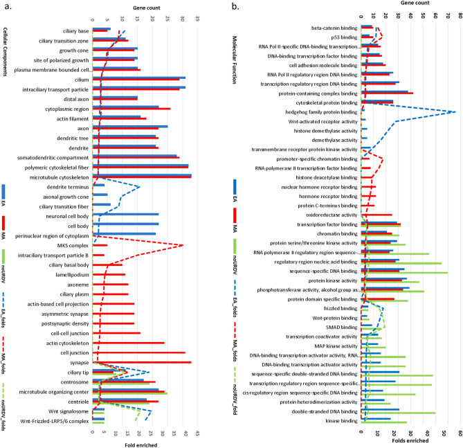
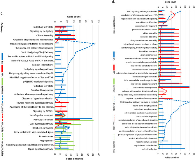
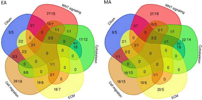
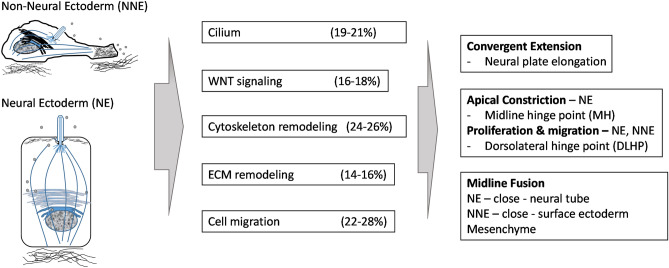
Myelomeningocele (MMC) affects one in 1000 newborns annually worldwide and each surviving child faces tremendous lifetime medical and caregiving burdens. Both genetic and environmental factors contribute to disease risk but the mechanism is unclear. This study examined 506 MMC subjects for ultra-rare deleterious variants (URDVs, absent in gnomAD v2.1.1 controls that have Combined Annotation Dependent Depletion score ≥ 20) in candidate genes either known to cause abnormal neural tube closure in animals or previously associated with human MMC in the current study cohort. Approximately 70% of the study subjects carried one to nine URDVs among 302 candidate genes. Half of the study subjects carried heterozygous URDVs in multiple genes involved in the structure and/or function of cilium, cytoskeleton, extracellular matrix, WNT signaling, and/or cell migration. Another 20% of the study subjects carried heterozygous URDVs in candidate genes associated with gene transcription regulation, folate metabolism, or glucose metabolism. Presence of URDVs in the candidate genes involving these biological function groups may elevate the risk of developing myelomeningocele in the study cohort.


Conflict of interest statement

The authors declare no competing interests.

Figures

Figure 1
Figure 1
Neural tube formation involves a series of biological processes including mechanochemical signal sensing from the environment to orchestrate cell proliferation, cytoskeleton remodeling, and migration of neural and non-neural ectodermal cells. (a) Neural plate cells undergo convergent-extension to facilitate posterior-anterior elongation of the neural tube. Neuroectodermal cells (NE) migrate to the midline in response to extracellular signals received by cilia in NE cells to establish and maintain polarity. (b) Midline NE cells undergo apical constriction via a biological process involving actomyosin cytoskeleton remodeling to form the midline hinge point (MHP). NE cell and non-neuroectodermal (NNE) cells continue proliferation and migration during the neural tube closing process upon sensing signals in the extracellular matrix (ECM). NE cells at the dorsolateral hinge point (DLHP) undergo proliferation, migration dorsally, and reshaping to form the DLHP bringing the NE/NNE edges at the dorsal midline together. Advancing migration and proliferation of NNE and mesenchymal cells facilitate ectodermal layer extension when dorsal midline cells’ protrusions meet and join at the closing point. The process also brings NE cells together to establish cell junctions and close the NT.
Figure 2
Figure 2
Distributions of URDVs Identified by WES. (a) Chart shows count of URDVs in NTD candidate gene(s) identified per exome of MM subjects varied from 0 to 8. In this study, the number of EA MMC subjects is 254 and the number of MA MMC subjects is 252. Approximately 25–30% subjects had no URDVs in NTD candidate genes, around 35% had one URDV, and the remaining had more than one. (b) Shows counts of URDVs discovered per NTD candidate gene ranged from one to nine with nearly 65% genes containing only one. Total number of URDV-containing NTD candidate genes found in EA and MA MMC subjects are 202 and 203 respectively.
Figure 3
Figure 3
URDV density per Kb coding sequence of NTD candidate genes. The majority of 302 NTD candidate genes show URDV/KbCDS density less than one. The x-axis showed the gene names ordered by the URDV/Kb CDS from highest (4.1 for CTNNBP1) to lowest (1.28, CYP26C1). Dashed line (- - - -) showed the URDV/Kb CDS ratio of the gene and dotted line (…….) showed the length of CDS in Kb. Count of URDV in EA showed in blue bar and MA showed in red bar. Approximately half of the 64 genes had URDVs found in both EA and MA. One-third of the 64 genes have ≥ 2 URDVs/Kbp CDS.
Figure 4
Figure 4
Results of ontology and pathway enrichment analysis of candidate genes with and without URDVs identified. Enrichment analysis of (a) cellular components, (b) molecular functions, (c) pathways, and (d) biological processes, were performed using online tools ToppFun and ToppCluster. Count of URDV-containing genes in ontology class for EA (blue) and MA (red) MMC subjects and genes without URDVs (green) were shown with bar charts. Enrichment folds were represented with dashed lines for URDV-containing genes in EA (blue), URDV-containing genes in MA (red) and genes without URDVs (green). Enrichment comparisons passed Bonferroni correction. Detail descriptions of ontology, gene names, fold enrichment, and Bonferroni corrected P values can be found in Supplementary Table 4.
Figure 4
Figure 4
Results of ontology and pathway enrichment analysis of candidate genes with and without URDVs identified. Enrichment analysis of (a) cellular components, (b) molecular functions, (c) pathways, and (d) biological processes, were performed using online tools ToppFun and ToppCluster. Count of URDV-containing genes in ontology class for EA (blue) and MA (red) MMC subjects and genes without URDVs (green) were shown with bar charts. Enrichment folds were represented with dashed lines for URDV-containing genes in EA (blue), URDV-containing genes in MA (red) and genes without URDVs (green). Enrichment comparisons passed Bonferroni correction. Detail descriptions of ontology, gene names, fold enrichment, and Bonferroni corrected P values can be found in Supplementary Table 4.
Figure 5
Figure 5
URDV containing candidate genes involved in multiple ontology groups. The Venn diagrams showed the relation and distribution of UTDV count and NTD candidate genes count to five ontology groups for EA (a) and MA (b). The number of URDVs and number of genes classified to cilium, WNT signaling, cytoskeleton, extracellular matrix (ECM) and cell migration are shown as fraction (count of URDV/count of gene). Genes classified to multiple categories were shown in the overlapped subset compartments between categories. Details on genes within each segment can be found in Supplementary Table 5.
Figure 6
Figure 6
Summary of proportion of study subjects with URDV-containing NTD candidate genes constituting the components of cilium structure and function, WNT-signaling, cytoskeleton remodeling, extracellular matrix (ECM) remodeling and cell migration. URDVs were identified in the DNAs extracted from blood lymphocytes and expected to be present in all body cell types including both neural and non-neural ectodermal cells (NE and NNE respectively). Identifying the URDVs is the first of many steps leading to the understanding of the genetic mechanisms of human MMC development. Cross interaction of genes within and between the five groups is anticipated with many genes assigned to more than one of these groups. Presence of multiple genes with URDVs in one subject provide basis for testing potential gene–gene interaction that may interrupt cell proliferation, or convergent-extension, or apical constriction, or midline fusion or a combination of these processes leading to MMC.

References

    1. Nikolopoulou E, Galea GL, Rolo A, Greene NDE, Copp AJ. Neural tube closure: Cellular, molecular and biomechanical mechanisms. Development. 2017;144:552–566. doi: 10.1242/dev.145904. - DOI - PMC - PubMed
    1. Veland IR, Lindbæk L, Christensen ST. Linking the primary cilium to cell migration in tissue repair and brain development. Bioscience. 2014;64:1115–1125. doi: 10.1093/biosci/biu179. - DOI - PMC - PubMed
    1. Juriloff D, Harris M. Insights into the etiology of mammalian neural tube closure defects from developmental, genetic and evolutionary studies. J. Dev. Biol. 2018;6:22. doi: 10.3390/jdb6030022. - DOI - PMC - PubMed
    1. Ray HJ, Niswander LA. Grainyhead-like 2 downstream targets act to suppress epithelial-to-mesenchymal transition during neural tube closure. Development. 2016;143:1192–1204. doi: 10.1242/dev.129825. - DOI - PMC - PubMed
    1. Gustavsson P, et al. Increased expression of Grainyhead-like-3 rescues spina bifida in a folate-resistant mouse model. Hum. Mol. Genet. 2007;16:2640–2646. doi: 10.1093/hmg/ddm221. - DOI - PubMed

Publication types