. 2021 Feb 11;11(1):3663.
doi: 10.1038/s41598-021-83330-w.

Detection of involved margins in breast specimens with X-ray phase-contrast computed tomography

Affiliations

Detection of involved margins in breast specimens with X-ray phase-contrast computed tomography

Lorenzo Massimi et al. Sci Rep. .

Abstract

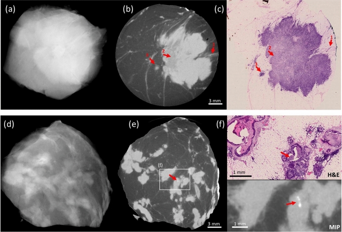
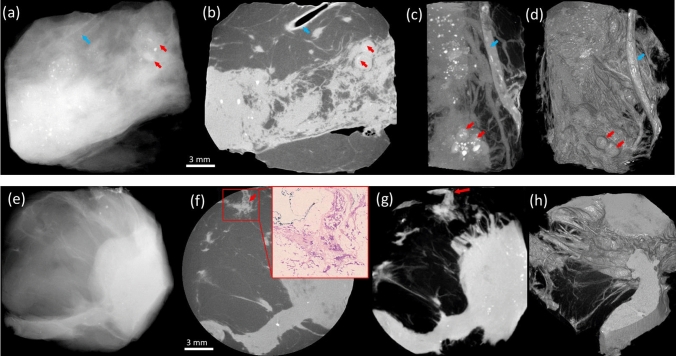
Margins of wide local excisions in breast conserving surgery are tested through histology, which can delay results by days and lead to second operations. Detection of margin involvement intraoperatively would allow the removal of additional tissue during the same intervention. X-ray phase contrast imaging (XPCI) provides soft tissue sensitivity superior to conventional X-rays: we propose its use to detect margin involvement intraoperatively. We have developed a system that can perform phase-based computed tomography (CT) scans in minutes, used it to image 101 specimens approximately half of which contained neoplastic lesions, and compared results against those of a commercial system. Histological analysis was carried out on all specimens and used as the gold standard. XPCI-CT showed higher sensitivity (83%, 95% CI 69-92%) than conventional specimen imaging (32%, 95% CI 20-49%) for detection of lesions at margin, and comparable specificity (83%, 95% CI 70-92% vs 86%, 95% CI 73-93%). Within the limits of this study, in particular that specimens obtained from surplus tissue typically contain small lesions which makes detection more difficult for both methods, we believe it likely that the observed increase in sensitivity will lead to a comparable reduction in the number of re-operations.


Conflict of interest statement

Hawker, Smit, Astolfo, Larkin, Haig and Bate are, or were at the time the research was carried out, Nikon employees. Waltham is a Nikon consultant. Endrizzi, Munro and Olivo are named inventors on patents owned by UCL protecting the technology used to obtain the described results. All other authors have no competing/conflicts of interest.

Figures

Figure 1
Figure 1
Examples of the imaging performance of XPCI-CT (b,e) compared to conventional specimen radiography (a,d) and benchmarked against histopathology (c,f). The top row focuses on the similarity between the XPCI-CT slice in (b) and the histological slice in (c). Arrow 1 indicates margin involvement, arrow 2 a variation in density in the internal structure of the tumour mass, arrow 3 tumour-induced inflammation. All this is confirmed by the histological slice in (c), and hardly visible in the conventional image in (a). The bottom row focuses on the detection of small calcifications, a key feature in DCIS. These are undetectable in (d), detected in (e), enhanced in the maximum intensity projection (MIP) image at the bottom of (f), and confirmed by histopathology in the top part of (f). The scale bar [shown in (b) and (e)] is the same for all images apart from (f), which has its own scale. Red arrows in (e) and (f) indicate the microcalcifications.
Figure 2
Figure 2
Versatility of representation offered by the XPCI-CT approach. The top row focuses on calcified DCIS, also detectable in the conventional image (a). However, the clear delineation of the enlarged ducts in the XPCI slice (b) should be noted (red arrows), which can be followed along their length through successive slices. Alternatively, MIPs can be used (c), or more sophisticated 3D rendering approaches such as solid casting followed by segmentation (e.g. fat tissue was segmented out in panel d, allowing the visualisation of an entire blood vessel and calcifications therein, blue arrow). The bottom row shows an involved margin, less clearly visible in the conventional image (e) and highlighted by an arrow in the XPCI slice (f), where the inset shows confirmation by histopathology, and in the MIP in (g). The 3D rendering in (h) allows visualising edge of the tumour (a metaplastic carcinoma), characterised by diffuse invasion along fibrous strands.
Figure 3
Figure 3
Size distribution of 248 consecutive WLEs examined at London’s Barts Hospital (a, vertical; b, horizontal).
Figure 4
Figure 4
XPCI-CT slices of a 5 cm diameter WLE specimen acquired with an overall scan time of 15 min (a) and 10 min (b). Zoomed-up regions of interest show images on the same scale as Figs. 1 and 2.
Figure 5
Figure 5
The breadboard XPCI-CT system. (a) A photograph of the system, with flat panel detector, the two masks in aluminium frames and the X-ray source (on a separate table) recognisable from left to right. (b) A schematic of the system (seen from above, not to scale), showing how mask M1 splits the beam into multiple beamlets before the sample, and how these hit the edges of an aperture on the detector mask M2. The way in which small deviations of the beamlets lead to a change of detected intensity in some pixels is also schematized.

References

    1. Early and locally advanced breast cancer: diagnosis and management. NICE 2018 Recommendation, NG101 NICE Guidance, UK. https://www.nice.org.uk/guidance/ng101/chapter/Recommendations. Accessed June 2019.
    1. Tang SS-K, et al. Current margin practice and effect on re-excision rates following the publication of the SSO-ASTRO consensus and ABS consensus guidelines: a national prospective study of 2858 women undergoing breast-conserving therapy in the UK and Ireland. Eur. J. Cancer. 2017;84:315–324. doi: 10.1016/j.ejca.2017.07.032. - DOI - PubMed
    1. John ES, et al. Diagnostic accuracy of intraoperative techniques for margin assessment in breast cancer surgery. Ann. Surg. 2017;265:300–310. doi: 10.1097/SLA.0000000000001897. - DOI - PubMed
    1. Pisano ED. Current status of full-field digital mammography. Radiology. 2000;214:26–28. doi: 10.1148/radiology.214.1.r00ja5226. - DOI - PubMed
    1. Kong K, et al. Towards intra-operative diagnosis of tumours during breast conserving surgery by selective-sampling Raman micro-spectroscopy. Phys. Med. Biol. 2014;59:6141–6152. doi: 10.1088/0031-9155/59/20/6141. - DOI - PubMed

Publication types