. 2021 Jan 26:11:539086.
doi: 10.3389/fimmu.2020.539086. eCollection 2020.

Experimental Trypanosoma cruzi Infection Induces Pain in Mice Dependent on Early Spinal Cord Glial Cells and NFκB Activation and Cytokine Production

Affiliations

Experimental Trypanosoma cruzi Infection Induces Pain in Mice Dependent on Early Spinal Cord Glial Cells and NFκB Activation and Cytokine Production

Sergio M Borghi et al. Front Immunol. .

Abstract

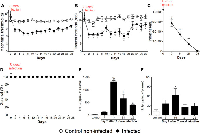
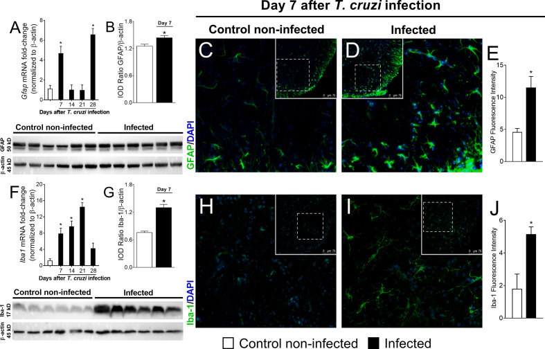
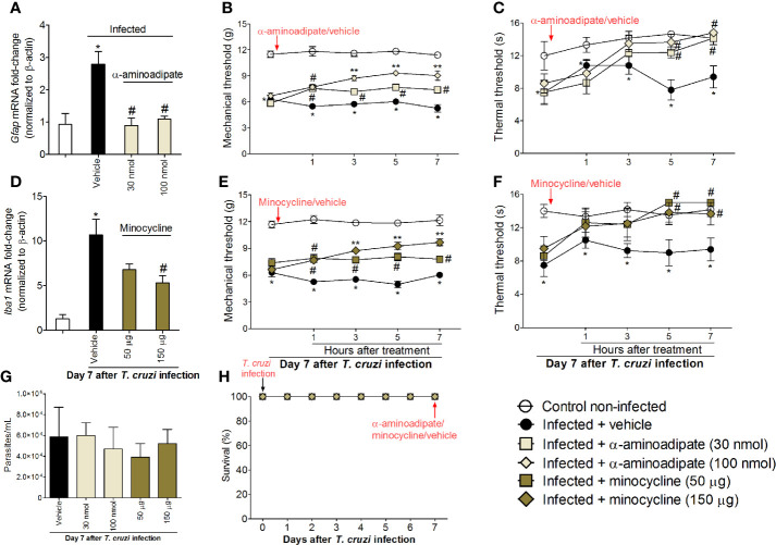
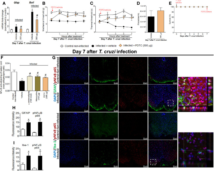
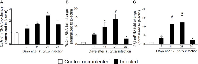
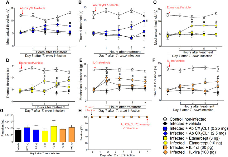
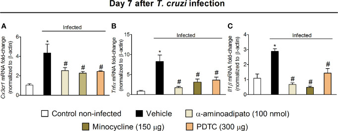
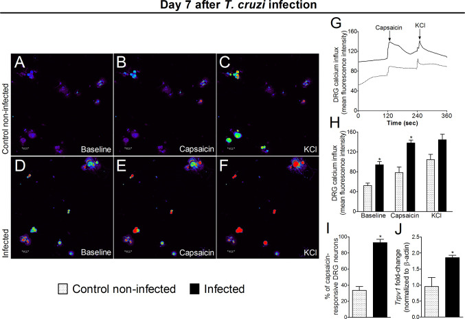
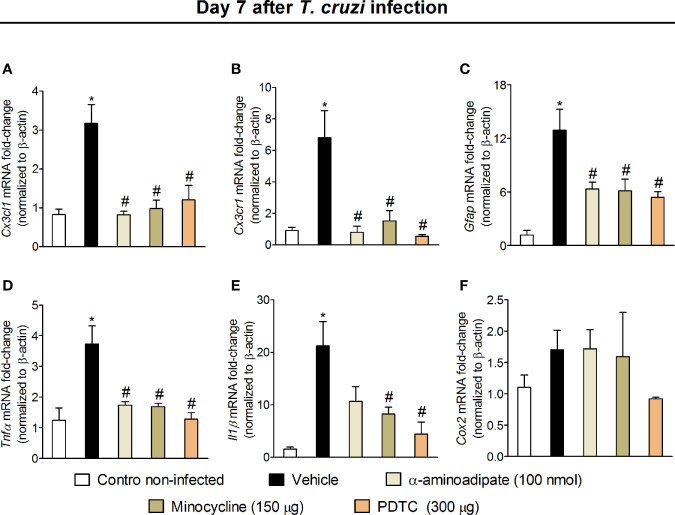
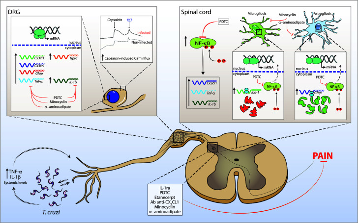
The neglected tropical infirmity Chagas disease (CD) presents high mortality. Its etiological agent T. cruzi is transmitted by infected hematophagous insects. Symptoms of the acute phase of the infection include fever, fatigue, body aches, and headache, making diagnosis difficult as they are present in other illnesses as well. Thus, in endemic areas, individuals with undetermined pain may be considered for CD. Although pain is a characteristic symptom of CD, its cellular and molecular mechanisms are unknown except for demonstration of a role for peripheral TNF-α in CD pain. In this study, we evaluate the role of spinal cord glial cells in experimental T. cruzi infection in the context of pain using C57BL/6 mice. Pain, parasitemia, survival, and glial and neuronal function as well as NFκB activation and cytokine/chemokine production were assessed. T. cruzi infection induced chronic mechanical and thermal hyperalgesia. Systemic TNF-α and IL-1β peaked 14 days postinfection (p.i.). Infected mice presented increased spinal gliosis and NFκB activation compared to uninfected mice at 7 days p.i. Glial and NFκB inhibitors limited T. cruzi-induced pain. Nuclear phosphorylated NFκB was detected surrounded by glia markers, and glial inhibitors reduced its detection. T. cruzi-induced spinal cord production of cytokines/chemokines was also diminished by glial inhibitors. Dorsal root ganglia (DRG) neurons presented increased activity in infected mice, and the production of inflammatory mediators was counteracted by glial/NFκB inhibitors. The present study unveils the contribution of DRG and spinal cord cellular and molecular events leading to pain in T. cruzi infection, contributing to a better understanding of CD pathology.

Keywords: NFκB; Trypanosoma cruzi; cytokine; glial cells; pain.


Conflict of interest statement

The authors declare that the research was conducted in the absence of any commercial or financial relationships that could be construed as a potential conflict of interest.

Figures

Figure 1
Figure 1
Experimental T. cruzi infection induces chronic pain. Mechanical hyperalgesia (A), thermal hyperalgesia (B), blood parasitemia (C), survival (D), and plasma levels of TNF-α (E) and IL-1β (F) were determined. Hyperalgesia tests were evaluated for 28 days p.i. every 2 days. Blood parasitemia and cytokine plasma levels were evaluated 7–28 days p.i. every 7 days. Survival rate was monitored daily over the model. Results are presented as mean ± SEM of six mice per group per experiment and are representative of two separated experiments. *p < 0.05 compared to control noninfected mice; fp < 0.05 compared to days 21 and 28; ωp < 0.05 compared to day 28; Ψ p < 0.05 compared to all groups; ┴ p < 0.05 compared to control noninfected mice and day 7 (two-way ANOVA followed by Tukey’s posttest for panels (A, B); and one-way ANOVA followed by Tukey’s posttest for panels (C, E, F). Dashed lines in panels (E, F) delimits the sensitivity of kits used for analysis.
Figure 2
Figure 2
Experimental T. cruzi infection induces spinal cord astrocytes (A–E) and microglial (F–J) activation. Gfap and Iba1 mRNA expression was determined in control noninfected and infected mice 7–28 days p.i. every 7 days by RT-qPCR (A, F, respectively). At day 7 p.i. (peak of Gfap and Iba1 mRNA expression), Western blot analysis of the spinal cord was performed to confirm GFAP (B) and Iba-1 (G) expression. Next, 7-day spinal cord samples were stained with antibodies for astrocytes (C–E) and microglia (H–J) (GFAP and Iba-1, respectively; green) and regular nucleus (DAPI, blue) detection. Representative immunostainings of the spinal cord of control noninfected and infected mice are shown in panels (C, D, H, I), respectively (20x magnification, scale bar 75 μm with zoom). Panels (E, J) show the percentage of GFAP and Iba-1 fluorescence intensity in each experimental group, respectively. Results are presented as mean ± SEM of six (A, B, F, G) or four mice (E, J) per group per experiment and are representative of two separate experiments. *p < 0.05 compared to control noninfected mice (one-way ANOVA followed by Tukey’s posttest).
Figure 3
Figure 3
Targeting spinal cord glial cells with α-aminoadipate and minocycline intrathecal treatments inhibits T. cruzi–induced hyperalgesia. Panels (A–C) show RT-qPCR data for Gfap mRNA expression and mechanical and thermal hyperalgesia after vehicle and α-aminoadipate treatments (30 and 100 nmol) at the 7th day p.i. Mensuration of hyperalgesia occurred 1–7 h after the treatments. Panels (D–F) show RT-qPCR data for Iba1 mRNA expression and mechanical and thermal hyperalgesia after vehicle and minocycline treatments (50 and 150 μg) at the 7th day p.i. Panel G presents blood parasitemia at the 7th day p.i. 7 h after the treatments. Panel H presents survival rates during the experimental protocol. Results are presented as mean ± SEM of six mice per group per experiment and are representative of two separate experiments. *p < 0.05 compared to control noninfected mice; # p < 0.05 compared to infected mice treated with vehicle; **p < 0.05 compared to infected mice treated with the lowest doses of α-aminoadipate and minocycline (one-way ANOVA followed by Tukey’s posttest for panels A, D; and two-way ANOVA followed by followed by Tukey’s posttest for panels B, C, E, F).
Figure 4
Figure 4
Targeting spinal cord NFκB with PDTC intrathecal treatment inhibits T. cruzi–induced glial activation and hyperalgesia as well as targeting glial cells with α-aminoadipate and minocycline reduced spinal cord NFκB activation. Panels (A–C) show RT-qPCR data for Gfap and Iba1 mRNA expression and mechanical and thermal hyperalgesia after vehicle and PDTC treatments (300 μg) at the 7th day p.i. Panel (D) presents blood parasitemia at the 7th day p.i. 7 h after the treatments. Panel (E) presents survival rates during the experimental protocol. The effects of targeting spinal cord glial cells with α-aminoadipate and minocycline upon NFκB activation at the 7th day p.i. 7 h after the treatments are presented in panel (F). Representative immunostainings of triple immunofluorescence (DAPI/pNFκB p65/GFAP and DAPI/pNFκB p65/Iba-1) of the spinal cord of control noninfected and infected mice are shown in (G) (20x magnification, scale bar 50 μm). 3-D images with zoom demonstrating pNFκB p65 staining surrounded by GFAP or Iba-1 are shown in inserts. Panels H and I demonstrate the fluorescence intensity (%) of GFAP and pNFκB p65, and Iba-1, and pNFκB p65, respectively. Results are presented as mean ± SEM of six (A–F) or four mice (G–I) per group per experiment and are representative of two separate experiments. *p < 0.05 compared to control noninfected mice; # p < 0.05 compared to infected mice treated with vehicle (one-way ANOVA followed by Tukey’s posttest for panels (A, F); and two-way ANOVA followed by followed by Tukey’s posttest for panels B, C).
Figure 5
Figure 5
Experimental T. cruzi infection induces spinal cord Cx3cr1 (A), Tnfα (B), and Il1β (C) mRNA expression. Time-course expressions of mRNA were evaluated 7–28 days p.i. every 7 days. Results are presented as mean ± SEM of six mice per group per experiment and are representative of two separate experiments. *p < 0.05 compared to control noninfected mice; #p < 0.05 compared to infected mice treated with vehicle (one-way ANOVA followed by Tukey’s posttest).
Figure 6
Figure 6
Targeting spinal cord CX3CL1, TNF-α, and IL-1β with neutralizing antibody anti-CX3CL1, etanercept, and IL-1ra intrathecal treatments, respectively, inhibits T. cruzi–induced hyperalgesia. Mechanical hyperalgesia (A, C, E) and thermal hyperalgesia (B, D, F) were evaluated at the 7th day p.i., after vehicle, neutralizing antibody anti-CX3CL1 (0.25 and 2.5 mg), etanercept (3 and 10 ng), and IL-1ra (30 and 300 pg) intrathecal treatments, 1–7 h after the treatments. Panel (G) presents blood parasitemia at the 7th day p.i. 7 h after the treatments. Panel (H) presents survival rates during the experimental protocol. Results are presented as mean ± SEM of six mice per group per experiment and are representative of two separate experiments. *p < 0.05 compared to control non-infected mice; # p < 0.05 compared to infected mice treated with vehicle (two-way ANOVA followed by followed by Tukey’s posttest).
Figure 7
Figure 7
Targeting spinal cord glial cells and NFκB with α-aminoadipate, minocycline, and PDTC intrathecal treatment inhibits T. cruzi–induced spinal cord Cx3cr1 (A), Tnfα (B), and Il1β (C) increased mRNA expression. Evaluations were performed at the 7th day p.i. after vehicle, α-aminoadipate (100 nmol), minocycline (150 μg), and PDTC (300 μg) intrathecal treatments, 7 h after the treatments. Results are presented as mean ± SEM of six mice per group per experiment and are representative of two separate experiments. *p < 0.05 compared to control noninfected mice; # p < 0.05 compared to infected mice treated with vehicle (one-way ANOVA followed by Tukey’s posttest).
Figure 8
Figure 8
Experimental T. cruzi infection induces the activation of DRG neurons. Seven days after the infection, DRGs were dissected for calcium imaging using Fluo‐4AM (AI) and mRNA expression by RT‐qPCR (J). Panels (AF) display representative fields of DRG neurons from control noninfected (A, C) and infected (D–F) mice. Panels (A, D): baseline fluorescence (first column); panels (B, E) fluorescence after capsaicin (second column); and panels (C, F) after KCl control (third column). Panel G displays the mean fluorescence intensity traces of calcium influx from the representative DRG fields (AF) throughout the 6 min of recording. The representative traces show that DRG neurons of infected mice presented higher calcium levels in the baseline than those DRG neurons of control noninfected mice. Panel (H) shows the mean fluorescence intensity of calcium influx of the baseline (0‐s mark) and that following the stimulus, either capsaicin (120‐s mark, TRPV1 agonist) or KCl (240‐s mark, activates all neurons). Panels (I, J) shows the capsaicin-responsive DRG cells and RT‐qPCR data, demonstrating that infected mice present an increased percentage of responsive cells and Trpv1 mRNA expression, respectively. Results are expressed as mean ± SEM; n = 4 DRG plates (each plate is a neuronal culture pooled from six mice) per group per experiment, and RT‐qPCR used n = 6 DRG per group per experiment and are representative of two separate experiments. *p < 0.05 compared to control noninfected mice (one‐way ANOVA followed by Tukey’s posttest).
Figure 9
Figure 9
Effect of intrathecal treatment with α-aminoadipate, minocycline, and PDTC against T. cruzi-induced DRG Cx3cl1 (A), Cx3cr1 (B), Gfap (C), Tnfα (D), Il1β (E), and Cox2 (F) mRNA expression. Evaluations were performed at the 7th day p.i. after vehicle, α-aminoadipate (100 nmol), minocycline (150 μg), and PDTC (300 μg) intrathecal treatments, 7 h after the treatments. Results are presented as mean ± SEM of six mice per group per experiment and are representative of two separate experiments. * p < 0.05 compared to control noninfected mice; # p < 0.05 compared to infected mice treated with vehicle (one-way ANOVA followed by Tukey’s posttest).
Figure 10
Figure 10
Schematic proposition for T. cruzi infection-induced hyperalgesia-related mechanisms in mice. T. cruzi infection induces upregulation of systemic levels of TNF-α and IL-1β (and yet undetermined hyperalgesic mediators). The interface between T. cruzi parasites and pro-inflammatory cytokines may sensitize nociceptor neurons in peripheral tissue initiating nociceptive neurotransmission. Infection activates DRG cells and promotes increase in mRNA expression of Cx3cl1, Cx3cr1, Gfap, Tnf-α, Il-1β, and Trpv1 at this site. In the spinal cord, T. cruzi infection leads to the activation of NFκB and increases mRNA expression of Cx3cr1, Tnf-α, and Il-1β. NFκB activation accounts for the gliosis in the spinal cord. These neuroinflammatory events contribute to central sensitization, resulting in increased pain in infected animals. In DRG, treatments with α-aminoadipate, minocycline, and PDTC inhibit the increased mRNA expression of hyperalgesic molecules with the exception of α-aminoadipate that did not inhibit the increase of Il-1β mRNA expression. In the spinal cord, α-aminoadipate, minocycline, and PDTC inhibit glial- and NFκB-dependent activities as well as increased mRNA expression of hyperalgesic molecules Cx3cr1, Tnf-α, and Il-1β. Confirming the data of DRG and spinal cord hyperalgesic molecule expression, spinal treatments with Ab anti-CX3CL1, etanercept, IL-1ra, α-aminoadipate, minocycline, and PDTC inhibit T. cruzi-induced hyperalgesia in infected animals.

Similar articles

Cited by

References

    1. Dutra WO, Menezes CA, Magalhaes LM, Gollob KJ. Immunoregulatory networks in human Chagas disease. Parasite Immunol (2014) 36(8):377–87. 10.1111/pim.12107 - DOI - PMC - PubMed
    1. Steverding D. The history of Chagas disease. Parasit Vectors (2014) 7:317. 10.1186/1756-3305-7-317 - DOI - PMC - PubMed
    1. da Silva RV, Malvezi AD, Augusto Lda S, Kian D, Tatakihara VL, Yamauchi LM, et al. Oral exposure to Phytomonas serpens attenuates thrombocytopenia and leukopenia during acute infection with Trypanosoma cruzi. PloS One (2013) 8(7):e68299. 10.1371/journal.pone.0068299 - DOI - PMC - PubMed
    1. Teixeira MM, Gazzinelli RT, Silva JS. Chemokines, inflammation and Trypanosoma cruzi infection. Trends Parasitol (2002) 18(6):262–5. 10.1016/s1471-4922(02)02283-3 - DOI - PubMed
    1. Silva JS, Vespa GN, Cardoso MA, Aliberti JC, Cunha FQ. Tumor necrosis factor alpha mediates resistance to Trypanosoma cruzi infection in mice by inducing nitric oxide production in infected gamma interferon-activated macrophages. Infect Immun (1995) 63(12):4862–7. 10.1128/IAI.63.12.4862-4867.1995 - DOI - PMC - PubMed

Publication types

MeSH terms