. 2021 Jan 26:2021:4536132.
doi: 10.1155/2021/4536132. eCollection 2021.

Kiwi Root Extract Inhibits the Development of Endometriosis in Mice by Downregulating Inflammatory Factors

Affiliations

Kiwi Root Extract Inhibits the Development of Endometriosis in Mice by Downregulating Inflammatory Factors

Tingting Liao et al. Evid Based Complement Alternat Med. .

Abstract

Purpose: To determine whether the kiwi root extract inhibits the development of endometriosis in mice by suppressing inflammatory factors.

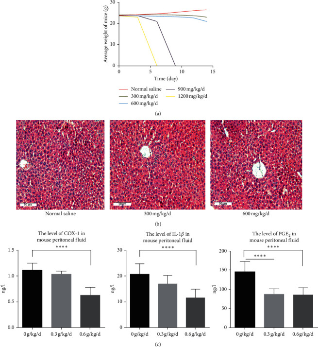
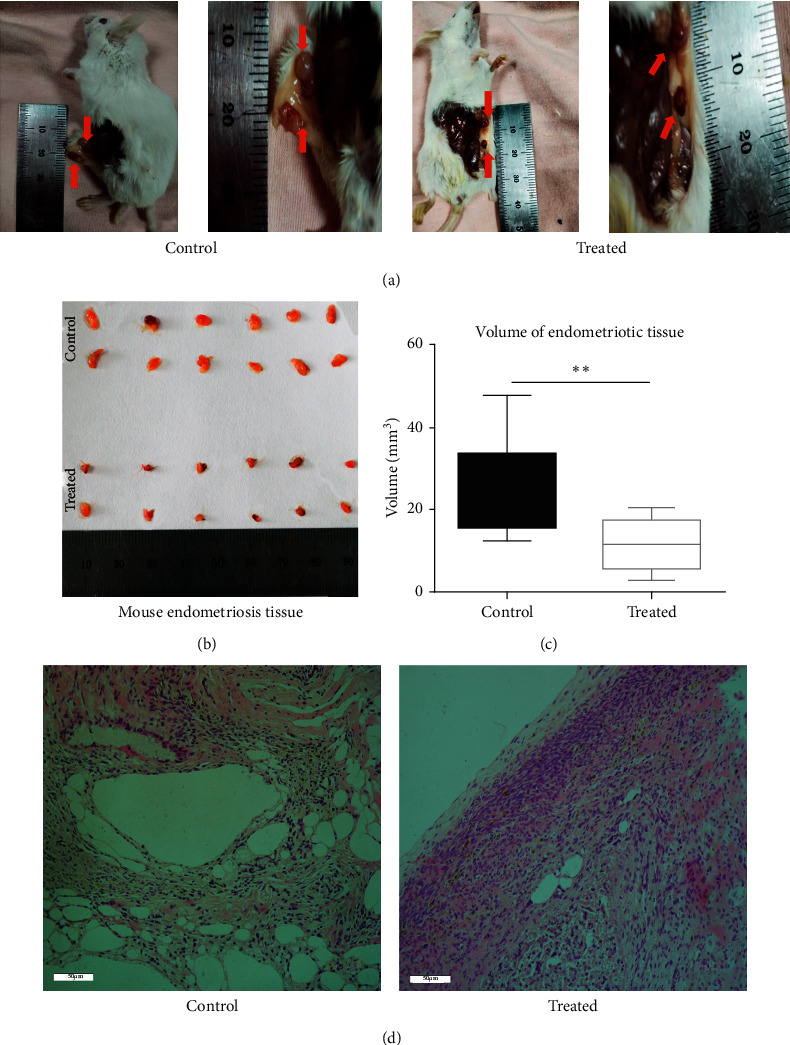
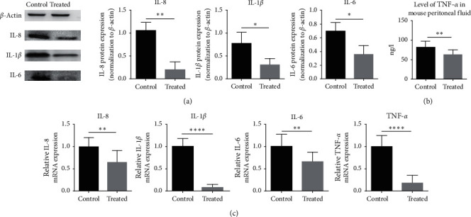
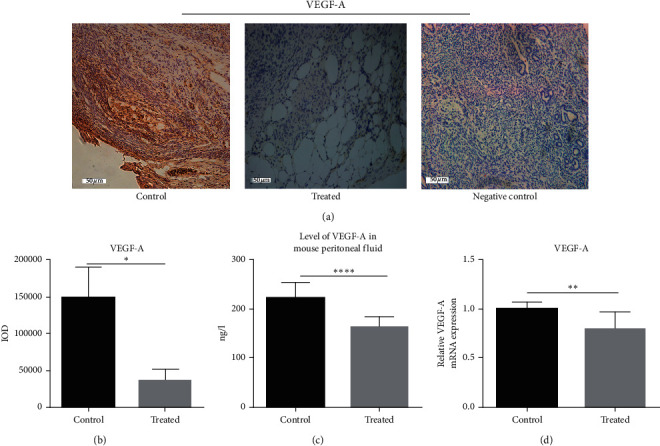
Materials and methods: The mouse model of endometriosis was induced by surgery after which the mice were continuously injected with the drug for 14 days. On the 14th day, the mice were sacrificed, and the peritoneal fluid was obtained for enzyme-linked immunosorbent assay. Endometrial ectopic tissue was weighed and analyzed by tissue immunochemistry, RT-PCR, western blotting, and gelatin zymography experiment.

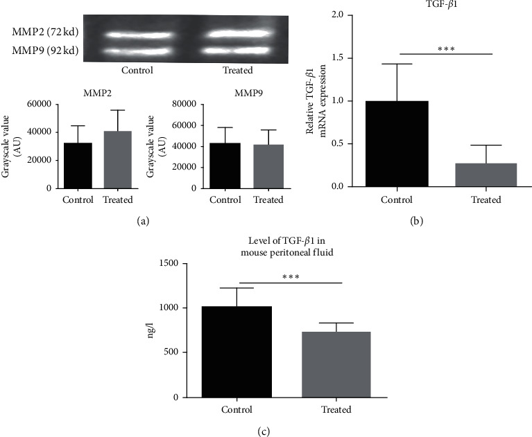
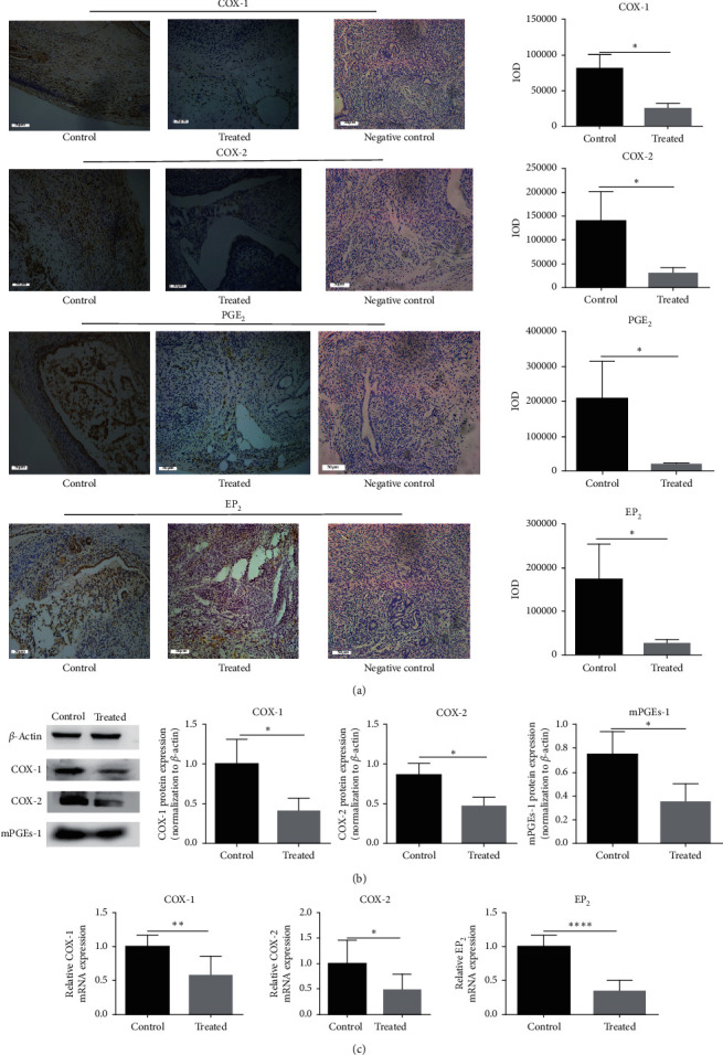
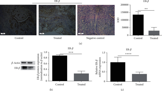
Results: Kiwi root extract significantly reduced endometriotic lesion volume and downregulated the proinflammatory cytokines IL-6, IL-8, IL-1β, and TNF-α, as well as the angiogenic factor VEGF-A. It also inhibited the mRNA and protein expression of COX-1 and COX-2, IL-6, TGF-β1, EP2 receptor, and ER-β in endometriotic lesions but did not affect the expression of MMP-9 and MMP-2.

Conclusions: Kiwi root extract could significantly inhibit the growth of surgery-induced endometriosis in mice. Our results suggest that the kiwi root extract may inhibit the development and progression of ectopic endometrium through disruption of neovascularization and reducing inflammation, which may be beneficial in treating this common gynecological disease.


Conflict of interest statement

The authors declare that they have no conflicts of interest.

Figures

Figure 1
Figure 1
The changes in indicators of each group of mice after treatment with the kiwi root extract. (a) The picture of the average body weight of each group of animals after drug injection over time. (b) Representative liver hematoxylin-eosin staining images of surviving mice. (c) The relative expression level of COX-1, IL-1β, and PGE2 protein in 0 g/kg/d, 0.3 g/kg/d, and 0.6 g/kg/d groups of mice was detected by ELISA.
Figure 2
Figure 2
Treatment with the kiwi root extract reduces the growth of endometriosis. (a) Picture of representative mice in the control group and the treated group. (b) Endometriosis tissues of the control group (control) and treated group (treated) were taken out of mice. (c) Endometriotic tissue volume, as shown in the picture; the endometriosis tissue volume of the control group (24.44 ± 3.219) is higher than the endometriosis tissue volume of the treated group (11.66 ± 1.813) (P = 0.002). (d) Representative hematoxylin-eosin staining images of the mouse endometriosis control group and treated group taken with 20× objective lens.
Figure 3
Figure 3
The expression of IL-6, IL-8, IL-1β, and TNF-α mRNA and protein in the endometriotic tissue of different groups. (a) The relative expression level of IL-6, IL-8, and IL-1β protein was detected by western blot. (b) The relative expression level of TNF-α protein was detected by ELISA. (c) The relative expression level of IL-6, IL-8, IL-1β, and TNF-α mRNA was detected by RT-PCR. Data are expressed as mean ± standard deviation from triplicate experiments (P < 0.05, ∗∗P < 0.01, ∗∗∗P < 0.001, and ∗∗∗∗P < 0.0001).
Figure 4
Figure 4
The expression of VEGF-A mRNA and protein in the endometriotic tissue of different groups. (a) Representative immunohistochemical images of mouse endometriosis control group and treated group taken with 20× objective lens (×200). The scale bar represents 50 microns. (b) The relative expression level of VEGF-A protein was detected by immunohistochemical staining. (c) The relative expression level of VEGF-A protein was detected by ELISA. Data are expressed as mean ± standard deviation from triplicate experiments. (d) The relative expression level of VEGF-A mRNA was detected by RT-PCR (P < 0.05, ∗∗P < 0.01, ∗∗∗P < 0.001, and ∗∗∗∗P < 0.0001).
Figure 5
Figure 5
The expression of TGF-β1, MMP2, and MMP9 mRNA and protein in endometriotic tissue of different groups. (a) The relative expression level of MMP2 and MMP9 protein was detected by gelatin zymography. Data are expressed as mean ± standard deviation from triplicate experiments. (b) The relative expression level of TGF-β1 mRNA was detected by RT-PCR. (c) The relative expression level of TGF-β1 protein was detected by ELISA (P < 0.05, ∗∗P < 0.01, ∗∗∗P < 0.001, and ∗∗∗∗P < 0.0001).
Figure 6
Figure 6
The expression of COX-1, COX-2, and EP2 mRNA and the expression of COX-1, COX-2, mPGEs, EP2, and PGE2 protein in endometriotic tissue of different groups. (a) The relative expression level of COX-1, COX-2, PGE2, and EP2 protein was detected by immunohistochemical staining (×200); the scale bar represents 50 microns. (b) The relative expression level of COX-1, COX-2, and mPGEs protein was detected by western blot. Data are expressed as mean ± standard deviation from triplicate experiments. (c) The relative expression level of COX-1, COX-2, and EP2 mRNA was detected by RT-PCR (P < 0.05, ∗∗P < 0.01, ∗∗∗P < 0.001, and ∗∗∗∗P < 0.0001).
Figure 7
Figure 7
The expression of ER-β mRNA and protein in the endometriotic tissue of different groups. (a) The relative expression level of ER-β protein was detected by immunohistochemical staining (×200); the scale bar represents 50 microns. (b) The relative expression level of ER-β protein was detected by western blot. Data are expressed as mean ± standard deviation from triplicate experiments. (c) The relative expression level of ER-β mRNA was detected by RT-PCR.

Similar articles

References

    1. Joseph S., Mahale S. D. Endometriosis Knowledgebase: A Gene-Based Resource on Endometriosis. Oxford, UK: 2019. - PMC - PubMed
    1. Yu J., Chen L. H., Zhang B., Zheng Q. M. The modulation of endometriosis by lncrna Malat1 via nf-kappab/inos. European Review for Medical and Pharmacological Sciences. 2019;23(10):4073–4080. - PubMed
    1. Harada T., Iwabe T., Terakawa N. Role of cytokines in endometriosis. Fertility and Sterility. 2001;76(1):1–10. doi: 10.1016/s0015-0282(01)01816-7. - DOI - PubMed
    1. Huhtinen K., Ståhle M., Perheentupa A., Poutanen M. Estrogen biosynthesis and signaling in endometriosis. Molecular and Cellular Endocrinology. 2012;358(2):146–154. doi: 10.1016/j.mce.2011.08.022. - DOI - PubMed
    1. Woo J.-H., Yang Y.-I., Ahn J.-H., Choi Y. S., Choi J.-H. Interleukin 6 secretion from alternatively activated macrophages promotes the migration of endometriotic epithelial cells†. Biology of Reproduction. 2017;97(5):660–670. doi: 10.1093/biolre/iox118. - DOI - PubMed

LinkOut - more resources