

Mycoplasma pneumoniae pneumonia with pulmonary embolism: A study on pediatric cases in Jilin province of China
- PMID: 33574906
- PMCID: PMC7818525
- DOI: 10.3892/etm.2021.9634
Mycoplasma pneumoniae pneumonia with pulmonary embolism: A study on pediatric cases in Jilin province of China
Abstract
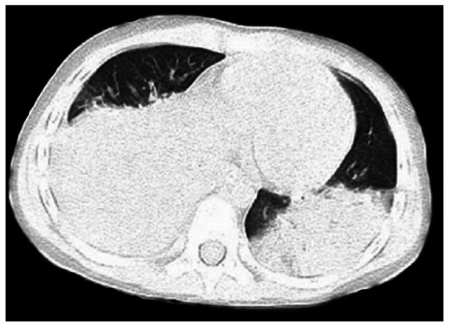
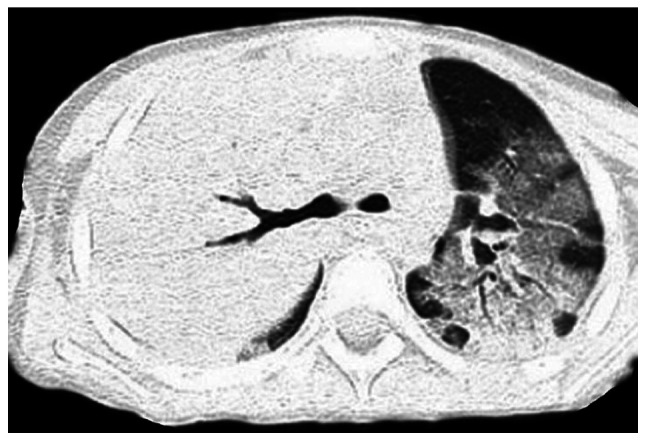
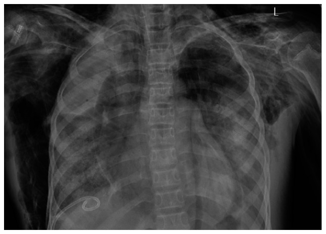
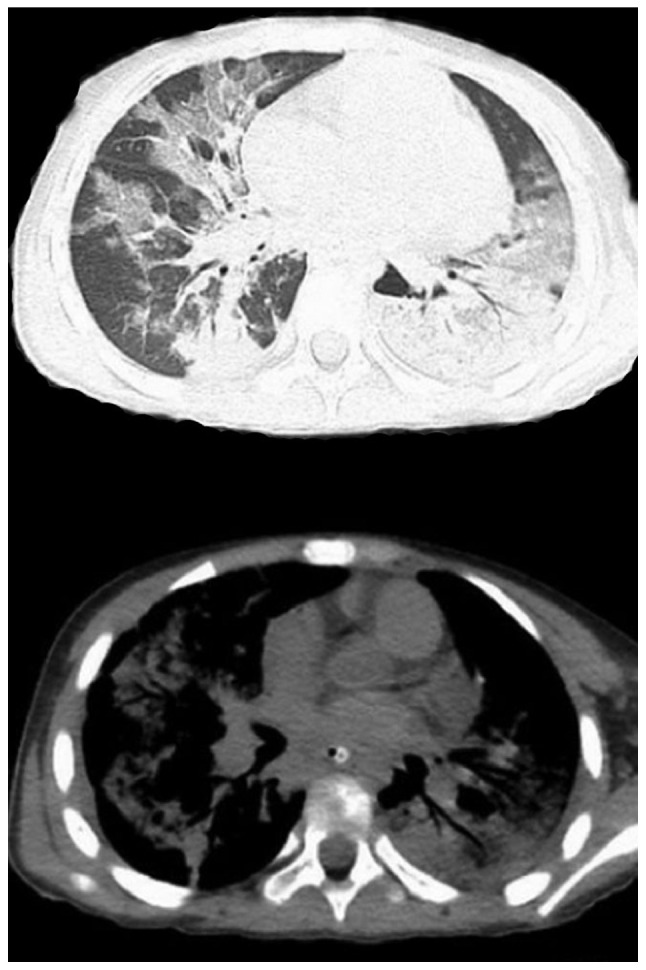
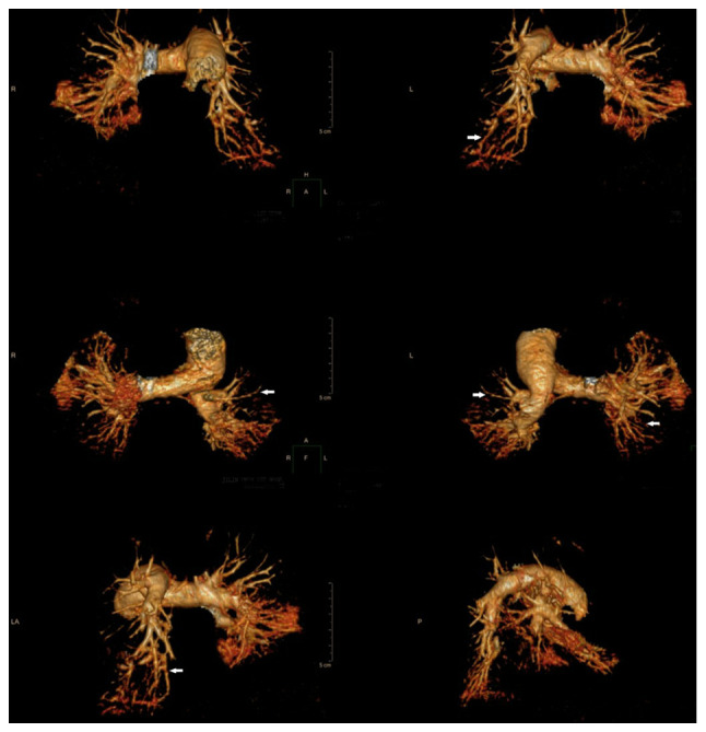
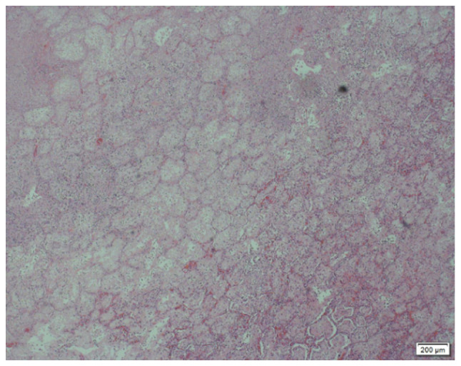
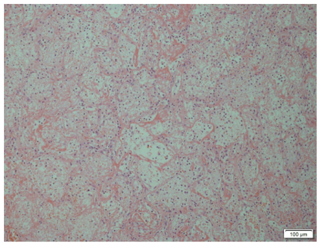
Mycoplasma is one of the most common pathogens causing community-acquired pneumonia in pediatric patients. In recent years, the number of refractory or severe cases with drug resistance has been gradually increasing and cases that developed embolism after Mycoplasma pneumoniae (M. pneumoniae) infection have been reported. The present study retrospectively analyzed the clinical features, diagnosis and treatment of M. pneumoniae pneumonia (MPP) combined with pulmonary embolism (PE) in a series of 7 cases encountered between January 1st, 2016 to August 1st, 2019 at the Department of Pediatric Intensive Care Unit of The First Hospital of Jilin University (Changchun, China). Combined with relevant Chinese and international studies published during the last two decades, a comprehensive analysis was performed. All of the pediatric patients of the present study had fever, cough and dyspnea respiratory symptoms at onset and the disease progressed rapidly. Thereafter, PE was confirmed by a series of examinations. Pulmonary CT indicated patchy inflammations and significantly elevated D-dimer levels, accompanied by positive anticardiolipin antibodies. Furthermore, a filling defect in the pulmonary artery branch was observed on CT pulmonary angiography (CTPA) examination. In 2 cases, the condition was improved with anti-infection and anticoagulation treatment with low-molecular-weight heparin and warfarin, respectively, and the pulmonary embolism disappeared after 3-4 months. A total of 5 cases, who were not responsive to the drug treatment, underwent surgical resection. During the operation, the local tissues were determined to be infarcted and the pathological diagnosis was consistent with pulmonary infarction. Among the 5 cases, 2 died of Acute Respiratory Distress Syndrome at 3-8 days after the operation. The remaining patients underwent 6-12 months of follow-up and respiratory rehabilitation and their quality of life is now good. In conclusion, compared with healthy individuals, pediatric patients with critical MPP have an elevated risk of embolism. It is necessary to be vigilant regarding whether MMP is combined with PE and perform timely CTPA examination. Early detection, early treatment and surgical intervention (if necessary) may significantly reduce the risk of mortality and disability.
Keywords: computed tomographic pulmonary angiography; mycoplasma pneumonia; pediatric patients; pulmonary embolism; refractory mycoplasma pneumonia.
Copyright: © Sheng et al.
Figures
Similar articles
-
Characteristics and Outcomes of Mycoplasma Pneumoniae Pneumonia Associated with Pulmonary Embolism and Necrotizing Pneumonia in Children.Infect Drug Resist. 2024 May 17;17:1961-1969. doi: 10.2147/IDR.S459626. eCollection 2024. Infect Drug Resist. 2024. PMID: 38779350 Free PMC article.
-
A retrospective study of the clinical characteristics of 9 children with pulmonary embolism associated with Mycoplasma pneumoniae pneumonia.BMC Pediatr. 2023 Jul 20;23(1):370. doi: 10.1186/s12887-023-04188-7. BMC Pediatr. 2023. PMID: 37474910 Free PMC article.
-
[Clinical analysis of pulmonary embolism in a child with Mycoplasma pneumoniae pneumonia].Zhonghua Er Ke Za Zhi. 2012 Feb;50(2):151-4. Zhonghua Er Ke Za Zhi. 2012. PMID: 22455642 Chinese.
-
Clinical Features of Severe or Fatal Mycoplasma pneumoniae Pneumonia.Front Microbiol. 2016 Jun 1;7:800. doi: 10.3389/fmicb.2016.00800. eCollection 2016. Front Microbiol. 2016. PMID: 27313568 Free PMC article. Review.
-
Clinical features, risk factors and treatment of fulminant Mycoplasma pneumoniae pneumonia: a review of the Japanese literature.J Infect Chemother. 2014 Mar;20(3):181-5. doi: 10.1016/j.jiac.2013.09.009. Epub 2013 Dec 11. J Infect Chemother. 2014. PMID: 24462437 Review.
Cited by
-
A Rare Case of Severe Hemolytic Anemia and Pulmonary Embolism Secondary to Mycoplasma pneumoniae Infection.J Med Cases. 2022 Mar;13(3):119-124. doi: 10.14740/jmc3866. Epub 2022 Mar 5. J Med Cases. 2022. PMID: 35356396 Free PMC article.
-
Characteristics and Outcomes of Mycoplasma Pneumoniae Pneumonia Associated with Pulmonary Embolism and Necrotizing Pneumonia in Children.Infect Drug Resist. 2024 May 17;17:1961-1969. doi: 10.2147/IDR.S459626. eCollection 2024. Infect Drug Resist. 2024. PMID: 38779350 Free PMC article.
-
Serum expression of ESM-1 and Syndecan-1 and its relationship with disease severity in children with Mycoplasma pneumoniae pneumonia.Ital J Pediatr. 2025 Aug 5;51(1):247. doi: 10.1186/s13052-025-02105-5. Ital J Pediatr. 2025. PMID: 40764585 Free PMC article.
-
Case Report: Refractory Mycoplasma pneumoniae pneumonia complicated by pulmonary embolism and infarction in a child.Front Pediatr. 2025 Jun 23;13:1608233. doi: 10.3389/fped.2025.1608233. eCollection 2025. Front Pediatr. 2025. PMID: 40625386 Free PMC article.
-
Risk factors for delayed radiographic resolution in children with refractory Mycoplasma pneumoniae pneumonia.J Int Med Res. 2021 May;49(5):3000605211015579. doi: 10.1177/03000605211015579. J Int Med Res. 2021. PMID: 34034536 Free PMC article.
References
-
- Tashiro M, Fushimi K, Kawano K, Takazono T, Saijo T, Yamamoto K, Kurihara S, Imamura Y, Miyazaki T, Yanagihara K, et al. Comparison of efficacy of antimicrobial agents among hospitalized patients with Mycoplasma pneumoniae pneumonia in Japan during large epidemics of macrolide-resistant M. pneumoniae infections: A nationwide observational study. Clin Infect Dis. 2017;65:1837–1842. doi: 10.1093/cid/cix695. - DOI - PubMed
LinkOut - more resources
Full Text Sources
Other Literature Sources