

Euryops arabicus Promotes Healing of Excised Wounds in Rat Skin: Emphasis on Its Collagen-Enhancing, Antioxidant, and Anti-Inflammatory Activities
- PMID: 33574987
- PMCID: PMC7857909
- DOI: 10.1155/2021/8891445
Euryops arabicus Promotes Healing of Excised Wounds in Rat Skin: Emphasis on Its Collagen-Enhancing, Antioxidant, and Anti-Inflammatory Activities
Retraction in
-
Retracted: Euryops arabicus Promotes Healing of Excised Wounds in Rat Skin: Emphasis on Its Collagen-Enhancing, Antioxidant, and Anti-Inflammatory Activities.Oxid Med Cell Longev. 2023 Oct 4;2023:9840217. doi: 10.1155/2023/9840217. eCollection 2023. Oxid Med Cell Longev. 2023. PMID: 37830037 Free PMC article.
Abstract
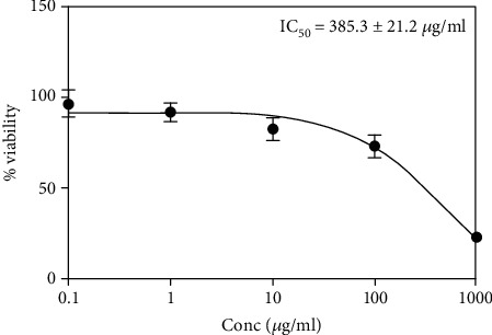
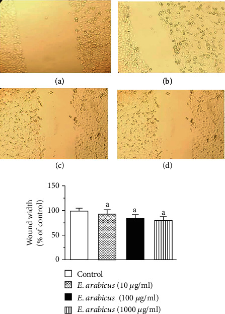
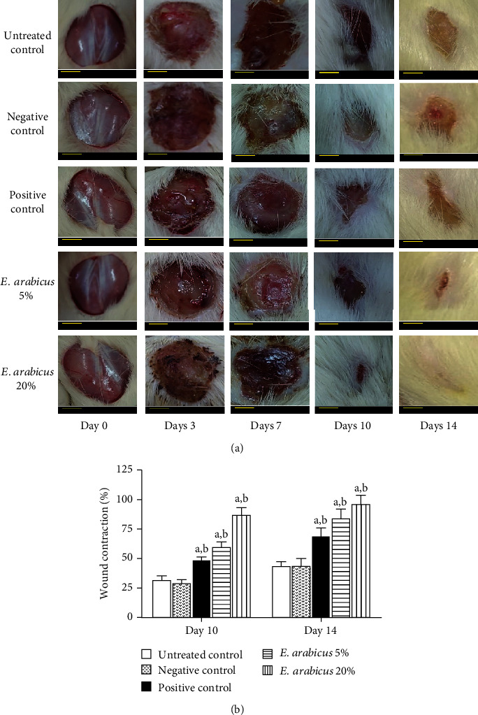
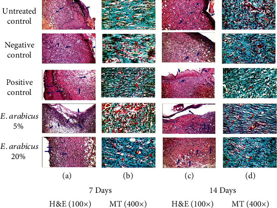
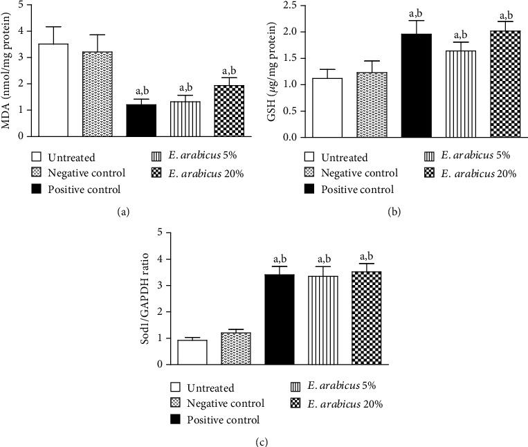
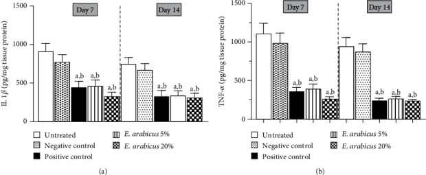
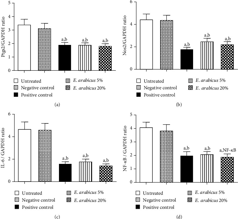
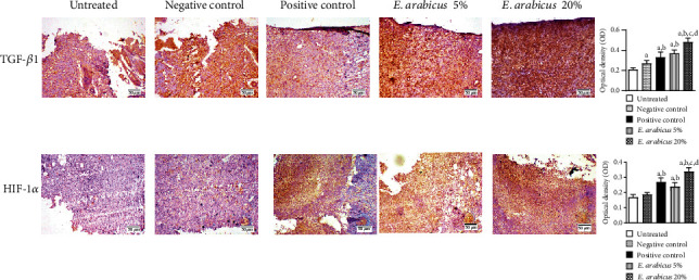
Euryops arabicus Steud (E. arabicus) belongs to the family Asteraceae. It has several uses in folk medicine in the Arabian Peninsula. The current study aimed at evaluating the wound healing properties of the E. arabicus extract in rats. Primarily, E. arabicus successfully accelerated cell migration in vitro and it also showed no signs of dermal toxicity. Topical application of E. arabicus extract (5% or 20%) expedited healing of excised skin in rats. Histological examinations indicated that E. arabicus shortened epithelization period, stimulated fibroblast activity, and increased collagen deposition in wound tissues. The plant extract exerted antioxidant activity as evidenced by inhibition of GSH depletion and MDA accumulation and enhanced mRNA expression of Sod1 in wound tissues collected at the end of the experiment. Further, E. arabicus inhibited the rise of TNF-α and IL-1β in the skin wound region. The anti-inflammatory was confirmed by the observed down regulation of Ptgs2, Nos2, IL-6, and NF-κB mRNA expression. In addition, the extract enhanced the expression of TGF-β1 and HIF-1α in wounded skin tissues as indicated immunohistochemically. Conclusively, E. arabicus expedites excision wound healing in rats. Collagen-enhancing, anti-inflammatory, and antioxidant properties mediate the observed wound healing activity. These findings might contribute to our understanding of the ethnobotanical use of E. arabicus in wounds.
Copyright © 2021 Ahmed Abdel-Lateff et al.
Conflict of interest statement
The authors have no conflict of interest to declare.
Figures
References
-
- MacDonald J. Global initiative for wound and lymphoedema care (GIWLC) Journal of Lymphoedema. 2009;4:92–95.
Publication types
MeSH terms
Substances
LinkOut - more resources
Full Text Sources
Other Literature Sources
Medical
Research Materials
Miscellaneous
