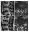
Magnetic resonance imaging findings of redundant nerve roots of the cauda equina
- PMID: 33574992
- PMCID: PMC7852347
- DOI: 10.4329/wjr.v13.i1.29
Magnetic resonance imaging findings of redundant nerve roots of the cauda equina
Abstract
Background: Redundant nerve roots (RNRs) of the cauda equina are often a natural evolutionary part of lumbar spinal canal stenosis secondary to degenerative processes characterized by elongated, enlarged, and tortuous nerve roots in the superior and/or inferior of the stenotic segment. Although magnetic resonance imaging (MRI) findings have been defined more frequently in recent years, this condition has been relatively under-recognized in radiological practice. In this study, lumbar MRI findings of RNRs of the cauda equina were evaluated in spinal stenosis patients.
Aim: To evaluate RNRs of the cauda equina in spinal stenosis patients.
Methods: One-hundred and thirty-one patients who underwent lumbar MRI and were found to have spinal stenosis between March 2010 and February 2019 were included in the study. On axial T2-weighted images (T2WI), the cross-sectional area (CSA) of the dural sac was measured at L2-3, L3-4, L4-5, and L5-S1 levels in the axial plane. CSA levels below 100 mm2 were considered stenosis. Elongation, expansion, and tortuosity in cauda equina fibers in the superior and/or inferior of the stenotic segment were evaluated as RNRs. The patients were divided into two groups: Those with RNRs and those without RNRs. The CSA cut-off value resulting in RNRs of cauda equina was calculated. Relative length (RL) of RNRs was calculated by dividing the length of RNRs at mid-sagittal T2WI by the height of the vertebral body superior to the stenosis level. The associations of CSA leading to RNRs with RL, disc herniation type, and spondylolisthesis were evaluated.
Results: Fifty-five patients (42%) with spinal stenosis had RNRs of the cauda equina. The average CSA was 40.99 ± 12.76 mm2 in patients with RNRs of the cauda equina and 66.83 ± 19.32 mm2 in patients without RNRs. A significant difference was found between the two groups for CSA values (P < 0.001). Using a cut-off value of 55.22 mm2 for RNRs of the cauda equina, sensitivity, specificity, positive predictive value (PPV), and negative predictive value (NPV) values of 96.4%, 96.1%, 89.4%, and 98.7% were obtained, respectively. RL was 3.39 ± 1.31 (range: 0.93-6.01). When the extension of RNRs into the superior and/or inferior of the spinal canal stenosis level was evaluated, it was superior in 54.5%, both superior and inferior in 32.8%, and inferior in 12.7%. At stenosis levels leading to RNRs of the cauda equina, 29 disc herniations with soft margins and 26 with sharp margins were detected. Disc herniation type and spondylolisthesis had no significant relationship with RL or CSA of the dural sac with stenotic levels (P > 0.05). As the CSA of the dural sac decreased, the incidence of RNRs observed at the superior of the stenosis level increased (P < 0.001).
Conclusion: RNRs of the cauda equina are frequently observed in patients with spinal stenosis. When the CSA of the dural sac is < 55 mm2, lumbar MRIs should be carefully examined for this condition.
Keywords: Cauda equina; Dural sac; Lumbar spine; Magnetic resonance imaging; Redundant nerve roots; Spinal stenosis.
©The Author(s) 2021. Published by Baishideng Publishing Group Inc. All rights reserved.
Conflict of interest statement
Conflict-of-interest statement: All authors declare no conflicts-of-interest related to this article.
Figures


Similar articles
-
[Study on related influencing factors on the occurrence of redundant sign in the cauda equina in lumbar spinal stenosis].Zhongguo Gu Shang. 2024 Aug 25;37(8):824-7. doi: 10.12200/j.issn.1003-0034.20201408. Zhongguo Gu Shang. 2024. PMID: 39183009 Chinese.
-
Redundant nerve roots of the cauda equina in lumbar spinal canal stenosis, an MR study on 500 cases.Eur Spine J. 2015 Oct;24(10):2315-20. doi: 10.1007/s00586-015-4059-y. Epub 2015 Jun 14. Eur Spine J. 2015. PMID: 26071946
-
Congenital lumbar spinal stenosis: a prospective, control-matched, cohort radiographic analysis.Spine J. 2005 Nov-Dec;5(6):615-22. doi: 10.1016/j.spinee.2005.05.385. Spine J. 2005. PMID: 16291100 Clinical Trial.
-
The clinical significance of redundant nerve roots of the cauda equina in lumbar spinal stenosis patients: A systematic literature review and meta-analysis.Clin Neurol Neurosurg. 2018 Nov;174:40-47. doi: 10.1016/j.clineuro.2018.09.001. Epub 2018 Sep 4. Clin Neurol Neurosurg. 2018. PMID: 30205275
-
Nursing Review Section of Surgical Neurology International Part 2: Lumbar Spinal Stenosis.Surg Neurol Int. 2017 Jul 7;8:139. doi: 10.4103/sni.sni_150_17. eCollection 2017. Surg Neurol Int. 2017. PMID: 28781916 Free PMC article. Review.
Cited by
-
Redundant nerve roots indicate higher degree of stenosis in lumbar spine stenotic patients.Acta Neurol Belg. 2023 Oct;123(5):1781-1787. doi: 10.1007/s13760-022-02040-w. Epub 2022 Aug 7. Acta Neurol Belg. 2023. PMID: 35934759
-
Clinical Features and Efficacy Analysis of Redundant Nerve Roots.Front Surg. 2021 Nov 1;8:628928. doi: 10.3389/fsurg.2021.628928. eCollection 2021. Front Surg. 2021. PMID: 34790693 Free PMC article.
-
The prevalence of redundant nerve roots in standing positional MRI decreases by half in supine and almost to zero in flexed seated position: a retrospective cross-sectional cohort study.Neuroradiology. 2022 Nov;64(11):2191-2201. doi: 10.1007/s00234-022-03047-z. Epub 2022 Sep 9. Neuroradiology. 2022. PMID: 36083504 Free PMC article.
-
MRI parameters predict central lumbar spinal stenosis combined with redundant nerve roots: a prospective MRI study.Front Neurol. 2024 May 27;15:1385770. doi: 10.3389/fneur.2024.1385770. eCollection 2024. Front Neurol. 2024. PMID: 38859971 Free PMC article.
References
-
- Cressman MR, Pawl RP. Serpentine myelographic defect caused by a redundant nerve root. Case report. J Neurosurg. 1968;28:391–393. - PubMed
-
- Nogueira-Barbosa MH, Savarese LG, Herrero CFPS, Defino HLA. Redundant nerve roots of the cauda equina: review of the literature. Radiol Bras. 2012;45:155–159.
-
- Hakan T, Celikoğlu E, Aydoseli A, Demir K. The redundant nerve root syndrome of the Cauda equina. Turk Neurosurg. 2008;18:204–206. - PubMed
-
- Savarese LG, Ferreira-Neto GD, Herrero CF, Defino HL, Nogueira-Barbosa MH. Cauda equina redundant nerve roots are associated to the degree of spinal stenosis and to spondylolisthesis. Arq Neuropsiquiatr. 2014;72:782–787. - PubMed
-
- Ono A, Suetsuna F, Irie T, Yokoyama T, Numasawa T, Wada K, Toh S. Clinical significance of the redundant nerve roots of the cauda equina documented on magnetic resonance imaging. J Neurosurg Spine. 2007;7:27–32. - PubMed
LinkOut - more resources
Full Text Sources
Research Materials

